
Tricuspid valve disorders are common although more often of functional nature. Organic tricuspid valve disease can be either similar to that which affects the mitral valve or unique to it. Tricuspid valve disorders are mostly congenital and rheumatic although carcinoid, anorexi- genic valvulopathy, infective endocarditis, myxomatous degeneration also afflict this valve. Functional tricuspid valve disorders secondary to long standing pacing leads in the right ventricle are subject of recent interest. Functional disorders are usually secondary to left-sided disease and help in assessment of pulmonary arterial and the right ventricular pressures. Both functional and organic disorders have impact on prognosis that can be altered by appropriate management. The most common cause of tricuspid regurgitation is not a primary disease but rather an impaired valve coaptation caused by a dilation of the right ventricle and/or of the tricuspid annulus. The tricuspid valve is a complex structure that, unlike the aortic and mitral valve, is not possible to visualize in one cross-sectional view using either transthoracic or transesophageal two-dimensional echocardiography (2DE; i.e. imaging all three leaflets and their attachment in the annulus simultaneously). Conversely, threedimensional echocardiography (3DE) allows users to visualize the whole tricuspid valve apparatus from any perspective and is currently gaining prominence.
FUNCTIONAL MORPHOLOGY OF THE TRICUSPID VALVE
The tricuspid valve complex consists of (Figs 7.1 to 7.4):
• Three leaflets (anterior, posterior and septal)
• The chordae tendinae
• Two or three discrete papillary muscles (large variation)
• Fibrous tricuspid annulus
• Right atrial and right ventricular myocardium Successful valve function depends on the integrity and
coordination of these components.1-3
The anterior leaflet is the largest, whereas the posterior leaflet is notable for the presence of multiple scallops. The septal leaflet is the smallest and arises medially directly from the tricuspid annulus above the interventricular septum. The anterior papillary muscle provides chordae to the anterior and posterior leaflets, and the medial papillary muscle provides chordae to the posterior and septal leaflets.3
The septal wall gives chordae to the anterior and septal leaflets (there is no formal septal papillary muscle as with the anterior and posterior (medial) papillary muscles). In addition, there may be accessory chordal attachments to the right ventricular free wall and to the moderator band. These multiple chordal attachments are important mediators of tricuspid valve regurgitation (TR), as they impair proper leaflet coaptation in the setting of right ventricular dysfunction and dilation (Fig. 7.5).

Fig. 7.1 : Schematic diagram showing tricuspid valve apparatus laid open longitudinally.

Fig. 7.2 : Schema showing en face view of the tricuspid annulus with leaflets and posterior relationships.

Fig. 7.3 : 2D echocardiographic four-chamber view (left panel) compared with anatomical specimen (right panel). (AL: Anterior leaflet; SL: Septal leaflet; MS: Membranous interventricular septum; AML: Anterior mitral leaflet).
The septal aspect of the tricuspid annulus is considered to be analogous to the intertrigonal portion of the mitral annulus in that it is relatively spared from annular dilation. Because of this property, tricuspid annular sizing
algorithms have been based on the dimension of the base of the septal leaflet.
Tricuspid valve is the most apically placed valve with the largest orifice. Position of septal attachment of the

Fig. 7.4 : 3D echocardiographic en face view (left panel) compared with anatomical specimen (right panel) seen from above. (MV: Mitral valve; PL: Posterior leaflet; IVC: Inferior vena cava; SVC: Superior vena cava; RAA: Right atrial appendage).

Fig. 7.5 : Aberrant chords (right panel) contrasted with normal arrangement (left panel).

Fig. 7.6 : Attachment of the septal (anterior) mitral leaflet is superior (white arrow) to that of tricuspid septal leaflet (yellow arrow).
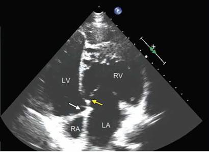
Fig. 7.7 : Atrioventricular discordance shown in four-chamber view. The right ventricle is identified by the position of the septal leaflet of the tricuspid valve as well as by the moderator band.
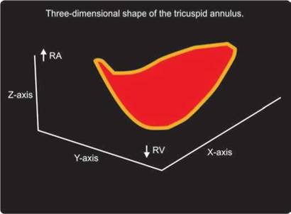
Fig. 7.8 : 3D shape of the tricuspid annulus which is elliptical and saddle-shaped.
valve compared to that (anterior mitral leaflet) on the left side helps in identification of the morphological right ventricle (Figs 7.6 and 7.7). The longitudinal height of this difference is about 10 mm.
The tricuspid annulus shows a nonplanar structure with an elliptical saddle-shaped pattern having two high points superiorly and two low points oriented inferiorly that is best seen in midsystole.4 The normal TV annulus is saddle shaped, with the highest points located in an anterior-posterior orientation and the lowest points in a medio-lateral orientation (Fig. 7.8).
The tricuspid annulus shape is not circular but oval both in normal-sized and in dilated annulus. Currently used tricuspid annulus diameters measured with 2DE (both measured in apical four-chamber view and in
parasternal short-axis view) systematically underestimate the actual tricuspid annulus size. Role of annular dilatation centers around the right ventricular (RV) free wall portion of the annulus as the primary lesion.
CONDITIONS AFFECTING TRICUSPID VALVE
Functional Tricuspid Regurgitation (FTR)
• Idiopathic FTR
• Hypertensive FTR
• Diastolic TR
Organic TR and TS (Anatomically abnormal valves)
• Rheumatic
• Infective endocarditis
• Ebstein's anomaly
• Floppy (prolapse)
• Congenital (non-Ebstein's)
• Carcinoid
• Trauma
• Connective tissue disorder (Marfan's)
• Rheumatoid arthritis
• Radiation therapy
The Carpentier's classification remains the most commonly used functional classification:5
Type I: leaflet perforation (infective endocarditis) or more frequently by annular dilatation
Type II: prolapse of one or more leaflets (tricuspid valve prolapse)
Type III: restricted motion as the consequence of rheumatic disease, significant calcifications, toxic valvulopathy, FTR.
REMODELLING IN TRICUSPID REGURGITATION
TR leads to RA and RV dilatation, a dilated and pulsatile inferior vena cava and hepatic vein, a dilated coronary sinus and systolic bowing of the interatrial septum toward the LA. Absence of these changes suggests milder degree of TR. Imaging the vena cava and its respiratory variation also provides an evaluation of RA pressure. Although not specific, a rapid anterior motion of the interventricular septum at the onset of systole (paradoxical ventricular septal motion) is a sign of RV volume overload due to severe TR.
Annulus diameter measured in apical four-chamber view (> 2.1 cm/M2) can both be the cause or effect of TR and also the threshold for intervention.6
One of the commonest observations during echocardiographic examinations is the presence of FTR. Nearly 50-60% of young adults exhibit mild TR and its prevalence increases with age. Up to 15% of normal people have moderate TR. Usually, substantial redundancy of leaflet tissue prevents TR in normal tricuspid valves (ratio leaflets/annular length > 1.45; Fig. 7.9). Tricuspid leaflets length (septal + anterior leaflets) can be easily measured and ratio to systolic annulus diameter is calculated to assess valvular coverage of annulus in systole.
FTR is characterized by structurally normal leaflets and is due to the deformation of the valvulo-ventricular
complex.6 While mild FTR is frequent and usually benign, patients with severe FTR may develop progressive ventricular dysfunction and incur increased mortality.
FTR is dependent on annular dilatation, with significant TR occurring with > 40% dilatation. A central zone of leaflet malcoaptation is consistent with annular dilatation demonstrating the dependence on the 3 leaflets coapting simultaneously (Fig. 7.10).
FTR often appears in conjunction with left-sided valve disease and left ventricular (LV) dysfunction despite the presence of a structurally normal tricuspid valve. ttis is usually associated with elevated right ventricular systolic pressure.
Idiopathic FTR (due to ageing, atrial fibrillation etc) is characterized by (Fig. 7.11):
• Dilated annulus
• Incomplete valve coverage
• Conical right ventricular shape with basal dilatation only
• No leaflet tethering
• Decreased RV systolic function
Hypertensive FTR is characterized by (Figs 7.12 and 7.13):
• Annular dilatation
• Spherical or elliptical RV dilatation
• Leaflet tethering
• Apparently normal RV systolic function due to volume overload.
Hypertensive FTR is predominantly due to valve deformation with tenting and only modest annular enlargement. Valvular tenting and leaflet tethering are linked to RV elongation and deformation (Fig. 7.13).
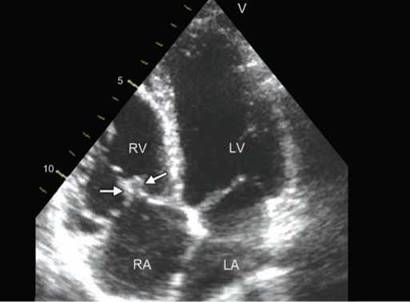
Fig. 7.9: Apical four-chamber view showing coaptation of the tricuspid valve (arrows). Note striking coaptation distance due to redundancy of the leaflets.
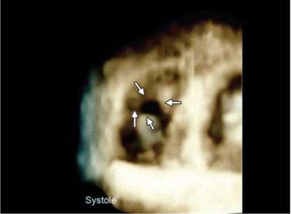
Fig. 7.10: Dilated annulus causing central malcoaptation responsible for tricuspid valve regurgitation (TR) (arrows). A 3DE en face view in systole.
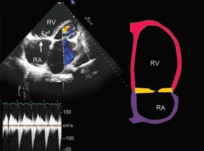
Fig. 7.11: Idiopathic functional tricuspid regurgitaion characterized by incomplete coaptation without leaflet tethering (2DE picture on left side and the schematic diagram on the right side).
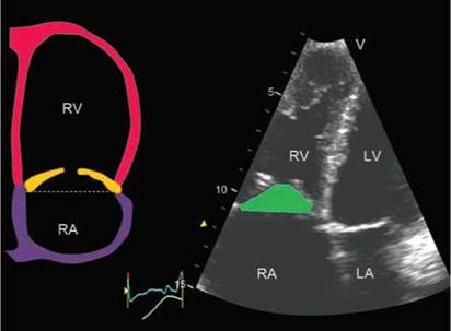
Fig. 7.12: Hypertensive functional tricuspid regurgitaion is characterized by leaflet tethering (left panel) with definite tenting height and area (right panel).

Fig. 7.13: Hypertensive functional tricuspid regurgitation in mitral stenosis (MS) showing symmetrical leaflet tethering and tenting height of 12 mm.
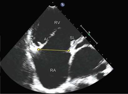
Fig. 7.14: Apical four-chamber view showing dilated annulus with elongated anterior leaflet as a compensatory mechanism.
With FTR, the annulus becomes larger, more planar and circular. Anterior leaflet is responsible for compensating for an increase in orifice area as a result of annular dilatation.7 Patients with annular dilatation but no TR have longer leaflets, specifically the anterior leaflet (Fig. 7.14).
Diastolic TR can occur in certain conditions, e.g., heart block, atrial flutter, severe pulmonary regurgitation, and restrictive cardiomyopathy, due to reversal of pressure gradient between RA and RV during diastole (Figs 7.15 and 7.16).
ORGANIC TRICUSPID VALVE DISORDERS
Tricuspid valve is examined in different views to obtain complete anatomic information. Parasternal long-axis view of the RV inflow is obtained by tilting the probe inferomedially and rotating it slightly clockwise from the parasternal long-axis view of the LV. This incidence reveals the anterior tricuspid leaflet (near the aortic valve) and the posterior tricuspid leaflet. Parasternal short-axis view, at the level of the aortic valve, apical four-chamber view and subcostal four-chamber view visualize the septal and the anterior tricuspid leaflets (Fig. 7.17).

Fig. 7.15: Diastolic TR (arrows) along with systolic TR in presence of first degree AV block.
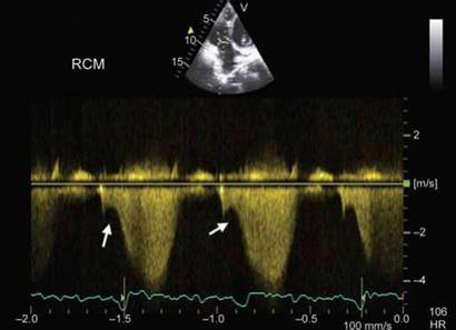
Fig. 7.16: Diastolic TR (arrows) along with systolic TR in restrictive cardiomyopathy and atrial fibrillation.

Fig. 7.17: Various views used to study the tricuspid valve.

Fig. 7.18: Apical four-chamber view showing rheumatic tricuspid and mitral stenosis (left panel) and tricuspid regurgitation (left panel). Thickening of both leaflets with doming is obvious.
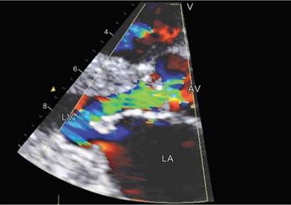
Fig. 7.19: Para-sternal long axis view in diastole showing doming of the mitral valve with narrowed orifice. There is thickening of the aortic valve with significant aortic regurgitation.
Most patients with rheumatic tricuspid valve disease present with TR or a combination of stenosis and regurgitation.8 Rheumatic TS does not occur as an isolated lesion, but it often accompanies mitral and aortic valve disease (Figs 7.18 and 7.19).
Rheumatic involvement of the TV is less common (25%) than that of left-sided valves. Regurgitation is a consequence of deformity, shortening and retraction of one or more leaflets of the TV as well as shortening and fusion of the chordae tendinae and papillary muscles.9 the 2DE usually detects thickening and the distortion of the leaflets but cannot provide a comprehensive assessment of extension of valve apparatus involvement (Fig. 7.20).
the full-volume data set from apical approach usually provides a comprehensive assessment of the whole TV apparatus, which can be examined from different

Fig. 7.20: Modified RV inflow view showing tethered and thickened leaflets causing free flow TR (right panel).

Fig. 7.21: 3DE en face view from the ventricular aspect in diastole. Tricuspid leaflets are thickened and commissures are patent; the orifice is wide open compared to severe mitral stenosis with narrow orifice and thick leaflets.
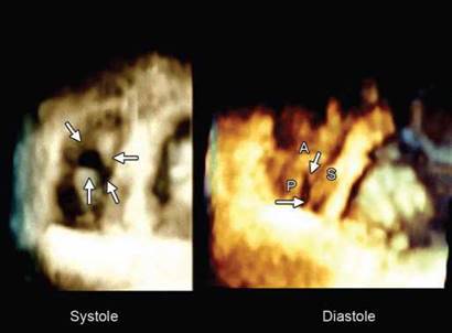
Fig. 7.22: 3DE en face view in systole showing regurgitant orifice and in diastole showing fusion of all three commissures with narrowed tricuspid diastolic orifice.

Fig. 7.23: Nodular thickening of the mitral valve leaflets as well as the septal leaflet of the tricuspid valve. A cleft is seen in the anterior tricuspid leaflet. Arrows point to the three commissures.
perspectives.10 the “en face” view of the TV obtained by RT-3DE allows the visualization of the commissural fusion, which is helpful to establish a correct diagnosis of rheumatic aetiology of TV regurgitation (Figs 7.21 to 7.23).
the 3D TEE produces an en face view of all the three valve leaflet/cusps simultaneously. ttis allows more detailed morphological assessment of each individual leaflet/cusp as well as coaptation between leaflets. Involvement of subvalvular apparatus is identified and better characterized. ttere is improved delineation of the
spatial relationship between valve, subvalvular apparatus and the endocardium of surrounding chambers (Fig. 7.23).
In the carcinoid disease, the valve appears thickened, fibrotic with markedly restricted motion during cardiac cycle11 (Fig. 7.24). the RT-3DE can show the regions of ineffective leaflet coaptation and the lack of commissural fusion.
Tricuspid valve infective endocarditis is rare and accounts for 5-10% of infective endocarditis.12 Its predispositions are congenital heart disease, insertion of
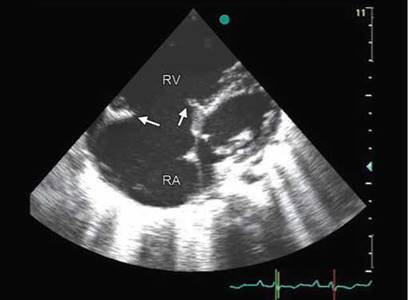
Fig. 7.24: Thickened and shortened tricuspid valve leaflets in Carcinoid heart.

Fig. 7.25: Vegetation on the tricuspid valve (arrow) in presence of central intravenous line.

Fig. 7.26: Large vegetations on ventricular surface of the tricuspid leaflets (arrow) in a patient on hemodialysis.

Fig. 7.27: Apical four-chamber view showing flail anterior leaflet (white arrow) with severe TR (right panel).
a central venous catheter, placement of a pacemaker or implantable defibrillator, a history of intravenous drug use and hemodialysis (Figs 7.25 and 7.26).
Pulmonary embolism occurs in 75-100% cases of tricuspid valve endocarditis.
Degenerative Tricuspid Valve Disease
Three types of tricuspid changes can be visualized:
1. Billowing valve
2. Prolapsing valve
3. Flail tricuspid valve
Tricuspid prolapse is generally associated with mitral valve prolapse and is defined as a midsystole posterior leaflet displacement beyond the annular plane. The coaptation line is behind the annular plane. Tricuspid prolapse most often involves the septal and anterior tricuspid leaflets. The most common phenotype of tricuspid prolapse is diffuse myxomatous degeneration (Barlow's disease). A flail tricuspid leaflet is observed when the free edge of a leaflet is completely reversed in the RA (Fig. 7.27), usually as a consequence of ruptured chordae (degenerative TR, infective endocarditis, trauma).

Fig. 7.28: Thickened mitral and tricuspid valve leaflets with normal thickness of chords in a case with degenerative MR.

Fig. 7.29: Apical four-chamber view showing large anterior leaflet and apically displaced septal leaflet of the tricuspid valve.

Fig. 7.30: Ebstein anomaly. Apical four-chamber view showing normal insertion of the anterior leaflet along the free wall and rudimentary displaced septal leaflet along the interventricular septum.

Fig. 7.31: Modified long axis view of the right heart structures showing displacement of posterior (along free wall) and the septal leaflet resulting in an atrialized chamber.
Fibroelastic degeneration also occurs in tricuspid valve, albeit much less common as the chamber pressures are low. However, it has morphological features and hemodynamic consequences similar to that seen in mitral valve (Fig. 7.28).
Ebstein anomaly is a congenital heart defect in which the septal leaflet of the tricuspid valve is displaced toward the apex of the right ventricle of the heart. the annulus of the valve is still in the normal position.13 However, the valve leaflets are, to a varying degree, attached to the walls and septum of the right ventricle. There is subsequent
“atrialization” of a portion of the morphologic right ventricle (which is then contiguous with the right atrium). This causes the right atrium to be large and the anatomic right ventricle to be small in size (Fig. 7.29).
Typically, there are anatomic abnormalities of the tricuspid valve, with enlargement of the anterior leaflet of the valve. the other leaflets are described as being plastered to the endocardium (Figs 7.30 and 7.31).
About 50% of individuals with Ebstein anomaly have an associated shunt between the right and left atria, either an atrial septal defect or a patent foramen ovale.

Fig. 7.32: Rudimentary tricuspid valve leaflets (arrows) with normal annular attachment and grossly incomplete coverage of the orifice.
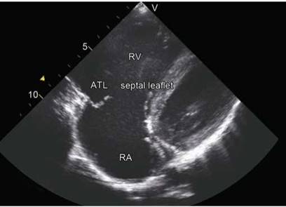
Fig. 7.33: Normally attached but thin and flail tricuspid valve leaflet causing unguarded orifice.

Fig. 7.34: Extreme form of tricuspid valve dysplasia showing markedly dilated annulus with hardly any leaflet tissue.

Fig. 7.35: Parasternal four-chamber view showing aneurysmally dilated right atrium, thrombus (arrow), spontaneous contrast and hardly any valvular tissue.
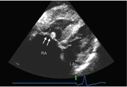
Fig. 7.36: Apical four-chamber view showing congenital tricuspid stenosis (arrows). The mitral valve is normal and presence of right ventricular hypertrophy is due to associated pulmonary stenosis.
Ebstein anomaly typically has TR as the hemodynamic lesion. However, rarely combined tricuspid regurgitation with tricuspid stenosis can also occur.
Non-Ebstein anomalies of the tricuspid valve due to tricuspid valve dysplasia are not rare.14,15 ttese are associated with TR and varying degree of right ventricular dysfunction and atrial fibrillation subsequently (Figs 7.32 to 7.35).
tricuspid valve stenosis
Tricuspid valve stenosis (TS) is an uncommon echocar- diographic and pathological lesion and is rarely diagnosed clinically. The common etiologies are:
• Rheumatic (99% of all)
• Congenital (Figs 7.36 to 7.38)

Fig. 7.37: Congenital parachute tricuspid valve causing TS.

Fig. 7.38: Congenital TS with hypoplastic tricuspid annulus. Concavity of the interatrial septum suggests raised right atrial pressure.
Fig. 7.39: 3DE en face view showing narrowed orifice of the tricuspid valve (on left side with) commissural fusion.
• Carcinoid
• Infective endocarditis
• Right atrial myxoma
• Anorexic drug abuse
• Postsurgical (restrictive annuloplasty)
Trans-tricuspid valve gradients are indicative of TS although there is no validated cutoff to define TS. Morphological features with reduced orifice, doming of the leaflets and commissural fusion usually suggest presence of TS, which are better appreciated by real-time 3D echocardiography (Fig. 7.39).

assessment of severity of tr
Color flow jet of TR can be used to estimate rough severity of TR like that in mitral regurgitation.16 However, the jet area is dependent upon several hemodynamic factors besides the machine settings. Hence it is not recommended to grade severity of TR based upon the jet area in the right atrium.17 However, if the jet is small and close to the leaflets, it is likely to be mild TR and if the color jet fills most of the right atrium and swirls, it is likely to be severe (Figs 7.40 and 7.41)
vena contracta
Vena contracta (VC) or the narrowest jet width just beyond the regurgitant orifice is a good index of severity of TR and is usually measured in apical four-chamber view. A VC width of > 7 mm suggests severe TR while < 6 mm is indicative of mild or moderate TR.18 Mild and moderate cannot be distinguished by width of VC (Fig. 7.42). Geometry of the regurgitant orifice is complex and 2D VC does not have good correlation with 3DE regurgitant orifice area. Proposed 3DE new criteria for estimating TR severity based on VC area or 3DE color Doppler regurgitant orifice are: < 0.5 cm2 for mild;
0.5-0.75 cm2 for moderate and > 0.75 cm2 for severe.19

Fig. 7.40: Color flow jet area of TR filling nearly 50% of the right atrium but no swirling although vena contracta is wide (13 mm) suggestive of severe TR.

Fig. 7.41: Large TR color Doppler jet area occupying > 2/3 of the right atrium. However in view of narrow vena contracta (4 mm) and small radius of PISA, it is unlikely to be severe.

Fig. 7.42: Apical four-chamber view showing TR color flow jet. Although VC is 10 mm (suggestive of severe TR). Effective regurgitant orifice area by PISA method is only 7 mm2 indicative of mild TR.
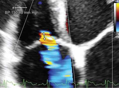
Fig. 7.43: PISA radius of 5 mm at aliasing velocity of 28.9 cm/sec should suggest mild TR although the vena contracta width is 7 mm.
proximal isovelocity surface area method
Proximal isovelocity surface area (PISA) method is rarely used in clinical practice and it is not sure if the regurgitant jet proximal to the orifice actually makes hemi-spheric shells in case of the tricuspid valve:
• It is usually performed in the four-chamber view.
• The area of interest is optimized by lowering imaging depth and the Nyquist limit to ~15-40 cm/s.
• Radius of the PISA is measured at midsystole using the first aliasing (Fig. 7.43).
• Qualitatively, a TR PISA radius > 9 mm at a Nyquist limit of 28 cm/s alerts to the presence of significant TR, whereas a radius of < 5 mm suggests mild TR17
• An effective regurgitant orifice area (ERO) of 3 40 mm2 or a regurgitant volume of з 45 mL indicates severe TR.
• There is a good chance of underestimation of the TR severity by this method (Figs 7.42 and 7.43).
anterograde velocity of tricuspid inflow
The severity of TR will affect the early tricuspid diastolic filling. In the absence of tricuspid stenosis, the peak

Fig. 7.44: Peak early diastolic tricuspid valve velocity of 200 cm/s (right panel) in a patient with no TS (left panel), suggesting severe TR.

Fig. 7.45: Normal hepatic vein flow pattern.
Fig. 7.46: Marked blunting of antegrade systolic waves (arrows) in hepatic vein in TR. Blunting lacks specificity.

Fig. 7.47: Dense CW Doppler envelope of severe TR.
E velocity increases in proportion to the degree of TR. Tricuspid inflow Doppler tracings are obtained at the tricuspid leaflet tips. A peak E velocity > 1 m/s suggests severe TR (Fig. 7.44).
hepatic vein flow in assessment of tr
In normal individuals, the pattern of flow velocity in hepatic veins consists of antegrade systolic (S), transient flow reversal as the TV annulus recoils at the end of systole, antegrade diastolic (D) and a retrograde A wave (AR) caused by atrial contraction (Fig. 7.45).
With increasing severity of TR, there is a decrease in hepatic vein systolic velocity. In severe TR, systolic flow

blunting or reversal occurs (Fig. 7.46). the sensitivity of flow reversal for severe TR is 80%. ttus the absence of systolic flow reversal does not rule out severe TR. Blunted systolic hepatic vein flow can be observed in case of abnormal right atrial and RV compliance, atrial fibrillation and elevated right atrial pressure from any cause. Blunting of hepatic flow may thus lack specificity.17
Continuous Wave Doppler Interrogation for TR Signal
• Density of the continuous wave (CW) envelope of the TR jet can be a guide to TR severity.
• A dense TR signal with a full envelope indicates more severe TR than a faint signal (Fig. 7.47).
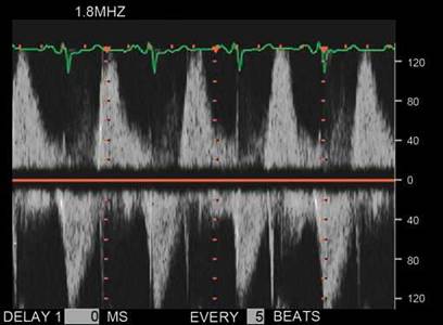
Fig. 7.48: Severe TR with triangular shape of the CW spectrum in systole due to elevated right atrial pressure. Early diastolic velocity of 120 cm/s and dense envelope suggest severe TR.
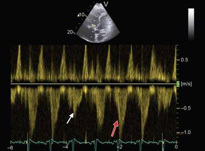
Fig. 7.49: Respiratory variation of TR signal with decrease in velocity during expiration (white arrow) compared to inspiration (red arrow).
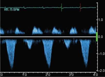
Fig. 7.50: CW Doppler signal alternans of TR.
• The CW Doppler envelope may be truncated (notch) with a triangular contour and an early peak velocity (blunt). This indicates elevated right atrial pressure or a prominent regurgitant pressure wave in the RA due to severe TR (Fig. 7.48).
Marked respiratory variation (decreased TR velocity with inspiration) suggests an elevated RA pressure and indirectly severe TR (Fig. 7.49).
Massive TR is often associated with a low jet velocity (< 2 m/s) as there is near equalization of RV and RA pressures (Figs 7.48 and 7.49).
The CW Doppler TR signal alternans is usually associated with severe TR (Fig. 7.50).
summary
Tricuspid valve regurgitation is likely to be mild when:
• No RA/RV dilatation and no malcoaptation are seen on 2DE
• There is small or narrow central color Doppler jet
• There is no annular dilatation
• Width of VC is < 7 mm
• PISA radius is < 5 mm at a Nyquist limit of 28 cm/sec
• There is normal hepatic vein flow during systole
• Tricuspid early diastolic velocity is < 100 cm/s
• There is a faint or incomplete CW Doppler envelope Tricuspid valve regurgitation is likely to be severe if,
in absence of other right-sided pathology, following signs
are present:
• Dilated right heart structures with paradoxically moving interventricular septum
• Large color Doppler jet area occupying > 50% of the right atrium with swirling of the jet and entry into the coronary sinus/superior vena cava and so forth
• Dense CW Doppler spectral envelope with triangular shape
• The CW velocity is < 200 cm/s with augmented respiratory variation
• Tricuspid flow E wave velocity is > 100 cm/s without evidence of obstruction
• Width of vena contracta is > 7 mm
• PISA radius is > 9 mm at Nyquist limit of 28 cm/s
• Regurgitant volume by PISA is > 45 mL
• Hepatic vein systolic wave flow reversal
• Obviously large patent orifice in systole with no coaptation.
The more of the above criteria are met, the more is the likelihood of accuracy. However, criteria 6-8 should be considered specific and others as supportive. From a therapeutic viewpoint, morphology of the tricuspid valve and geometry of the annulus are equally important.
references
1. Silver MD, Lam JH, Ranganathan N, et al. Morphology of the human tricuspid valve. Circulation. 1971;43:333-48.
2. Ho SY, Nihoyannopoulos P. Anatomy, echocardiography, and normal right ventricular dimensions. Heart. 2006;92 (Suppl. 1):i2-i13.
3. Rogers JH, Bolling SF. the tricuspid valve: current perspective and evolving management of tricuspid regurgitation. Circulation. 2009;119:2718-25.
4. Fukuda S, Saracino G, Matsumura Y, et al. ttree-dimen- sional geometry of the tricuspid annulus in healthy subjects and in patients with functional tricuspid regurgitation: a real-time, 3-dimensional echocardiographic study. Circulation. 2006;114 (Suppl):I-492-I-8.
5. Carpentier A, Deloche A, Hanania G, et al. Surgical management of acquired tricuspid valve disease. J ttorac Cardiovasc Surg. 1974;67:53-65.
6. Dreyfus GD, Corbi PJ, Chan KM, et al. Secondary tricuspid regurgitation or dilatation: which should be the criteria for surgical repair? Ann ttorac Surg. 2005;79: 127-32.
7. Ton-Nu TT, Levine RA, Handschumacher MD, et al. Geometric eterminants of functional tricuspid regurgitation: insights from 3-dimensional echocardiography. Circulation. 2006;114:143-9.
8. Manoharan S, Mohan JC, Sethi KK, Organic tricuspid involvement in rheumatic heart disease—a prospective study by two-dimensional echocardiography. Ind Heart J. 1986;38:60-3.
9. Deloche A, Guerinon J, Fabiani JN, et al. Anatomical study of rheumatic tricuspid valve diseases: application to the various valvuloplasties [in French]. Ann Chir ttorac Cardiovasc. 1973;12:343-9.
10. Badano LP, Agricola E, Perez de Isla L, et al. Evaluation of the tricuspid valve morphology and function by transthoracic real-time three-dimensional echocardiography. Eur J Echocardiogr. 2009;10:477-84.
11. Fox DJ, Khattar RS: Carcinoid heart disease: Presentation, diagnosis and management. Heart. 2004;90:1224-8
12. Morokuma H, Minato N, Kamohara K, et al. ttree surgical cases of isolated tricuspid valve infective endocarditis. Ann ttorac Cardiovasc Surg. 2010;16:134-8.
13. Paranon S, Acar P. Ebstein's anomaly of the tricuspid valve: from fetus to adult: congenital heart disease. Heart. 2008;94:237-43.
14. Mohan JC, Passey R, Arora R. Echocardiographic spectrum of congenitally unguarded tricuspid valve orifice and patent right ventricular outflow tract. Int J Cardiol. 2000;74:153-7.
15. Mohan JC, Tandon R. Congenital mitral and tricuspid stenosis presenting with cyanosis. Ind Heart J. 1986;38: 481-2.
16. Gonzalez-Vilchez F, Zarauza J, Vazquez de Prada JA, et al. Assessment of tricuspid regurgitation by Doppler color flow imaging: angiographic correlation. Int J Cardiol. 1994;44:275-83.
17. Lancellotti P, Moura L, Pierard LA, et al. European association of echocardiography recommendations for the assessment of valvular regurgitation. Eur J Echocardiogr. 2010;11:307-32.
18. Tribouilloy CM, Enriquez-Sarano M, Bailey KR, et al. Quantification of tricuspid regurgitation by measuring the width of the vena contracta with Doppler color flow imaging: a clinical study. J Am Coll Cardiol. 2000;36:472-8.
19. Velayudhan DE, Brown TM, Nanda NC, et al. Quantification of tricuspid regurgitation by live three-dimensional transthoracic echocardiographic measurements of vena contracta area. Echocardiography. 2006;23:793-800.