
Holly A. LaVoie1
(1)
Department of Cell Biology and Anatomy, University of South Carolina School of Medicine, Columbia, SC 29208, USA
Holly A. LaVoie
Email: holly.lavoie@uscmed.sc.edu
Abstract
Progesterone is the major functional steroid end product of the corpus luteum. In contrast to the ovarian follicle where mainly the theca cell layer could utilize cholesterol for de novo steroidogenesis, in the corpus luteum both the granulosa- and theca-derived luteal cells have this ability. This increased capacity for de novo steroidogenesis allows greater production of progesterone by the corpus luteum compared to the follicle. Luteinization, particularly of the follicular granulosa cells, is accompanied by a dramatic increase in the expression of genes and their corresponding proteins that mediate progesterone synthesis. The proteins include those involved in cholesterol transport, delivery of cholesterol into the inner mitochondria by steroidogenic acute regulatory protein, conversion of cholesterol to pregnenolone by the cytochrome P450 cholesterol side-chain cleavage complex, and conversion of pregnenolone to progesterone by 3-beta-hydroxysteroid dehydrogenase. Estrogen synthesis capability is lost in the corpora lutea of many species, but in some species such as primates and the pregnant rodent estrogen synthesis is reinitiated in luteal cells through renewed expression of aromatase. Androgen synthesis occurs in luteal cells of species where the corpus luteum makes estrogen and involves the enzymes cytochrome P450 17-alpha-hydroxylase/17, 20 lyase, and 17-beta-hydroxysteroid dehydrogenase. This chapter provides an overview of the hormonal and transcriptional regulation of the genes and proteins involved in luteal steroidogenesis.
Keywords
SteroidogenesisProgesteronePregnenoloneSteroidogenic acute regulatory protein (STARD1)Cytochrome P450 cholesterol side-chain cleavage enzyme (CYP11A1)CholesterolLow density lipoproteinHigh density lipoprotein3-Beta-hydroxysteroid dehydrogenase (HSD3B)Cytochrome P450 17-alpha-hydroxylase/1720 lyase (CYP17A1)17-Beta-hydroxysteroid dehydrogenase (HSD17B)Aromatase (CYP19A1)GranulosaThecaTranscription factorsGene promotersLuteinizing hormoneHuman chorionic gonadotropin START domain proteins
Steroidogenesis is the primary function of the corpus luteum , with progesterone being the major functional hormone. In some species estradiol is also produced by the corpus luteum. The high level of progesterone production is needed to maintain the uterine lining for implantation and conceptus development. Several species, including mice, rats, pigs, goats, and cows, require the corpus luteum to produce progesterone for most of (cow) or the entire pregnancy, whereas others such as primates (including humans) and sheep only require the corpus luteum for early pregnancy until the placenta is sufficiently developed to produce adequate steroid [1, 2].
During the last stages of follicular maturation , under the influence of the luteinizing hormone (LH) surge, and even before ovulation, the theca and granulosa cells of the ovulatory follicle start terminal differentiation or luteinization into luteal cells [3]. Before the LH surge, steroidogenesis in the dominant follicle(s) had the ultimate goal of making the steroid hormone estradiol. For most mammals, it is widely accepted that the theca cells utilize cholesterol for de novo synthesis of pregnenolone, which is then stepwise enzymatically converted into androgens . The androgens cross the basement membrane into the granulosa cell layer, where they are converted to estrogens by the actions of aromatase (CYP19A1) [4]. The LH surge shuts off this production of estrogen by decreasing aromatase. In most species aromatase production remains off for the luteal lifespan, whereas in other species such as primates and pregnant rodents, the corpus luteum reacquires the ability to make estrogen [5, 6]. A major event that happens during luteinization is that granulosa cells gain the ability for massive de novo steroidogenesis by upregulating the protein machinery for the delivery of cholesterol substrate into the mitochondria, and by upregulating the components of the cytochrome P450 cholesterol side-chain cleavage enzyme (P450scc, CYP11A1) complex, which converts cholesterol into the first steroid hormone pregnenolone [7]. Luteinized thecal cells retain these functions. The P450scc enzyme dictates the initial enzymatic control point for the conversion of cholesterol into pregnenolone. P450scc complexes with electron transfer proteins adrenodoxin and adrenodoxin reductase to carry out the complete conversion of cholesterol to pregnenolone, and thus appropriate levels of these P450scc partners must be present. 3-Beta-hydroxysteroid dehydrogenase (3beta-HSD/HSD3B) abundance must be increased above follicular levels to convert the augmented pregnenolone produced into progesterone. The transfer of cholesterol from the outer to the inner mitochondrial membrane, where the P450scc complex resides, is widely accepted to be the critical rate-limiting step in de novo steroidogenesis, and in ovarian follicles and luteal cells this task is carried out by steroidogenic acute regulatory protein (StAR or STARD1) [8, 9].
3.1 Cholesterol Substrate for De Novo Steroidogenesis
Unesterified cholesterol is the substrate for de novo steroidogenesis. For the rapid increase in progesterone production observed in the early corpus luteum to occur, this substrate must be abundant. Cholesterol for steroidogenesis can be obtained in several ways: de novo synthesis from acetate through the HMG CoA synthase/reductase pathway, from exogenous plasma low density lipoproteins (LDLs ) and high density lipoproteins (HDLs), and existing intracellular stores [10]. Cholesteryl esters stored in lipid droplets serve as a readily available source for the steroidogenic pathway, and the removal of the ester group to yield free cholesterol by hormone-sensitive lipase (also known as cholesterol ester hydrolase) is increased upon luteinization [11]. Cholesterol within the plasma membrane may be used when other sources are not readily available. De novo cholesterol synthesis is energetically expensive to cells and thus may only be significant when intracellular stores are depleted. Lipoproteins from plasma are accepted to be the primary source of cholesterol for steroidogenesis. Low density lipoproteins are a major source of sterol in larger mammals [12], and HDL serves as the major source in rodents [13]. Cholesterol esters from HDL enter the cell via the scavenger receptor type B class 1 (SR-B1) encoded by the SCARB1 gene. LDL binds its surface receptor (LDLR) and the complex is internalized, where cholesterol is ultimately liberated from the LDL particle in lysosomes. Recent work indicates that cholesterol esters associated with the LDL particle can be delivered inside the cell by SR-B1 also [14]. Both SR-B1 and LDL receptors are increased by gonadotropin signals, and both genes are increased by intracellular cholesterol depletion through activation of SREBP transcription factors [15, 16]. Cholesterol from these external sources can be stored as cholesteryl esters or move into the steroidogenic pathway. Data indicate that levels of SR-B1 and LDL receptor mRNA drop in the regressive corpus luteum compared to the functional corpus luteum, suggesting cholesterol uptake is diminished with luteal regression [17, 18].
Transport of cholesterol through the cytoplasm of the steroidogenic cell is still poorly understood. Vesicular and nonvesicular transport mechanisms exist, yet little is known about these modes of transport in ovarian cells [10]. A few cholesterol transport proteins have been hypothesized to move cholesterol through the cytoplasm to the outer mitochondria, but their individual contributions are not clear and even controversial. Nonspecific cholesterol transporters sterol carrier 2 (SCP2 ) and specific StAR-related lipid transfer (START) domain proteins may serve this purpose. START domain proteins possess a lipid-binding pocket similar to STARD1 [19]. In addition to STARD1, START domain protein 4 (STARD4) and 6 (STARD6) are the only other cholesterol-binding START domain family members localized to steroidogenic cells of the ovary including luteinized granulosa and/or luteal cells [20, 21]. As does SCP-2, STARD4 and STARD6 possess cholesterol transport properties and promote steroidogenesis in model cells [22–24], but whether this occurs naturally in ovarian steroidogenic cells has not been proven. SCP-2 is regulated by LH and estradiol in rodents [25, 26], whereas the regulation of STARD4 and STARD6 in ovarian cells is not yet known. As these proteins lack a mitochondrial targeting sequence they likely transport cholesterol between organelles randomly or by interacting with other proteins [19]. A requirement for SCP2, STARD4, or STARD6 in ovarian cholesterol transport has yet to be demonstrated experimentally. Moreover, the presence of multiple cytoplasmic cholesterol transporters suggests possibly functional redundancy.
Figure 3.1 summarizes the major steps and molecules participating in luteal cell steroidogenesis .
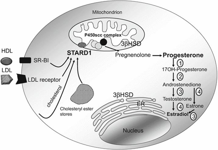
Fig. 3.1
Steroidogenesis in luteal cells . Cholesterol can be taken up from extracellular lipoprotein through the actions of the low density lipoprotein (LDL) receptor (LDL only) and scavenger receptor B1 (SR-B1, cholesteryl esters from HDL and LDL). Cholesterol can also be synthesized by the cell. Intracellular- and extracellular-derived cholesterol can be stored as cholesteryl esters. Liberated free cholesterol is transported to the outer mitochondrial membrane potentially by sterol carrier protein 2 (SCP2) and/or START domain proteins 4 and 6 (not shown). Steroidogenic acute regulatory protein (STARD1) sits on the outer mitochondrial membrane and serves to transfer cholesterol to the inner mitochondrial membrane, where the cytochrome P450 cholesterol side-chain (P450scc) cleavage complex resides. The P450scc enzyme partners with electron transfer proteins adrenodoxin and adrenodoxin reductase to convert cholesterol to the first steroid hormone pregnenolone. Pregnenolone is converted to progesterone by the actions of 3-beta-hydroxysteroid dehydrogenase (3βHSD), which has been localized in both mitochondria and the endoplasmic reticulum (ER). In species with luteal estradiol synthesis, steps 1–4 are present. The enzymes mediating these reactions needed for estradiol synthesis are 1 and 2, CYP17A1 (17-alpha-hydroxylase/17,20-lyase); 3, HSD17B (17-beta-hydroxysteroid dehydrogenase); 4, CYP19A1 (aromatase)
3.2 Regulation of the Genes Involved in Steroidogenesis
In vivo the main stimulus for the induction genes encoding the machinery needed for de novo synthesis of luteal steroids, namely, STARD1, CYP11A1, and HSD3B, is the mid-cycle LH surge [27]. Both mural granulosa and theca cells of the ovulatory follicle possess LH receptors. Activation of the seven-transmembrane G protein-coupled LH receptor by surge levels of the gonadotropin activates adenylate cyclase-generating cyclic AMP (cAMP), thereby activating protein kinase A (PKA), the major regulator of transcription factors targeting the genes governing the steroidogenic pathway [28, 29]. In addition, high levels of LH have also been shown to increase intracellular calcium levels, although the full implications of activating this secondary pathway on luteal steroidogenesis are unclear [30, 31]. Exogenous human chorionic gonadotropin (hCG) can also bind and activate the LH receptor [32], and it is frequently used in immature PMSG (pregnant mare serum gonadotropin)-primed animals to mimic the LH surge. Similarly, exogenous hGC is frequently used in assisted reproduction protocols for final follicle maturation. Downstream of PKA signaling there is activation of ERK signaling [29, 33] that can contribute to regulation of transcription of these genes as well. Protein kinase A typically activates cyclic AMP response-element-binding protein (CREB), and although CREB activation via its phosphorylation by PKA may be a major mechanism for activating genes involved in steroidogenesis in the follicle [34], its importance in the corpus luteum is reduced and other transcription factors tend to mediate PKA effects on the genes of the steroidogenic pathway. Among the major transcription factors involved in mediating PKA signaling are members of the NR5A, NR4A, GATA, Sp1, Activator Protein (AP), and CCAAT/enhancer binding protein (C/EBP) families [7].
Much of what we know about the transcriptional regulation of these steroidogenic genes comes from studies with primary cultures of luteinizing granulosa cells, with fewer studies being actually performed with luteal cell preparations. Next we summarize the hormonal regulation of the mRNA and protein levels and the transcriptional regulation of the major steps in steroidogenesis in luteal cells.
3.3 Regulation of StAR/STARD1
In most mammals examined, STARD1 mRNA and its protein are expressed in theca cells but are not significantly expressed in granulosa of healthy follicles before the LH surge [35–38]. Exogenous hGC given in lieu of LH to PMSG-primed follicles can mimic the LH surge, inducing ovulation and STARD1 mRNA and protein [36, 39]. In luteinizing granulosa cell cultures, STARD1 mRNA and protein can be increased by follicle-stimulating hormone (FSH) through PKA [40–42], although the relevance of this to corpus luteum function is uncertain because FSH receptors are typically downregulated with luteinization in vivo [43, 44]. Several other hormones and growth factors have been shown to inhibit or stimulate STARD1 mRNA and/or protein levels, most often by modulating the cellular response to gonadotropin or PKA signaling. Of those relevant to luteal function known to be stimulatory are estradiol, insulin-like growth factors (IGFs), insulin, and prostaglandin E2 (PGE2) [40, 42, 45–47]. Bone morphogenetic proteins (BMPs) tend to be inhibitory to STARD1 expression, as are tumor necrosis factor-alpha (TNFα), and activin A [48–53]. Prostaglandin F2-alpha (PGF2α) is inhibitory and may serve to reduce STARD1 at the onset of luteal regression [54–56]. Factors such as transforming growth factor-beta (TGFβ) and leptin have stimulatory or inhibitory actions depending on the context of the granulosa/luteal cell [57–60]. Active STARD1 exists as a phosphoprotein with PKA mediating phosphorylation [61]. There is evidence in ovine large luteal cells that STARD1 is phosphorylated by the high basal PKA activity in these cells [62]. The mechanism by which STARD1 works is not fully understood, but data support the model that STARD1 protein sits on the outer mitochondrial membrane to transfer cholesterol to the inner membrane [63]. Internalization of STARD1 protein into the mitochondria inactivates its activity [64].
The proximal 5′-flanking DNA of the STARD1 gene serves as the main promoter region and has transcription factor-binding sites that are highly conserved across species. Although many different types of transcription factors, including NR5A, GATA, NR4A, AP-1, Sp1/3, SREBP, Kruppel-like factors (KLFs), and forkhead proteins, have been shown to regulate the promoter [7], presented here are only those factors relevant to the corpus luteum.
Several different NR5A response elements that have the ability to bind steroidogenic factor 1 (SF-1/NR5A1) and liver receptor homologue 1 (LRH-1/NR5A2) reside in both the proximal STARD1 promoter region (first −150 bp) with others lying more distal between −900 and −3400 bp upstream of the transcriptional start site [65–67]. One of the proximal NR5A sites was shown to be specifically recruited in rodent luteal cells, although it was not utilized in less differentiated granulosa cells [66]. NR5A sites participate in both basal and cAMP-stimulated transcriptional activity [7]. In bovine luteal cells, beta-catenin interacts with LRH-1 to promote STARD1 transcription [68]. A member of the NR4A family Nur77 can also recognize the same NR5A response elements, and Nur77 reduction lowers STARD1 mRNA levels in theca cells [69]. It is unknown if Nur77 impacts STARD1 gene expression in luteal cells.
A conserved GATA consensus site (TTATCT) is located within the −70- to −55-bp region of the proximal STARD1 promoter of numerous species, and mutational analyses have demonstrated its importance to basal and/or cAMP-stimulated transactivity [7]. The transcription factors GATA4 and GATA6 are present in follicular cells and luteal cells, and both factors have been demonstrated to bind to the site [70]. There is some debate about the importance of GATA4 versus GATA6 to STARD1 promoter activity. Although both factors bind the STARD1 promoter GATA element in gel shift assays and overexpression of both recombinant factors can promote transactivation [71–73], most evidence indicates GATA4 as the major regulator of the promoter. This observation is complicated by the fact that GATA4 levels are typically lower with luteinization (especially in rodents) where GATA6 is strongly expressed [71, 74, 75]. To help clarify this point, one study in luteinizing pig granulosa cells showed that lowering GATA4 levels by RNAi actually increased STARD1 mRNA levels but not when GATA6 was also reduced, inferring that GATA6 has the potential to drive STARD1 gene expression when GATA4 levels drop [76]. These data support the idea that the ratio of GATA6 to GATA4 influences the contribution of GATA factors to STARD1 transcription in the corpus luteum.
Several C/EBP-binding sites exist in the proximal STARD1 promoter. Two of the sites are highly conserved whereas the sequence and location of other binding sites vary by location between species [7]. There is also overlap between C/EBP sites and CREB half-sites. Although CREB can associate with the promoter, studies in rodents showed that an ovulatory stimulus of hCG causes rapid C/EBPβ association with the promoter region, whereas CREB associates more slowly [77]. In addition, studies with several species of luteinized granulosa cells indicate C/EBP elements are needed for basal and cAMP responsiveness of the promoter [66, 72]. Further emphasizing the need for C/EBPs, cultured mouse granulosa with depleted C/EBPα and C/EBPβ have reduced induction of STARD1 mRNA in culture [78]. In rodent luteal cells, AP-1 family member Fra-2 displaces the CREB bound to the promoter of less mature granulosa [66]. In sum, although CREB can transactivate the promoter, in luteal cells C/EBPβ and Fra-2 occupy potential CREB-binding regions.
Other ovarian transcriptional regulators influence luteal STARD1 expression in a less clear manner. Forkhead transcription factor FOXO1 may act to repress the expression of STARD1 in granulosa cells before luteinization induced by the LH surge [79]. KLF4, -9, and -13 overexpression in luteinizing porcine granulosa cells reduced LH stimulated STARD1 promoter activity, yet KLF13 overexpression increased STARD1 mRNA levels [80]. There are no data yet as to whether KLF factors interact with the STARD1 promoter region in the context of the luteal cell.
3.4 Regulation of P450scc /CYP11A1
In many respects the regulation of CYP11A1 shares similarities to the regulation of STARD1. CYP11A1 mRNA and protein are present in follicular theca and some granulosa cells before the ovulatory LH surge; levels increase in granulosa cells during luteinization, are maintained at high levels in the functional corpus luteum, and drop off with regression [27]. The cow has a transient downregulation of CYP11A1 mRNA in the late preovulatory follicle [81]. CYP11A1 is constitutively expressed in the rodent corpus luteum once pregnancy is established [82]. Similar to STARD1, LH or exogenous hCG increases CYP11A1 mRNA and protein [27]. In luteinizing granulosa cell cultures, FSH, insulin, IGFs, epidermal growth factor, progesterone, PGE2, and prolactin act alone or in concert with gonadotropin to increase CYP11A1 mRNA [47, 83–88]. Repressors of CYP11A1 mRNA levels include TNFα, some BMPs, and luteolytic PGF2α [50, 51, 89–92]. TGFβ and activin A have mixed effects on CYP11A1 mRNA levels [57, 58].
The timing and abundance of CYP11A1 appears to be important for normal luteal function as transgenic mice overexpressing CYP11A1 have reduced progesterone production by the early corpus luteum [93]. In these transgenic mice, corpus luteum function is able to normalize by mid-pregnancy, suggesting a delayed luteinization of follicles.
There is fairly high homology between species within the first −100 bp upstream of the transcriptional start site of the CYP11A1 gene [7]. A proximal NR5A site in the cow promoter is active in basal and cAMP-driven activity in luteal cells [94, 95]. Similarly, rodent granulosa demonstrate NR5A site importance [96], and targeted reduction of LRH-1 in mice express reduced CYP11A1 mRNA in granulosa when given an ovulatory stimulus of hCG [97].
Overexpression of GATA can drive the promoter in nonovarian cells. and a GATA site at −475 to −470 of the rat promoter helps confer granulosa responsiveness to FSH [98]. However, in luteinizing pig granulosa cells there is little impact of reduction of GATA4/6 factors on CYP11A1 mRNA expression, suggesting GATA may not be important to CYP11A1 expression in luteal cells [76].
At least one functional Sp1 site exists in the proximal promoter region [7]. Bovine luteal extracts exhibit Sp1 and/or Sp3 binding, and this site is important for basal and cAMP-stimulated activity [95]. In pig luteinizing granulosa cells, the Sp1 site confers responsiveness to both IGF1 and cAMP stimuli [99].
CYP11A1 promoter activity is also influenced by the differentiation status of the cell. Demonstrating this, CREB binds to a CRE half-site in less differentiated rodent granulosa cells and is replaced by Fra-2 upon luteinization [98].
Similar to STARD1, forkhead factors and KLF factors may influence luteal expression. FOXO1 may serve to repress the expression of CYP11A1 in granulosa cells before luteinization [100]. KLF4, -9, and -13 overexpression in luteinizing porcine granulosa cells reduced CYP11A1 promoter activity and KLF13 overexpression decreased CYP11A1 mRNA levels as well [80].
3.5 Regulation of 3-Beta-HSD/HSD3B (1 or 2)
Depending on the species, one or more HSD3B genes for 3-beta-hydroxysteroid dehydrogenase/delta5 delta4-isomerase exist, and the numbering system between species varies. For example the human has two genes (HSD3B1 and HSD3B2), the mouse has six genes (hsd3b1-6), and the pig has one gene (HSD3B1) [101]. In humans the main ovarian gene expressed that mediates steroidogenesis is HSD3B2 and in other species HSD3B1 (hsd3b1 in rodent). In rat luteal cells, using electron microscopy immunoreactive HSD3B protein has been localized to both the smooth ER and the mitochondria [102]. The mRNA for the ovarian form of HSD3B is expressed in theca cells of developing follicles, appears in the granulosa cells of growing follicles even before the ovulatory period, and is widely distributed in functional luteal cells, falling off with luteal regression [27]. Similar to STARD1 and CYP11A1, LH (or exogenous hCG) is a potent inducer of HSD3B transcripts in granulosa cells during the periovulatory period [27]. Other positive regulators of ovarian HSD3B expression include FSH and IGF1 or PGE2 either alone or in combination with gonadotropin [42, 47]. TGFβ and prolactin affect HSD3B expression either positively or negatively depending on the cellular setting [57, 58, 88]. Several BMPs and luteolytic PGF2α are inhibitory to HSD3B mRNA expression [47, 48, 90, 92].
Most cellular studies of HSD3B promoter regulation have been performed in cells lines, and it is unclear how these studies relate to periovulatory and luteal transcriptional events. However, there have been a few studies with luteinized granulosa and luteal cells. NR5A sites have been identified in the human HSD3B gene 5′-flanking DNA [7]. Overexpression of SF-1 can drive HSD3B promoter activity in human granulosa tumor cells [103]. LRH-1 has been shown to bind two NR5A sites in luteinized granulosa cell extracts, and both sites contribute to transactivation with the more proximal site at −309 bp being most critical [104]. In contrast, in mice with granulosa cell-targeted loss of LRH-1, hsd3b1 mRNA levels following an ovulatory hCG stimulus are not affected [97]; however, there is always the possibility that SF-1 may substitute for LRH-1 in this setting.
There are at least four potential GATA-binding sites in the 5′-flanking DNA of the human HSD3B2 gene [7]. The GATA site at −196 bp relative to the transcriptional start site has been shown to stimulate promoter transactivation in nonovarian cells [105]. Studies in luteinizing pig granulosa cells indicate that a loss of GATA4 suppresses basal expression of HSD3B1 mRNA [76].
Nur77 may contribute to HSD3B gene expression in human theca cells [69], but the relevance of this factor to luteal cells is unknown.
3.6 Luteal Estrogen Synthesis
3.6.1 Regulation of Luteal Aromatase/CYP19A1
The CYP19A1 gene encodes aromatase, whose activity is required to convert androgens into estrogens. In the developing follicle, mural granulosa cells are the primary site for aromatase expression , which is driven mainly by FSH and cAMP/PKA signaling [6]. The LH surge downregulates granulosa aromatase [106]. In most large mammals, the CYP19A1 gene stays quiescent for the remainder of the corpus luteum lifespan. In the cow, downregulation of CYP19A1 by the LH surge is associated with silencing DNA methylation in the promoter 2 region in luteal cells [107]. This finding may extend to other species that lack luteal aromatase. In comparison, CYP19A1 is expressed robustly in the rodent corpus luteum of pregnancy and the primate corpus luteum, enabling luteal estradiol synthesis [106, 108, 109]. During the follicular phase, estradiol production and granulosa cell aromatase expression increase to reach a peak in the dominant follicle(s) [6]. In the human, mid- and late-luteal phase corpora lutea have the highest CYP19A1 mRNA expression levels compared to other follicular/luteal stages [109]. Granulosa cells utilize the ovarian-specific CYP19A1 promoter region to drive transcription in response to cAMP/PKA signals [110]. In the rat this region is located within −300 bp upstream of the gene and involves a cAMP-response element-like (CLS) sequence regulated by CREB, two NR5A sites that can bind SF-1 or LRH-1, and a GATA-4-binding site [110, 111]. In the luteal cell, the rat CLS site loses its transcription factor-binding ability and an AP3 site becomes recruited [111].
The human CYP19A1 promoter also has a functional CLS and NR5A region. When human CYPA19A1 promoter constructs are transfected into bovine luteal cells, both regions confer reporter gene activity [112]. The primate corpus luteum requires pituitary LH to maintain steroidogenesis during the luteal phase. In human cultured granulosa-lutein cells, LH increases aromatase mRNA and enzyme activity [113, 114], which infers that the human CYP19A1 promoter likely has some continued dependence on PKA signaling during the luteal phase.
A comparison of the CYP19A1 promoters of several species showed that cow and goat promoter constructs had minimal responsiveness to PKA activation in luteinizing granulosa cells, whereas the human and rat promoters were activated [115]. In addition, the responsiveness to overexpressed transcription factors LRH-1 and FOXL2 varied, providing a possible explanation for the species differences in luteal aromatase expression.
3.6.2 Regulation of Luteal P450c17/CYP17A1
In the follicle cytochrome P450 17-alpha-hydroxylase/17, 20 lyase (P450c17 or CYP17A1) is expressed in the thecal cell layer where it converts pregnenolone and progesterone-derived intermediates into androgens. This enzyme becomes downregulated with luteinization [116]. In the primate corpus luteum, theca lutein cells later reexpress this enzyme to provide the androstenedione and testosterone for estrogen synthesis [108]. Rodent CYP17A1 expression is increased by hGC treatment of luteal cells [117]. In the rodent, luteal androgen synthesis occurs in early pregnancy but the placenta serves as the main source of androgens for estradiol synthesis as pregnancy progresses [118].
3.6.3 Regulation of Luteal 17-Beta-HSD/HSD17B
More than a dozen types of 17-beta-hydroxysteroid dehydrogenases have been identified with various substrate specificities [119]. The main roles of ovarian HSD17B enzymes are to interconvert androgen forms and interconvert estrogen forms. In luteal cells, androstenedione is aromatized to estrone and then can be converted to estradiol by HSD17B. Alternatively, androstenedione can first be converted to testosterone by HSD17B and then aromatized to estradiol. HSD17B1 and HSD17B2 have been localized to luteal cells in humans [120]. HSD17B type 1 activity, which converts estrone to estradiol, predominates in human granulosa-luteal cells [121]. Mice null for hsd17b1 have increased estrone:estradiol and androstenedione:testosterone ratios, reduced progesterone, structural changes in corpora lutea, and are subfertile [122]. HSD17B7 serves to convert estrone to estradiol and is found in the corpora lutea of all mammalian species examined including rodents [123]. Expression of hsd17b1 disappears in the rodent corpus luteum, where hsd17b7 is strongly expressed [124]. Additionally, in rodents the hsd17b1 gene is upregulated by LH, whereas the hsd17b7 gene is increased by prolactin and repressed by LH/hCG. The promoter for rat hsd17b7 has a functional Sp1 site that regulates basal promoter activity and a NF-Y-binding site that mediates its inhibition by PKA [125]. Of note, HSD17B7 has also been demonstrated to mediate a reaction in cholesterol biosynthesis [119]. Deletion of hsd17B7 in mice is embryonic lethal because of deficiencies in cholesterol synthesis [126, 127]. These data infer that the HSD17B7 enzyme may have a dual purpose in the corpus luteum: cholesterol synthesis and estradiol synthesis.
3.7 Conclusions
Luteal progesterone biosynthesis depends on the coordinated upregulation and maintenance of the gene products of STARD1, CYP11A1 and its mitochondrial partner proteins, and HSD3B. Luteal estradiol synthesis by the corpus luteum occurs in a species-specific manner and involves CYP17A1, HSD17B, and CYP19A1. Most of the transcriptional studies performed have utilized various stages of luteinized granulosa cells in culture to study of upregulation of these genes during the periovulatory luteinization period. Comparison of the transcriptional activity of STARD1, CYP11A1, and CYP19A1 in granulosa cells and luteal cells of the rodent has demonstrated that the extent of cellular luteinization is critical for the recruitment of specific transcription factors. Figure 3.2 depicts a summary of putative regulators of STARD1, CYP11A1, HSD3B, and CYP19A1 by cellular differentiation status. Even though LH activates the genes for the major steroidogenic pathway molecules in the early corpus luteum, how the expression of these genes is maintained in the mature corpus luteum and that of pregnancy is an area that requires much more research.
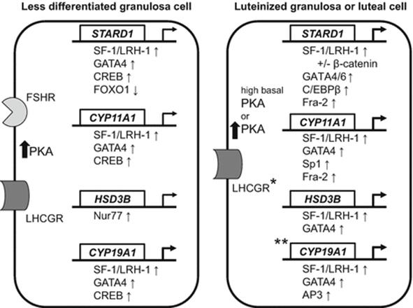
Fig. 3.2
Summary of the major transcription factors shown to regulate the STARD1, CYP11A1, HSD3B, and CYP19A1 genes in less differentiated granulosa cells or luteinized granulosa cells and luteal cells. * indicates that the presence of luteinizing hormone receptors (LHCGR) on luteal cells varies by steroidogenic luteal cell type and by species. ** indicates that the expression of CYP19A1 occurs in primate and pregnant rodent corpora lutea. Thin arrows indicate the transcription factor increases (↑) or decreases (↓) transactivation of the gene immediately above it in one or more species. Thick arrows indicate protein kinase A (PKA) is increased by activation of the follicle-stimulating hormone receptor (FSHR) and LHCGR by their respective ligands. In most cases PKA leads to increased transcriptional activity of the factors shown. Less differentiated granulosa cells refers to those that have not been luteinized in vivo by a gonadotropin surge or by culture conditions in vitro. STARD1 is likely repressed in vivo by FOXO1 or other unknown factor before the LH surge, as its mRNA is minimally expressed in the pre-surge maturing follicles of most species
References
1.
McCracken JA, Custer EE, Lamsa JC. Luteolysis: a neuroendocrine-mediated event. Physiol Rev. 1999;79:263–323.PubMed
2.
Magness RR. Maternal cardiovascular and other physiologic responses to the endocrinology of pregnancy. In: Endocrinology of pregnancy. Totowa: Humana Press; 1998. p. 507–39
3.
Richards JS, Russell DL, Robker RL, Dajee M, Alliston TN. Molecular mechanisms of ovulation and luteinization. Mol Cell Endocrinol. 1998;145:47–54.PubMed
4.
Gilling-Smith C, Franks S. Ovarian function in assisted reproduction. In: Leung PC, Adashi EY, editors. The ovary. 2nd ed. San Diego: Elsevier Academic Press; 2004. p. 473–88.
5.
Sanders SL, Stouffer RL. Localization of steroidogenic enzymes in macaque luteal tissue during the menstrual cycle and simulated early pregnancy: immunohistochemical evidence supporting the two-cell model for estrogen production in the primate corpus luteum. Biol Reprod. 1997;56:1077–87.PubMed
6.
Stocco C. Aromatase expression in the ovary: hormonal and molecular regulation. Steroids. 2008;73:473–87.PubMedPubMedCentral
7.
LaVoie HA, King SR. Transcriptional regulation of steroidogenic genes: STARD1, CYP11A1 and HSD3B. Exp Biol Med (Maywood). 2009;234:880–907.
8.
Stocco DM, Clark BJ. Regulation of the acute production of steroids in steroidogenic cells. Endocr Rev. 1996;17:221–44.PubMed
9.
Clark BJ, Wells J, King SR, Stocco DM. The purification, cloning, and expression of a novel luteinizing hormone-induced mitochondrial protein in MA-10 mouse Leydig tumor cells. Characterization of the steroidogenic acute regulatory protein (StAR). J Biol Chem. 1994;269:28314–22.PubMed
10.
Miller WL, Bose HS. Early steps in steroidogenesis: intracellular cholesterol trafficking. J Lipid Res. 2011;52:2111–35.PubMedPubMedCentral
11.
Tuckey RC, Stevenson PM. Cholesteryl esterase and endogenous cholesteryl ester pools in ovaries from maturing and superovulated immature rats. Biochim Biophys Acta. 1980;618:501–9.PubMed
12.
Grummer RR, Carroll DJ. A review of lipoprotein cholesterol metabolism: importance to ovarian function. J Anim Sci. 1988;66:3160–73.PubMed
13.
Khan I, Belanger A, Chen YD, Gibori G. Influence of high-density lipoprotein on estradiol stimulation of luteal steroidogenesis. Biol Reprod. 1985;32:96–104.PubMed
14.
Brodeur MR, Luangrath V, Bourret G, Falstrault L, Brissette L. Physiological importance of SR-BI in the in vivo metabolism of human HDL and LDL in male and female mice. J Lipid Res. 2005;46:687–96.PubMed
15.
Lopez D, McLean MP. Sterol regulatory element-binding protein-1a binds to cis elements in the promoter of the rat high density lipoprotein receptor SR-BI gene. Endocrinology. 1999;140:5669–81.PubMed
16.
Sekar N, Veldhuis JD. Involvement of Sp1 and SREBP-1a in transcriptional activation of the low density lipoprotein-receptor gene by insulin and luteinizing hormone in cultured porcine granulosa-luteal cells. Am J Physiol Endocrinol Metab. 2004;287:E128–35.PubMed
17.
Bishop CV, Bogan RL, Hennebold JD, Stouffer RL. Analysis of microarray data from the macaque corpus luteum; the search for common themes in primate luteal regression. Mol Hum Reprod. 2011;17:143–51.PubMed
18.
LaVoie HA, Benoit AM, Garmey JC, Dailey RA, Wright DJ, Veldhuis JD. Coordinate developmental expression of genes regulating sterol economy and cholesterol side-chain cleavage in the porcine ovary. Biol Reprod. 1997;57:402–7.PubMed
19.
Clark BJ. The mammalian START domain protein family in lipid transport in health and disease. J Endocrinol. 2012;212:257–75.PubMed
20.
Skiadas CC, Duan S, Correll M, Rubio R, Karaca N, Ginsburg ES, et al. Ovarian reserve status in young women is associated with altered gene expression in membrana granulosa cells. Mol Hum Reprod. 2012;18:362–71.PubMedPubMedCentral
21.
LaVoie HA, Whitfield NE, Shi B, King SR, Bose HS, Hui YY. STARD6 is expressed in steroidogenic cells of the ovary and can enhance de novo steroidogenesis. Exp Biol Med (Maywood) 2014;239:430–435.
22.
Chanderbhan R, Noland BJ, Scallen TJ, Vahouny GV. Sterol carrier protein 2. Delivery of cholesterol from adrenal lipid droplets to mitochondria for pregnenolone synthesis. J Biol Chem. 1982;257:8928–34.PubMed
23.
Rodriguez-Agudo D, Ren S, Wong E, Marques D, Redford K, Gil G, et al. Intracellular cholesterol transporter StarD4 binds free cholesterol and increases cholesteryl ester formation. J Lipid Res. 2008;49:1409–19.PubMedPubMedCentral
24.
Bose HS, Whittal RM, Ran Y, Bose M, Baker BY, Miller WL. StAR-like activity and molten globule behavior of StARD6, a male germ-line protein. Biochemistry. 2008;47:2277–88.PubMed
25.
McLean MP, Puryear TK, Khan I, Azhar S, Billheimer JT, Orly J, et al. Estradiol regulation of sterol carrier protein-2 independent of cytochrome P450 side-chain cleavage expression in the rat corpus luteum. Endocrinology. 1989;125:1337–44.PubMed
26.
Rennert H, Amsterdam A, Billheimer JT, Strauss III JF. Regulated expression of sterol carrier protein 2 in the ovary: a key role for cyclic AMP. Biochemistry. 1991;30:11280–5.PubMed
27.
King SR, LaVoie HA. Gonadal transactivation of STARD1, CYP11A1 and HSD3B. Front Biosci. 2012;17:824–46.
28.
Davis JS, Weakland LL, West LA, Farese RV. Luteinizing hormone stimulates the formation of inositol trisphosphate and cyclic AMP in rat granulosa cells. Evidence for phospholipase C generated second messengers in the action of luteinizing hormone. Biochem J. 1986;238:597–604.PubMedPubMedCentral
29.
Salvador LM, Maizels E, Hales DB, Miyamoto E, Yamamoto H, Hunzicker-Dunn M. Acute signaling by the LH receptor is independent of protein kinase C activation. Endocrinology. 2002;143:2986–94.PubMed
30.
Flores JA, Aguirre C, Sharma OP, Veldhuis JD. Luteinizing hormone (LH) stimulates both intracellular calcium ion ([Ca2+]i) mobilization and transmembrane cation influx in single ovarian (granulosa) cells: recruitment as a cellular mechanism of LH-[Ca2+]i dose response. Endocrinology. 1998;139:3606–12.PubMed
31.
Davis JS, West LA, Weakland LL, Farese RV. Human chorionic gonadotropin activates the inositol 1,4,5-trisphosphate-Ca2+ intracellular signalling system in bovine luteal cells. FEBS Lett. 1986;208:287–91.PubMed
32.
Casarini L, Lispi M, Longobardi S, Milosa F, La MA, Tagliasacchi D, et al. LH and hCG action on the same receptor results in quantitatively and qualitatively different intracellular signalling. PLoS ONE. 2012;7, e46682.PubMedPubMedCentral
33.
Fan HY, Liu Z, Shimada M, Sterneck E, Johnson PF, Hedrick SM, et al. MAPK3/1 (ERK1/2) in ovarian granulosa cells are essential for female fertility. Science. 2009;324:938–41.PubMedPubMedCentral
34.
Mukherjee A, Park-Sarge OK, Mayo KE. Gonadotropins induce rapid phosphorylation of the 3′,5′-cyclic adenosine monophosphate response element binding protein in ovarian granulosa cells. Endocrinology. 1996;137:3234–45.PubMed
35.
Kiriakidou M, McAllister JM, Sugawara T, Strauss III JF. Expression of steroidogenic acute regulatory protein (StAR) in the human ovary. J Clin Endocrinol Metab. 1996;81:4122–8.PubMed
36.
Ronen-Fuhrmann T, Timberg R, King SR, Hales KH, Hales DB, Stocco DM, et al. Spatio-temporal expression patterns of steroidogenic acute regulatory protein (StAR) during follicular development in the rat ovary. Endocrinology. 1998;139:303–15.PubMed
37.
Bao B, Garverick HA. Expression of steroidogenic enzyme and gonadotropin receptor genes in bovine follicles during ovarian follicular waves: a review. J Anim Sci. 1998;76:1903–21.PubMed
38.
Bonnet A, Le Cao KA, Sancristobal M, Benne F, Robert-Granie C, Law-So G, et al. In vivo gene expression in granulosa cells during pig terminal follicular development. Reproduction. 2008;136:211–24.PubMed
39.
Mizutani T, Sonoda Y, Minegishi T, Wakabayashi K, Miyamoto K. Molecular cloning, characterization and cellular distribution of rat steroidogenic acute regulatory protein (StAR) in the ovary. Life Sci. 1997;61:1497–506.PubMed
40.
Balasubramanian K, LaVoie HA, Garmey JC, Stocco DM, Veldhuis JD. Regulation of porcine granulosa cell steroidogenic acute regulatory protein (StAR) by insulin-like growth factor I: synergism with follicle-stimulating hormone or protein kinase A agonist. Endocrinology. 1997;138:433–9.PubMed
41.
Pescador N, Houde A, Stocco DM, Murphy BD. Follicle-stimulating hormone and intracellular second messengers regulate steroidogenic acute regulatory protein messenger ribonucleic acid in luteinized porcine granulosa cells. Biol Reprod. 1997;57:660–8.PubMed
42.
Eimerl S, Orly J. Regulation of steroidogenic genes by insulin-like growth factor-1 and follicle-stimulating hormone: differential responses of cytochrome p450 side-chain cleavage, steroidogenic acute regulatory protein, and 3beta-hydroxysteroid dehydrogenase/isomerase in rat granulosa cells. Biol Reprod. 2002;67:900–10.PubMed
43.
Zhang YM, Roy SK. Downregulation of follicle-stimulating hormone (FSH)-receptor messenger RNA levels in the hamster ovary: effect of the endogenous and exogenous FSH. Biol Reprod. 2004;70:1580–8.PubMed
44.
Lapolt PS, Tilly JL, Aihara T, Nishimori K, Hsueh AJ. Gonadotropin-induced up- and down-regulation of ovarian follicle-stimulating hormone (FSH) receptor gene expression in immature rats: effects of pregnant mare’s serum gonadotropin, human chorionic gonadotropin, and recombinant FSH. Endocrinology. 1992;130:1289–95.PubMed
45.
Townson DH, Wang XJ, Keyes PL, Kostyo JL, Stocco DM. Expression of the steroidogenic acute regulatory protein in the corpus luteum of the rabbit: dependence upon the luteotropic hormone, estradiol-17 beta. Biol Reprod. 1996;55:868–74.PubMed
46.
Sekar N, Garmey JC, Veldhuis JD. Mechanisms underlying the steroidogenic synergy of insulin and luteinizing hormone in porcine granulosa cells: joint amplification of pivotal sterol-regulatory genes encoding low-density lipoprotein (LDL) receptor, steroidogenic acute regulatory (StAR) protein and cytochrome P450 side-chain cleavage (P450scc) enzyme. Mol Cell Endocrinol. 2000;159:25–35.PubMed
47.
Rekawiecki R, Nowik M, Kotwica J. Stimulatory effect of LH, PGE2 and progesterone on StAR protein, cytochrome P450 cholesterol side chain cleavage and 3beta hydroxysteroid dehydrogenase gene expression in bovine luteal cells. Prostaglandins Other Lipid Mediat. 2005;78:169–84.PubMed
48.
Brankin V, Quinn RL, Webb R, Hunter MG. Evidence for a functional bone morphogenetic protein (BMP) system in the porcine ovary. Domest Anim Endocrinol. 2005;28:367–79.PubMed
49.
Miyoshi T, Otsuka F, Suzuki J, Takeda M, Inagaki K, Kano Y, et al. Mutual regulation of follicle-stimulating hormone signaling and bone morphogenetic protein system in human granulosa cells. Biol Reprod. 2006;74:1073–82.PubMed
50.
Otsuka F, Moore RK, Shimasaki S. Biological function and cellular mechanism of bone morphogenetic protein-6 in the ovary. J Biol Chem. 2001;276:32889–95.PubMed
51.
Pierre A, Pisselet C, Dupont J, Mandon-Pepin B, Monniaux D, Monget P, et al. Molecular basis of bone morphogenetic protein-4 inhibitory action on progesterone secretion by ovine granulosa cells. J Mol Endocrinol. 2004;33:805–17.PubMed
52.
Chen YJ, Feng Q, Liu YX. Expression of the steroidogenic acute regulatory protein and luteinizing hormone receptor and their regulation by tumor necrosis factor alpha in rat corpora lutea. Biol Reprod. 1999;60:419–27.PubMed
53.
Chang HM, Cheng JC, Klausen C, Taylor EL, Leung PC. Effects of recombinant activins on steroidogenesis in human granulosa-lutein cells. J Clin Endocrinol Metab. 2014;99:E1922–32.PubMed
54.
Sandhoff TW, McLean MP. Repression of the rat steroidogenic acute regulatory (StAR) protein gene by PGF2alpha is modulated by the negative transcription factor DAX-1. Endocrine. 1999;10:83–91.PubMed
55.
Juengel JL, Meberg BM, Turzillo AM, Nett TM, Niswender GD. Hormonal regulation of messenger ribonucleic acid encoding steroidogenic acute regulatory protein in ovine corpora lutea. Endocrinology. 1995;136:5423–9.PubMed
56.
Chung PH, Sandhoff TW, McLean MP. Hormone and prostaglandin F2 alpha regulation of messenger ribonucleic acid encoding steroidogenic acute regulatory protein in human corpora lutea. Endocrine. 1998;8:153–60.PubMed
57.
Chen YJ, Lee MT, Yao HC, Hsiao PW, Ke FC, Hwang JJ. Crucial role of estrogen receptor-alpha interaction with transcription coregulators in follicle-stimulating hormone and transforming growth factor beta1 up-regulation of steroidogenesis in rat ovarian granulosa cells. Endocrinology. 2008;149:4658–68.PubMed
58.
Zheng X, Price CA, Tremblay Y, Lussier JG, Carriere PD. Role of transforming growth factor-beta1 in gene expression and activity of estradiol and progesterone-generating enzymes in FSH-stimulated bovine granulosa cells. Reproduction. 2008;136:447–57.PubMed
59.
Minegishi T, Tsuchiya M, Hirakawa T, Abe K, Inoue K, Mizutani T, et al. Expression of steroidogenic acute regulatory protein (StAR) in rat granulosa cells. Life Sci. 2000;67:1015–24.PubMed
60.
Ruiz-Cortes ZT, Martel-Kennes Y, Gevry NY, Downey BR, Palin MF, Murphy BD. Biphasic effects of leptin in porcine granulosa cells. Biol Reprod. 2003;68:789–96.PubMed
61.
Arakane F, King SR, Du Y, Kallen CB, Walsh LP, Watari H, et al. Phosphorylation of steroidogenic acute regulatory protein (StAR) modulates its steroidogenic activity. J Biol Chem. 1997;272:32656–62.PubMed
62.
Bogan RL, Niswender GD. Constitutive steroidogenesis in ovine large luteal cells may be mediated by tonically active protein kinase A. Biol Reprod. 2007;77:209–16.PubMed
63.
Bose HS, Lingappa VR, Miller WL. The steroidogenic acute regulatory protein, StAR, works only at the outer mitochondrial membrane. Endocr Res. 2002;28:295–308.PubMed
64.
Stocco DM, Sodeman TC. The 30-kDa mitochondrial proteins induced by hormone stimulation in MA-10 mouse Leydig tumor cells are processed from larger precursors. J Biol Chem. 1991;266:19731–8.PubMed
65.
Sugawara T, Kiriakidou M, McAllister JM, Kallen CB, Strauss III JF. Multiple steroidogenic factor 1 binding elements in the human steroidogenic acute regulatory protein gene 5′-flanking region are required for maximal promoter activity and cyclic AMP responsiveness. Biochemistry. 1997;36:7249–55.PubMed
66.
Yivgi-Ohana N, Sher N, Melamed-Book N, Eimerl S, Koler M, Manna PR, et al. Transcription of steroidogenic acute regulatory protein (STAR) in the rodent ovary and placenta: alternative modes of cyclic adenosine 3′,5′-monophosphate dependent and independent regulation. Endocrinology. 2009;150:977–89.PubMed
67.
Mizutani T, Yazawa T, Ju Y, Imamichi Y, Uesaka M, Inaoka Y, et al. Identification of a novel distal control region upstream of the human steroidogenic acute regulatory protein (star) gene that participates in SF-1 dependent chromatin architecture. J Biol Chem. 2010;285:28240–51.PubMedPubMedCentral
68.
Roy L, McDonald CA, Jiang C, Maroni D, Zeleznik AJ, Wyatt TA, et al. Convergence of 3′,5′-cyclic adenosine 5′-monophosphate/protein kinase A and glycogen synthase kinase-3beta/beta-catenin signaling in corpus luteum progesterone synthesis. Endocrinology. 2009;150:5036–45.PubMedPubMedCentral
69.
Li M, Xue K, Ling J, Diao FY, Cui YG, Liu JY. The orphan nuclear receptor NR4A1 regulates transcription of key steroidogenic enzymes in ovarian theca cells. Mol Cell Endocrinol. 2010;319:39–46.PubMed
70.
LaVoie HA. The role of GATA in mammalian reproduction. Exp Biol Med (Maywood). 2003;228:1282–90.
71.
Gillio-Meina C, Hui YY, LaVoie HA. GATA-4 and GATA-6 transcription factors: expression, immunohistochemical localization, and possible function in the porcine ovary. Biol Reprod. 2003;68:412–22.PubMed
72.
LaVoie HA, Singh D, Hui YY. Concerted regulation of the porcine steroidogenic acute regulatory protein gene promoter activity by follicle-stimulating hormone and insulin-like growth factor I in granulosa cells involves GATA-4 and CCAAT/enhancer binding protein beta. Endocrinology. 2004;145:3122–34.PubMed
73.
Silverman E, Eimerl S, Orly J. CCAAT enhancer-binding protein beta and GATA-4 binding regions within the promoter of the steroidogenic acute regulatory protein (StAR) gene are required for transcription in rat ovarian cells. J Biol Chem. 1999;274:17987–96.PubMed
74.
LaVoie HA, McCoy GL, Blake CA. Expression of the GATA-4 and GATA-6 transcription factors in the fetal rat gonad and in the ovary during postnatal development and pregnancy. Mol Cell Endocrinol. 2004;227:31–40.PubMed
75.
Heikinheimo M, Ermolaeva M, Bielinska M, Rahman NA, Narita N, Huhtaniemi IT, et al. Expression and hormonal regulation of transcription factors GATA-4 and GATA-6 in the mouse ovary. Endocrinology. 1997;138:3505–14.PubMed
76.
Hui YY, LaVoie HA. GATA4 reduction enhances cyclic AMP-stimulated steroidogenic acute regulatory protein mRNA and progesterone production in luteinized porcine granulosa cells. Endocrinology. 2008;149:5557–67.PubMedPubMedCentral
77.
Hiroi H, Christenson LK, Chang L, Sammel MD, Berger SL, Strauss III JF. Temporal and spatial changes in transcription factor binding and histone modifications at the steroidogenic acute regulatory protein (StAR) locus associated with StAR transcription. Mol Endocrinol. 2004;18:791–806.PubMed
78.
Fan HY, Liu Z, Johnson PF, Richards JS. CCAAT/enhancer-binding proteins (C/EBP)-{alpha} and -{beta} are essential for ovulation, luteinization, and the expression of key target genes. Mol Endocrinol. 2011;25:253–68.PubMed
79.
Pisarska MD, Bae J, Klein C, Hsueh AJ. Forkhead l2 is expressed in the ovary and represses the promoter activity of the steroidogenic acute regulatory gene. Endocrinology. 2004;145:3424–33.PubMed
80.
Natesampillai S, Kerkvliet J, Leung PC, Veldhuis JD. Regulation of Kruppel-like factor 4, 9, and 13 genes and the steroidogenic genes LDLR, StAR, and CYP11A in ovarian granulosa cells. Am J Physiol Endocrinol Metab. 2008;294:E385–91.PubMed
81.
Voss AK, Fortune JE. Levels of messenger ribonucleic acid for cholesterol side-chain cleavage cytochrome P-450 and 3 beta-hydroxysteroid dehydrogenase in bovine preovulatory follicles decrease after the luteinizing hormone surge. Endocrinology. 1993;132:888–94.PubMed
82.
Goldring NB, Durica JM, Lifka J, Hedin L, Ratoosh SL, Miller WL, et al. Cholesterol side-chain cleavage P450 messenger ribonucleic acid: evidence for hormonal regulation in rat ovarian follicles and constitutive expression in corpora lutea. Endocrinology. 1987;120:1942–50.PubMed
83.
Winters TA, Hanten JA, Veldhuis JD. In situ amplification of the cytochrome P-450 cholesterol side-chain cleavage enzyme mRNA in single porcine granulosa cells by IGF-1 and FSH acting alone or in concert. Endocrine. 1998;9:57–63.PubMed
84.
Mamluk R, Greber Y, Meidan R. Hormonal regulation of messenger ribonucleic acid expression for steroidogenic factor-1, steroidogenic acute regulatory protein, and cytochrome P450 side-chain cleavage in bovine luteal cells. Biol Reprod. 1999;60:628–34.PubMed
85.
Pescador N, Stocco DM, Murphy BD. Growth factor modulation of steroidogenic acute regulatory protein and luteinization in the pig ovary. Biol Reprod. 1999;60:1453–61.PubMed
86.
Rusovici R, Hui YY, LaVoie HA. Epidermal growth factor-mediated inhibition of follicle-stimulating hormone-stimulated StAR gene expression in porcine granulosa cells is associated with reduced histone H3 acetylation. Biol Reprod. 2005;72:862–71.PubMed
87.
Swan CL, Agostini MC, Bartlewski PM, Feyles V, Urban RJ, Chedrese PJ. Effects of progestins on progesterone synthesis in a stable porcine granulosa cell line: control of transcriptional activity of the cytochrome p450 side-chain cleavage gene. Biol Reprod. 2002;66:959–65.PubMed
88.
Stocco C, Callegari E, Gibori G. Opposite effect of prolactin and prostaglandin F(2 alpha) on the expression of luteal genes as revealed by rat cDNA expression array. Endocrinology. 2001;142:4158–61.PubMed
89.
Veldhuis JD, Garmey JC, Urban RJ, Demers LM, Aggarwal BB. Ovarian actions of tumor necrosis factor-alpha (TNF alpha): pleiotropic effects of TNF alpha on differentiated functions of untransformed swine granulosa cells. Endocrinology. 1991;129:641–8.PubMed
90.
Otsuka F, Yamamoto S, Erickson GF, Shimasaki S. Bone morphogenetic protein-15 inhibits follicle-stimulating hormone (FSH) action by suppressing FSH receptor expression. J Biol Chem. 2001;276:11387–92.PubMed
91.
Li XM, Juorio AV, Murphy BD. Prostaglandins alter the abundance of messenger ribonucleic acid for steroidogenic enzymes in cultured porcine granulosa cells. Biol Reprod. 1993;48:1360–6.PubMed
92.
Tian XC, Berndtson AK, Fortune JE. Changes in levels of messenger ribonucleic acid for cytochrome P450 side-chain cleavage and 3 beta-hydroxysteroid dehydrogenase during prostaglandin F2 alpha-induced luteolysis in cattle. Biol Reprod. 1994;50:349–56.PubMed
93.
Chien Y, Cheng WC, Wu MR, Jiang ST, Shen CK, Chung BC. Misregulated progesterone secretion and impaired pregnancy in Cyp11a1 transgenic mice. Biol Reprod. 2013;89:91.PubMed
94.
Liu Z, Simpson ER. Molecular mechanism for cooperation between Sp1 and steroidogenic factor-1 (SF-1) to regulate bovine CYP11A gene expression. Mol Cell Endocrinol. 1999;153:183–96.PubMed
95.
Liu Z, Simpson ER. Steroidogenic factor 1 (SF-1) and SP1 are required for regulation of bovine CYP11A gene expression in bovine luteal cells and adrenal Y1 cells. Mol Endocrinol. 1997;11:127–37.PubMed
96.
Clemens JW, Lala DS, Parker KL, Richards JS. Steroidogenic factor-1 binding and transcriptional activity of the cholesterol side-chain cleavage promoter in rat granulosa cells. Endocrinology. 1994;134:1499–508.PubMed
97.
Duggavathi R, Volle DH, Mataki C, Antal MC, Messaddeq N, Auwerx J, et al. Liver receptor homolog 1 is essential for ovulation. Genes Dev. 2008;22:1871–6.PubMedPubMedCentral
98.
Sher N, Yivgi-Ohana N, Orly J. Transcriptional regulation of the cholesterol side chain cleavage cytochrome P450 gene (CYP11A1) revisited: binding of GATA, cyclic adenosine 3′,5′-monophosphate response element-binding protein and activating protein (AP)-1 proteins to a distal novel cluster of cis-regulatory elements potentiates AP-2 and steroidogenic factor-1-dependent gene expression in the rodent placenta and ovary. Mol Endocrinol. 2007;21:948–62.PubMed
99.
Urban RJ, Shupnik MA, Bodenburg YH. Insulin-like growth factor-I increases expression of the porcine P-450 cholesterol side chain cleavage gene through a GC-rich domain. J Biol Chem. 1994;269:25761–9.PubMed
100.
Liu Z, Rudd MD, Hernandez-Gonzalez I, Gonzalez-Robayna I, Fan HY, Zeleznik AJ, et al. FSH and FOXO1 regulate genes in the sterol/steroid and lipid biosynthetic pathways in granulosa cells. Mol Endocrinol. 2009;23:649–61.PubMedPubMedCentral
101.
Rasmussen MK, Ekstrand B, Zamaratskaia G. Regulation of 3beta-hydroxysteroid dehydrogenase/delta(5)-delta(4) isomerase: a review. Int J Mol Sci. 2013;14:17926–42.PubMedPubMedCentral
102.
Pelletier G, Li S, Luu-The V, Tremblay Y, Belanger A, Labrie F. Immunoelectron microscopic localization of three key steroidogenic enzymes (cytochrome P450(scc), 3 beta-hydroxysteroid dehydrogenase and cytochrome P450(c17)) in rat adrenal cortex and gonads. J Endocrinol. 2001;171:373–83.PubMed
103.
Havelock JC, Smith AL, Seely JB, Dooley CA, Rodgers RJ, Rainey WE, et al. The NGFI-B family of transcription factors regulates expression of 3beta-hydroxysteroid dehydrogenase type 2 in the human ovary. Mol Hum Reprod. 2005;11:79–85.PubMed
104.
Peng N, Kim JW, Rainey WE, Carr BR, Attia GR. The role of the orphan nuclear receptor, liver receptor homologue-1, in the regulation of human corpus luteum 3beta-hydroxysteroid dehydrogenase type II. J Clin Endocrinol Metab. 2003;88:6020–8.PubMed
105.
Martin LJ, Taniguchi H, Robert NM, Simard J, Tremblay JJ, Viger RS. GATA factors and the nuclear receptors, steroidogenic factor 1/liver receptor homolog 1, are key mutual partners in the regulation of the human 3beta-hydroxysteroid dehydrogenase type 2 promoter. Mol Endocrinol. 2005;19:2358–70.PubMed
106.
Hickey GJ, Krasnow JS, Beattie WG, Richards JS. Aromatase cytochrome P450 in rat ovarian granulosa cells before and after luteinization: adenosine 3′,5′-monophosphate-dependent and independent regulation. Cloning and sequencing of rat aromatase cDNA and 5′ genomic DNA. Mol Endocrinol. 1990;4:3–12.PubMed
107.
Vanselow J, Spitschak M, Nimz M, Furbass R. DNA methylation is not involved in preovulatory down-regulation of CYP11A1, HSD3B1, and CYP19A1 in bovine follicles but may have a role in permanent silencing of CYP19A1 in large granulosa lutein cells. Biol Reprod. 2010;82:289–98.PubMed
108.
Sasano H, Okamoto M, Mason JI, Simpson ER, Mendelson CR, Sasano N, et al. Immunolocalization of aromatase, 17 alpha-hydroxylase and side- chain-cleavage cytochromes P-450 in the human ovary. J Reprod Fertil. 1989;85:163–9.PubMed
109.
Doody KJ, Lorence MC, Mason JI, Simpson ER. Expression of messenger ribonucleic acid species encoding steroidogenic enzymes in human follicles and corpora lutea throughout the menstrual cycle. J Clin Endocrinol Metab. 1990;70:1041–5.PubMed
110.
Fitzpatrick SL, Richards JS. Identification of a cyclic adenosine 3′,5′-monophosphate-response element in the rat aromatase promoter that is required for transcriptional activation in rat granulosa cells and R2C leydig cells. Mol Endocrinol. 1994;8:1309–19.PubMed
111.
Stocco C, Kwintkiewicz J, Cai Z. Identification of regulatory elements in the Cyp19 proximal promoter in rat luteal cells. J Mol Endocrinol. 2007;39:211–21.PubMed
112.
Michael MD, Michael LF, Simpson ER. A CRE-like sequence that binds CREB and contributes to cAMP-dependent regulation of the proximal promoter of the human aromatase P450 (CYP19) gene. Mol Cell Endocrinol. 1997;134:147–56.PubMed
113.
McAllister JM, Mason JI, Byrd W, Trant JM, Waterman MR, Simpson ER. Proliferating human granulosa-lutein cells in long term monolayer culture: expression of aromatase, cholesterol side-chain cleavage, and 3 beta-hydroxysteroid dehydrogenase. J Clin Endo Metab. 1990;71:26–33.
114.
Tapanainen J, McCamant S, Orava M, Ronnberg L, Martkainen H, Vihko R, et al. Regulation of steroid and steroid sulfate production and aromatase activity in cultured human granulosa-luteal cells. J Steroid Biochem Mol Biol. 1991;39:19–25.PubMed
115.
Sahmi F, Nicola ES, Zamberlam GO, Goncalves PD, Vanselow J, Price CA. Factors regulating the bovine, caprine, rat and human ovarian aromatase promoters in a bovine granulosa cell model. Gen Comp Endocrinol. 2014;200:10–7.PubMed
116.
Nimz M, Spitschak M, Furbass R, Vanselow J. The pre-ovulatory luteinizing hormone surge is followed by down-regulation of CYP19A1, HSD3B1, and CYP17A1 and chromatin condensation of the corresponding promoters in bovine follicles. Mol Reprod Dev. 2010;77:1040–8.PubMed
117.
McLean MP, Nelson SE, Billheimer JT, Gibori G. Differential capacity for cholesterol transport and processing in large and small rat luteal cells. Endocrinology. 1992;131:2203–12.PubMed
118.
Jackson JA, Albrecht ED. The development of placental androstenedione and testosterone production and their utilization by the ovary for aromatization to estrogen during rat pregnancy. Biol Reprod. 1985;33:451–7.PubMed
119.
Saloniemi T, Jokela H, Strauss L, Pakarinen P, Poutanen M. The diversity of sex steroid action: novel functions of hydroxysteroid (17beta) dehydrogenases as revealed by genetically modified mouse models. J Endocrinol. 2012;212:27–40.PubMed
120.
Zhang Y, Word RA, Fesmire S, Carr BR, Rainey WE. Human ovarian expression of 17 beta-hydroxysteroid dehydrogenase types 1, 2, and 3. J Clin Endocrinol Metab. 1996;81:3594–8.PubMed
121.
Blomquist CH, Bealka DG, Hensleigh HC, Tagatz GE. A comparison of 17 beta-hydroxysteroid oxidoreductase type 1 and type 2 activity of cytosol and microsomes from human term placenta, ovarian stroma and granulosa-luteal cells. J Steroid Biochem Mol Biol. 1994;49:183–9.PubMed
122.
Hakkarainen J, Jokela H, Pakarinen P, Heikela H, Katkanaho L, Vandenput L, et al. Hydroxysteroid (17beta)-dehydrogenase 1-deficient female mice present with normal puberty onset but are severely subfertile due to a defect in luteinization and progesterone production. FASEB J. 2015;29(9):3806–16.
123.
Parmer TG, McLean MP, Duan WR, Nelson SE, Albarracin CT, Khan I, et al. Hormonal and immunological characterization of the 32 kilodalton ovarian-specific protein. Endocrinology. 1992;131:2213–21.PubMed
124.
Stocco C, Telleria C, Gibori G. The molecular control of corpus luteum formation, function, and regression. Endocr Rev. 2007;28:117–49.PubMed
125.
Risk M, Shehu A, Mao J, Stocco CO, Goldsmith LT, Bowen-Shauver JM, et al. Cloning and characterization of a 5′ regulatory region of the prolactin receptor-associated protein/17{beta}hydroxysteroid dehydrogenase 7 gene. Endocrinology. 2005;146:2807–16.PubMed
126.
Shehu A, Mao J, Gibori GB, Halperin J, Le J, Devi YS, et al. Prolactin receptor-associated protein/17beta-hydroxysteroid dehydrogenase type 7 gene (Hsd17b7) plays a crucial role in embryonic development and fetal survival. Mol Endocrinol. 2008;22:2268–77.PubMedPubMedCentral
127.
Jokela H, Rantakari P, Lamminen T, Strauss L, Ola R, Mutka AL, et al. Hydroxysteroid (17beta) dehydrogenase 7 activity is essential for fetal de novo cholesterol synthesis and for neuroectodermal survival and cardiovascular differentiation in early mouse embryos. Endocrinology. 2010;151:1884–92.PubMed