
Heather Talbott1 and John S. Davis1, 2
(1)
Departments of Obstetrics and Gynecology and Biochemistry and Molecular Biology, University of Nebraska Medical Center, Omaha, Nebraska 68198-3255, USA
(2)
Veterans Affairs Nebraska-Western Iowa Health Care System, Omaha, NE 68105, USA
Heather Talbott
Email: heather.talbott@unmc.edu
John S. Davis (Corresponding author)
Email: jsdavis@unmc.edu
Abstract
This review focuses on recent advances in the understanding of metabolic processes used by the corpus luteum to control steroidogenesis and other cellular functions. The corpus luteum has abundant lipid droplets that are believed to store cholesteryl esters and triglycerides. Recent studies in other tissues indicate that cytoplasmic lipid droplets serve as platforms for cell signaling and interactions with other organelles. Lipid droplets are also critical organelles for controlling cellular metabolism. Emerging evidence demonstrates that LH via activation of the cAMP and the protein kinase A (PKA) signaling pathway stimulates the phosphorylation and activation of hormone-sensitive lipase (HSL), an enzyme that hydrolyzes cholesteryl esters stored in lipid droplets to provide cholesterol for steroidogenesis and fatty acids for utilization by mitochondria for energy production. The energy sensor AMP-activated protein kinase (AMPK) can inhibit steroidogenesis by interrupting metabolic pathways that provide cholesterol to the mitochondria or the expression of genes required for steroidogenesis. In addition to lipid droplets, autophagy also contributes to the regulation of the metabolic balance of the cell by eliminating damaged organelles and providing cells with essential nutrients during starvation. Autophagy in luteal cells is regulated by signaling pathways that impact AMPK activity and lipid droplet homeostasis. In summary, a number of signaling pathways converge on luteal lipid droplets to regulate steroidogenesis and metabolism. Knowledge of metabolic pathways in luteal cells is fundamental to understanding events that control the function and lifespan of the corpus luteum.
Keywords
Corpus luteumMetabolismLipid dropletsProtein kinase AAMP-activated protein kinaseHormone-sensitive lipaseSteroidogenesisProgesteroneAutophagy
4.1 Introduction
Recent research has provided great insight into mechanisms contributing to corpus luteum formation, function, and regression. Many of these studies have focused on changes in gene expression and protein expression and activity . The availability of new techniques for metabolomics, lipidomics, and proteomics has renewed interest in determining how cellular metabolic events control steroidogenesis. Specifically, there is an interest in understanding how lipids are stored and utilized during the lifespan of the corpus luteum. One of the notable features observed during luteal development is the acquisition of cytoplasmic lipid droplets (LDs). These unique organelles are surrounded by a phospholipid monolayer that coats a core of neutral lipids including cholesteryl esters and triglycerides. Lipid droplets have been most extensively studied in adipocytes and preadipocytes for their pivotal role in energy conservation and homeostasis [1, 2]; however, LDs have been observed in nearly all cell types, from prokaryotes [3] to hepatocytes [4], cardiac myocytes [5], macrophages [6], and steroid-secreting cells [7, 8]. In many of these cells, LDs are a sign of pathological stress because of an overabundance of environmental lipids (e.g., the foamy macrophage seen in atherosclerotic lesions [6]). However, LD formation and presence in steroidogenic tissues such as the ovarian follicle and corpus luteum appear to be nonpathological and required for healthy, fully functional steroidogenic ovarian cells.
4.2 Lipid Droplets
Recent reviews point to cytoplasmic LDs as critical mediators of metabolic health and disease [1, 9, 10]. Intracellular LDs store triglycerides and cholesteryl esters as reservoirs for energetic substrates (fatty acids) or cholesterol for membrane biosynthesis or sterol production [11, 12]. They also serve to protect cells from lipotoxicity [13]. Key to understanding LD size and activity is the presence or absence of specific LD coat proteins [14]. The family of perilipin (PLIN) proteins serves as LD coat proteins and organizing centers for enzymes and transporters in lipid metabolism [15–17]. The PLIN family of proteins is composed of PLIN1 (perilipin), PLIN2 (adipophilin or ADRP), PLIN3 (previously Tip47), PLIN4 (previously S3-12), and PLIN5 (previously OXPAT). PLIN1 and PLIN4 are highly expressed in white adipose [16] whereas PLIN2, PLIN3, and PLIN4 are widely expressed; although PLIN2 is abundant in liver and PLIN5 is found in oxidative tissues such as heart and brown adipose [18]. Plin1-null mice have a distinct phenotype of reduced fat mass, increased lipolysis, and increased β-oxidation [19]. Plin2-null mice are resistant to high-fat diet-induced obesity [20], and Plin3compensates for the loss of Plin2 in these mice [21]. Inactivation of Plin4 downregulates Plin5 and reduces cardiac lipid accumulation in mice [22]. It seems, therefore, that the level of PLIN proteins in specific cell types regulates lipolysis in target tissues. Reports in the monkey [23] and mouse [24] indicate that the ovary expresses PLIN2, a LD coat protein associated with cholesteryl ester storage [25]. We have found that the bovine corpus luteum predominately expresses PLIN2 and PLIN3 mRNA with low levels of PLIN1, a different pattern of PLINs when compared to adipose tissue (Fig. 4.1a). Bovine large and small luteal cells express comparable levels of PLIN2 and PLIN3 mRNA but different levels of PLIN1 and PLIN4 mRNA (Talbott, Krauss, and Davis, unpublished data). Exactly how the LD-associated PLINs impact luteal LDs and steroidogenesis are subjects of current investigation.
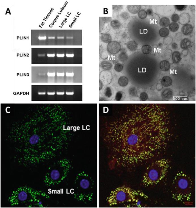
Fig. 4.1
Large and small bovine luteal cells express lipid droplet (LD) coat proteins and have unique LDs. Panel a: Expression of the PLIN family of LD coat proteins in bovine white adipose tissue, corpus luteum, and centrifugal elutriation-enriched large and small luteal cells (LC). Reverse transcriptase-polymerase chain reaction (RT-PCR) analysis of mRNA isolated from bovine fat and luteal tissue. Panel b: Electron microscopy of lipid droplets (LD) and mitochondria (Mt) in a bovine luteal cell. Panels c and d: Small and large luteal cells were stained with Bodipy 493/503 (Molecular Probes, 10 μg/ml) to detect neutral lipids (green). Nuclei: DAPI (blue). Cells in Panel d were were also immuno-labeled with with adipocyte triglyceride lipase (red) showing colocalization with the LDs and the difference in LD morphology between small and large luteal cells
Hormone-sensitive lipase (HSL) is a key cytosolic enzyme in the regulation of lipid stores in adipocytes that translocates to the LD in response to catecholamine stimulation [26–28]. A current view of the mechanisms regulating lipolysis in adipose tissue suggests that the LD-associated PLIN1 coats the LD and functions as a scaffold in the regulation of lipolysis [16, 29, 30]. Under basal conditions, PLIN1 acts as a barrier to the hydrolysis of lipids within the LD by preventing access of adipocyte triglyceride lipase (ATGL) and HSL, the major lipases in adipose cells. Following β-adrenergic stimulation of cAMP and protein kinase A (PKA) signaling, PLIN1 and HSL are phosphorylated, which leads to the movement of HSL from the cytosol to the LD [31]. The phosphorylation of HSL facilitates its association with the LD and with lipid substrates [32], permitting lipid hydrolysis to proceed. Phosphorylation of HSL by PKA occurs on multiple sites, including Ser‐563 and Ser‐660, which stimulate catalytic activity and translocation of HSL to LDs [33–36]. Phosphorylation of HSL also occurs at Ser-565, a non‐PKA site, which is a negative regulator of HSL activity and is believed to be mutually exclusive with phosphorylation on the Ser‐563 site [37]. Thus, hormonal cues that signal for elevations in systemic energy stimulate PKA to phosphorylate HSL, which contributes to lipolysis to maintain energy homeostasis.
The presence of both PLIN coat proteins [38] and HSL [39] in the ovary suggests that LH via a cAMP/PKA signaling pathway may regulate the phosphorylation of PLINs and HSL to hydrolyze cholesteryl esters stored in luteal LDs to produce substrate for progesterone synthesis . Studies with HSL-null mice revealed that knockout of HSL resulted in decreased steroidogenesis in the adrenals and inhibited sperm production in the testis [40, 41]. These findings suggest that HSL is involved in the intracellular processing and availability of cholesterol for adrenal and gonadal steroidogenesis. Manna et al. [42] recently reported that activation of the PKA pathway in MA-10 mouse Leydig cells enhanced expression of HSL and its phosphorylation at Ser-563 and Ser-660. Inhibition of HSL activity suppressed cAMP-induced progesterone synthesis and resulted in increased cholesteryl ester levels in MA-10 cells. Also of interest is a report [43] demonstrating an interaction between StAR and HSL in the rat adrenal following treatment with ACTH. Furthermore, the coexpression of StAR and HSL resulted in elevated HSL activity and mitochondrial cholesterol content. These observations suggest that the proteins that produce and transport cholesterol may colocalize in LDs and mitochondria . Furthermore, we have observed that mitochondria are closely associated with cytoplasmic LDs in bovine luteal cells (Fig. 4.1b) indicating that luteal LDs and mitochondria may interact to facilitate steroidogenesis. Although the evidence points to an important role for HSL in steroidogenesis, there is little information concerning the LD and the events that control these early steps in ovarian steroidogenesis [44].
Despite the renewed interest in cytoplasmic LD as platforms for cell signaling , interactions with other organelles, and metabolic control [45, 46], few studies have characterized the protein and lipid composition of the LD. The LD proteome has been characterized to varying degrees in a few mammalian tissues or cell lines (mouse mammary epithelial cells [47] and 3T3-L1 adipocytes [48, 49], rat liver and mouse muscle tissue [50, 51], and human cell lines [52–54]). Khor et al. [55] compared the proteome of LDs from rat granulosa cells treated in vitro with either high density lipoproteins or fatty acids to enrich cytoplasmic LDs with cholesteryl esters or triacylglycerides, respectively. When comparing the LD proteomes 278 proteins were common to the LDs prepared from either treatment. These proteins included PLIN2 and were similar to other studies on LD proteomes. They also identified 61 proteins unique to the cholesteryl ester-rich LDs and 40 unique proteins unique to triacylglycerol-rich LDs. Notably, they identified Hsd3b1, vimentin, and voltage-dependent anion channel (Vdac1) proteins enriched in the cholesteryl ester-rich LDs. Recent reports on the proteomic analysis of LD isolated from the mouse Leydig tumor cell line MLTC-1 [56] and mouse testes [57] also revealed the presence of PLIN family proteins and enzymes involved in the synthesis of steroid hormones. Despite the recent work on characterization of the LD proteome in various tissues, there is still a lack of information about the protein composition of luteal LDs and the effects of hormones or metabolic alterations on luteal LD properties. In our studies (Talbott, Krauss, and Davis, unpublished) the LDs isolated from bovine luteal tissue predominantly contain PLIN2 and PLIN3 coat proteins, as well as HSL, HSD3B1, CYP11A1, and StAR. Collectively, these studies indicate that the LD may serve as a novel hormonally responsive platform that is essential for steroidogenesis.
Comprehensive analysis of the lipid composition of LDs in other tissues is just beginning to be evaluated [58]. The protein composition of LDs, particularly the PLIN family of LD coat proteins, is believed to influence the type of lipids stored in LDs and metabolic activity of tissues [1, 59]. The lipid composition of ovarian LDs and the effects of hormones on the lipids contained therein are currently unknown. Our preliminary studies indicate that compared to granulosa and theca cells, the total lipid content of luteal cells is increased. Several studies reported the types and changes of lipids in the intact corpus luteum of rats [60], pigs [61, 62], sheep [63], and humans [64].These studies reported that cholesteryl esters and free fatty acids remain relatively constant during the functional phases of the luteal lifespan whereas triglycerides accumulated in the regressing corpus luteum. The increased lipid content of luteal cells is likely to be stored exclusively within the LDs; however, this remains to be shown experimentally. Additional studies are needed to determine the role and fate of lipids in LDs during both function and regression of the corpus luteum.
Bovine and ovine corpora lutea have two distinct steroidogenic cells , large and small luteal cells, with different abilities to produce progesterone [65–67]. The small luteal cells respond to LH with robust increases in progesterone secretion whereas the large luteal cells have a high basal rate of progesterone secretion and respond to LH with a comparatively modest fold increase in progesterone secretion. The luteal tissue of women, monkeys, pigs, and rodents also possess large and small luteal cells, although the basal and LH-stimulated progesterone secretion differ from the bovine corpus luteum [68]. Our preliminary data indicate that bovine large and small luteal cells have LDs with distinctive morphology. As indicated by BODIPY 493/503 staining of neutral lipids (green) and the LD protein adipocyte triglyceride lipase (ATGL), small luteal cells have large LDs, whereas large cells have abundant dispersed small LDs (Fig. 4.1c, d). Whether and how the LDs in either cell type contribute to the ability to respond to LH or to the basal rate of progesterone secretion is currently unknown. Studies in other tissues indicate that PKA-dependent phosphorylation of PLIN1 induces dispersion of clustered LDs in HEK293 cells , fibroblasts, and 3T3L1 adipocytes [69, 70]. Based on these findings it seems possible that the dispersed LDs observed in bovine large luteal cells may be the result of constitutive PKA activity reported to be present in large luteal cells [71].
Fatty acids (either synthesized de novo or provided by the hydrolysis of stored cholesteryl esters, triglycerides, or phospholipids) are essential for energy production and the synthesis of most lipids, including those found in membranes and lipids involved in cellular signaling. Despite their fundamental physiological importance, an oversupply of nonesterified fatty acids can be detrimental to cellular function [10]. Fatty acids are transported across the outer mitochondrial membrane by carnitine palmitoyltransferase I (CPT1A) , the rate-limiting step in fatty acid oxidation. Fatty acids are consumed by mitochondria through β-oxidation to produce acetyl-CoA, nicotinamide adenine dinucleotide (NADH), and flavin adenine dinucleotide (FADH2) for use in the electron transport chain to produce ATP [72]. The hydrolysis of cholesteryl esters by HSL liberates cholesterol and fatty acids (Fig. 4.2). The fatty acids are either re-esterified and stored in LDs or membranes or used for β-oxidation, producing reducing equivalents and acetyl-CoA for the citric acid cycle [72]. Although little is known about the role of fatty acid β-oxidation in luteal cells, recent studies indicate that fatty acid β-oxidation is a key in cumulus oocyte complex metabolism and oocyte maturation [73, 74]. These studies found that promoting β-oxidation with L-carnitine improved embryo development and that pharmacological inhibition of fatty acid β-oxidation with etomoxir, a CPT1A inhibitor, impaired oocyte maturation and embryo development. Steroidogenic tissues use glycolysis to support steroidogenesis [75]; however, it seems likely that the production of large quantities of progesterone by luteal cells would also require β-oxidation of fatty acids to provide the energy needed for optimal steroidogenesis under basal conditions, but this remains to be critically evaluated. It seems likely that large and small luteal cells may have different energy-processing requirements, based on the pronounced differences in the ability of large and small luteal cells to produce progesterone under basal and stimulated steroidogenesis. Our preliminary studies indicate that CPT1A mRNA expression in large luteal cells is 5.6 fold greater than in granulosa cells, whereas no difference in CPT1A mRNA expression was observed between theca and small luteal cells. These data support our idea that β-oxidation may be important in the metabolic regulation of large luteal cells. Given the intense interest in pathologies that result in lipid accumulation and conditions (i.e., obesity, diabetes, metabolic syndrome) that elevate free fatty acids and alter metabolism, understanding how LDs, glycolysis, and β-oxidation are regulated in the corpus luteum may provide clues for improving ovarian function, treating ovarian disorders, and enhancing fertility.
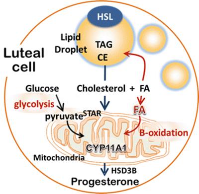
Fig. 4.2
Hormone-sensitive lipase (HSL) stimulates the hydrolysis of cholesteryl esters (CE) stored in lipid droplets to liberate cholesterol and fatty acids. The cholesterol is converted to pregnenolone by the cytochrome p450 side-chain cleavage enzyme (CYP11A1) in the mitochondria and subsequently converted by the enzyme 3β-hydroxy-steroid dehydrogenase (HSD3B) to progesterone. The released fatty acids (FA) are re-esterified and stored in the lipid droplets or used for energy production by mitochondrial β-oxidation
4.3 AMP-Activated Protein Kinase
The AMP-activated protein kinase (AMPK) is a master regulator of cellular metabolism [72, 76]. The AMPK complex is a heterotrimer consisting of an α-catalytic subunit and noncatalytic β- and γ-regulatory subunits [77]. Studies from a number of investigators demonstrate that AMPK is present in the oocyte, granulosa and theca cells of the follicle, as well as luteal cells (reviewed by Bertoldo et al. [78]). As its name suggests, AMPK is allosterically activated by adenosine monophosphate, AMP. The enzyme is activated by increases in AMP:ATP or ADP:ATP ratios , which occur when cellular energy status has been compromised by metabolic stresses that either interfere with ATP production or accelerate ATP consumption [79]. AMPK acts to restore energy homeostasis by activating alternate catabolic processes generating ATP while inhibiting energy-consuming processes , such as protein, carbohydrate, and lipid biosynthesis, as well as cell growth and proliferation (Fig. 4.3). AMPK acts via direct phosphorylation of metabolic enzymes and by longer-term effects via phosphorylation of transcription regulators [80, 81].
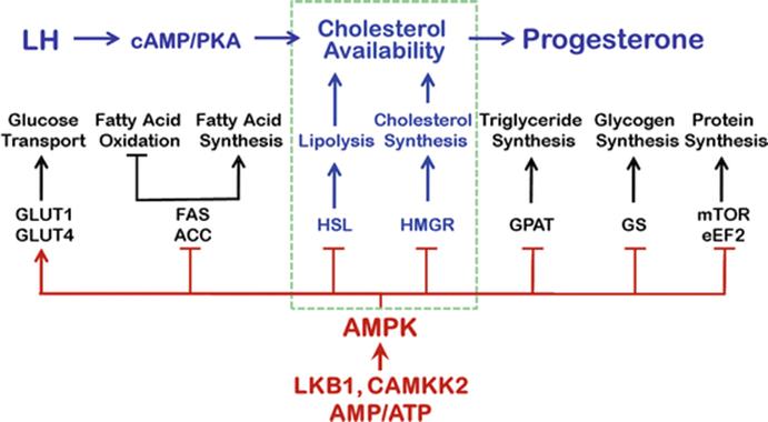
Fig. 4.3
Luteinizing hormone (LH) stimulates cAMP and protein kinase A (PKA) to activate proteins that will supply cholesterol for progesterone synthesis. The master metabolic regulator AMP-activated protein kinase (AMPK) is a highly conserved metabolic fuel gauge and can influence progesterone secretion by luteal cells. Elevations in AMP to ATP ratios stimulate AMPK to restore energy homeostasis by activating alternate catabolic processes generating ATP while inhibiting energy-consuming processes, that is, protein, carbohydrate, and lipid biosynthesis, as well as cell growth and proliferation. Activation of AMPK can disrupt steroidogenesis by phosphorylating and inhibiting hormone-sensitive lipase (HSL) and blocking HMGCR (3-hydroxy-3-methyl-glutaryl-coenzyme A reductase), the rate-controlling enzyme of the pathway that produces cholesterol. AMPK can be activated by the tumor suppressor kinase liver kinase B1 (LKB1) and the Ca2+/calmodulin activated protein kinase CaMKK2, which is activated when intracellular Ca2+ is increased by hormones such as PGF2α
AMPK can be activated by a number of synthetic allosteric effectors (A-769662, 991, MT 63–78) identified by Abbott Laboratories using high-throughput screens for AMPK. Other allosteric effectors are salicylate, the major breakdown product of aspirin, and pro-drugs: AICAR (5-amino-imidazole-4-carboxamide riboside) and C13, which are converted into AMP analogues following cellular uptake. For example, AICAR, a widely used AMPK activator, is taken into cells and then converted to the monophosphorylated derivative ZMP, which mimics the effect of AMP on both the allosteric activation of the kinase and inhibition of the dephosphorylation of Thr-172 on AMPK. Pharmacological AMPK activators (e.g., metformin, berberine, resveratrol, hydrogen peroxide) are typically viewed as metabolic poisons that inhibit ATP synthesis and stimulate AMPK indirectly by increasing cellular AMP levels [79]. Activation of AMPK by upstream kinases occurs by phosphorylation of a conserved threonine within the ‘activation loop’ of the kinase domain (Thr-172). The primary upstream kinases that phosphorylate Thr-172 are the tumor suppressor liver kinase B1 (LKB1) (also known as serine and threonine kinase 11 or STK11), and the calcium/calmodulin-dependent protein kinase kinase 2, CAMKK2. The latter is activated when intracellular Ca2+ is increased by the action of hormones.
AMPK likely controls multiple aspects of metabolism in ovarian cells . AMPK phosphorylates and inactivates acetyl-CoA carboxylase (ACC) and 3-hydroxy-3-methylglutaryl-CoA reductase (HMGCR), key enzymes involved in regulating de novo biosynthesis of fatty acids and cholesterol (Fig. 4.2). Activation of AMPK also blocks the activation of the mechanistic target of rapamycin (MTOR) and protein synthesis by phosphorylating the key regulatory proteins, raptor and tuberous sclerosis proteins [81]. Another immediate consequence of enhanced AMPK activity is the phosphorylation of HSL at Ser‐565, which precludes activation of HSL by PKA [82]. Conversely, conditions that stimulate PKA‐induced phosphorylation of HSL at Ser‐660 and Ser‐563 suppress the phosphorylation of HSL at the AMPK site Ser‐565. In vitro kinase assays using purified PKA and AMPK support the notion that phosphorylation of HSL at Ser‐563 and Ser‐565 is mutually exclusive. Thus, in steroidogenic tissues, activation of AMPK can inhibit HSL-mediated hydrolysis of cholesteryl esters and prevent the release of free cholesterol for steroidogenesis [83]. The observation that HSL is a key enzyme in adipocytes and steroidogenic cells strategically positions AMPK to control the expression of genes required for steroidogenesis and the availability of cholesterol for ovarian progesterone synthesis (Fig. 4.4).
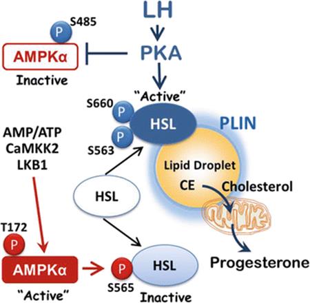
Fig. 4.4
Luteinizing hormone (LH) stimulates protein kinase A (PKA)-dependent phosphorylation on Ser-563 and Ser-660, resulting in activation of hormone-sensitive lipase (HSL), which hydrolyzes cholesteryl esters (CE) stored in lipid droplets (LD) to release cholesterol and fatty acids (FA). AMP-activated protein kinase (AMPK) suppresses the activation of HSL by phosphorylation of HSL on Ser-565. LH also inactivates AMPK by increasing AMPK phosphorylation on Ser-485 and reducing phosphorylation on Thr-172. The ability of LH to suppress AMPK and activate HSL ensures adequate cholesterol availability for progesterone synthesis
Reports from the DuPont Laboratory [84, 85] demonstrate that AMPK activators metformin and AICAR inhibit the secretion of progesterone or estradiol by granulosa cells in a manner dependent on the state of cellular differentiation and the species investigated [78, 84, 86]. In rat and bovine granulosa cells, AMPK activation induced by metformin reduced the expression of mRNA for key enzymes required for progesterone synthesis, HSD3B1, CYP11A1, and StAR [85, 87]. In the human KGN granulosa cell line (L. Huang, X. Hou and J.S. Davis, unpublished data), treatment with the AMPK activator metformin inhibited StAR expression and progesterone synthesis. In general, the studies in granulosa cells suggest that the reduction in steroidogenesis was a result of a reduction in the transcription of genes in the steroidogenic pathway . Other studies showed that metformin impairs proliferation of bovine granulosa cells and rat theca cells via mechanisms involving AMPK-mediated inhibition of MTOR signaling and protein synthesis [88–90].
Bowdridge et al. recently reported increases in the expression of AMPKα-, β-, and γ-subunits during the maturation of the bovine corpus luteum , with the exception of AMPKγ1- and γ2-subunits [91]. Other studies from the Flores Laboratory provide evidence for increased expression of genes encoding distinct protein kinase C isoforms and genes participating in Ca2+ homeostasis during luteal maturation [92]. Goravanahally et al. [93] reported that CAMKK2, a downstream target of Ca2+ and upstream regulator of AMPK, is also more highly expressed in mature bovine corpus luteum than in newly formed luteal tissue. It should be noted that two important physiological processes occur during this developmental period: (1) the corpus luteum develops maximal capacity for progesterone secretion and (2) the corpus luteum develops the capacity to undergo luteolysis in response to PGF2α. Based on the high rate of progesterone production during the mid-luteal phase and pregnancy, it seems likely that any factors that influence metabolic activity in steroidogenic cells would increase or decrease AMPK activity and impact steroid secretion. Hou et al. [94] reported that treatment of primary cultures of bovine luteal cells with AICAR rapidly increased AMPK activity and significantly reduced LH-stimulated MTOR activity and progesterone secretion. Additional findings in this report indicated that the response to AICAR was independent of MTOR since other experiments showed that inhibition of MTOR with rapamycin did not contribute to the reduction in LH-stimulated progesterone secretion. More recently, Bowdridge et al. [91] observed that treatment of bovine luteal tissue slices with either metformin or AICAR acutely reduced basal progesterone secretion. These results indicate that AMPK activators acutely inhibit luteal progesterone synthesis , suggesting that the energy status of luteal cells is an important regulator of steroidogenesis.
4.4 LH Inhibits AMPK
The C-terminal domains of AMPKα-subunit isoforms in vertebrates contain a serine/threonine-rich insert of 50–60 amino acids, the so-called ST loop [95]. Phosphorylation of the ST loop serves as a means for negative regulation of AMPK. The amino acid residues defining the ends of this loop are close to the Thr-172 residue and contain a number of regulatory phosphorylation sites. The best characterized of these sites is Ser-485 on the AMPKα1-subunit. The Ser-485 site is phosphorylated by the cyclic AMP-dependent protein kinase, PKA [96], or by Akt [97], which subsequently inhibits the phosphorylation of the AMPKα-subunit Thr-172 residue by upstream kinases, LKB1 or CaMKK2 [95]. The AMPK-α2 subunit contains a similar conserved ST loop, and phosphorylation of Ser-491 is likely to exert the same inhibitory effect, although Ser-491 is a poor substrate for Akt and appears to be modified by autophosphorylation [95]. Additionally, PKA can phosphorylate the Ser-173 residue (adjacent to Thr-172 within the activation loop), which can inhibit Thr-172 phosphorylation [98]. In a study using primary cultures of bovine luteal cells, Hou et al. reported that treatment with LH rapidly inhibited AMPK activity as evidenced by reduced AMPK Thr-172 phosphorylation and reduced phosphorylation of the AMPK substrate acetyl-CoA carboxylase [94]. Treatment with LH also increased phosphorylation of AMPK on Ser-485, which is associated with inhibition of AMPK activity [94].
In contrast to granulosa cells , bovine luteal cells contain the required steroidogenic machinery including HSL, which enables luteal cells to respond to LH or cAMP with rapid increases in progesterone synthesis. The increases in progesterone occur within 10–30 min [99–101] and precede the LH-induced increase in STAR expression, which is typically observed 2–4 h after treatment [102]. These changes are associated with reduced phosphorylation of HSL at the inhibitory AMPK phosphorylation site Ser-565 and increased phosphorylation of HSL at Ser-563 and -660, residues that are required for HSL activity (Krause, Talbott, Hou, and Davis, unpublished). Thus, the ability of LH to reduce AMPK activity may allow optimal LH- and PKA-dependent activation of HSL and provide cholesterol for the already existing steroidogenic machinery. An experimental model of the proposed interaction among PKA and AMPK regulation of HSL is shown in Fig. 4.4. Physiological conditions that increase the activity of AMPK require phosphorylation of the AMPK α-subunit on Thr-172 residues [103], leadings to the phosphorylation of the AMPK substrates ACC (Ser-79) and HSL (Ser-565), which could reduce the ability of luteal cells to provide cholesterol substrate in response to a pulse of LH. LH or PKA activators attenuate AMPK activity through modulation of at least two AMPK α-subunit phosphorylation sites, Thr-172 (reduced), and Ser-485 (increased). Reduced HSL phosphorylation by AMPK allows PKA to phosphorylate HSL on Ser-563 and Ser-660 resulting in increased HSL activity, which presumptively provides cholesterol for progesterone synthesis.
4.5 PGF2α Activates AMPK
Early studies established that PGF2α binds to and activates its cognate Gq protein-coupled receptor, the prostaglandin F receptor, PTGFR. This initial event provokes the rapid activation of phospholipase C, which leads to increases in both cytoplasmic Ca2+ and activation of protein kinase C. These early events contribute to the activation of additional protein kinase cascades such as the mitogen-activated protein kinases (ERK1/2, p38, and JNK) [104] that contribute to the induction of early-response genes such as FOS, JUN, EGR1, and ATF3 [105–108]. Although these early-response genes have been implicated in the luteolytic response to PGF2α, it is not clear how or whether they impact metabolic events in luteal cells. The developmental-specific expression of protein kinase C and CAMKK2 isoforms, proteins involved in Ca2+ homeostasis, and AMPK have been implicated in the cellular mechanisms of acquisition of luteolytic capacity by bovine corpus luteum [92, 93, 109]. Based on these observations it seems reasonable to predict that PGF2α could activate Ca2+/CAMKK2 pathways leading to the activation and phosphorylation of AMPK on Thr-172.
Bowdridge et al. [91] recently reported that PGF2α rapidly (2 min) and transiently stimulated the phosphorylation of AMPK on the Ser-485 site in dispersed bovine luteal cells . The response was prevented by treatment with STO-609, a CAMKK2 inhibitor. Treatment with STO-609 also prevented the modest inhibitory effect of PGF2α on progesterone synthesis in overnight incubations of dispersed luteal cells [91]. In recent studies using bovine large luteal cells, we have observed that PGF2α rapidly stimulates the phosphorylation of AMPK on the stimulatory Thr-172 residue as well as the inhibitory Ser-485 residue (Hou, Zhang, Talbott, and Davis, unpublished data). The phosphorylation of AMPK was coupled to the phosphorylation of the AMPK target ACC, indicating that AMPK was activated by PGF2α. The observation that PGF2α can target multiple sites on AMPK is consistent with findings that PGF2α activates multiple protein kinase pathways in luteal cells: pathways linked to calcium signaling, protein kinase C, mitogen-activated protein kinases, and MTOR signaling [110]. Although additional studies are needed to determine exactly how PGF2α regulates AMPK in luteal cells, it seems clear that activation of AMPK with pharmacological tools disrupts luteal progesterone synthesis (Fig. 4.5). Studies are also needed to determine whether AMPK is activated in vivo during natural and PGF2α-induced luteolysis. It is conceivable that changes in luteal blood flow, hypoxia, and the presence of inflammatory mediators all contribute to altering the metabolic status of steroidogenic luteal cells , resulting in the activation of AMPK and disrupting progesterone synthesis.
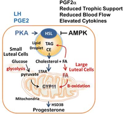
Fig. 4.5
LH and PGF2α have opposite effects on AMP-activated protein kinase (AMPK). LH-dependent activation of protein kinase A (PKA) activates hormone-sensitive lipase (HSL). In contrast, activation of AMPK blocks activation of HSL. LH-dependent stimulation of cellular metabolism regulates the use of glucose and fatty acids (FA) for optimal progesterone synthesis. Conditions that activate AMPK (hormones, cytokines, reduced nutrients, reduced blood flow, hypoxia, drugs, and environmental insults) reduce the ability of LH to provide cholesterol for progesterone synthesis
4.6 Autophagy
Autophagy plays an important role in cellular and tissue physiology [111–113]. The main function of autophagy is to protect cells against starvation by allowing cells to salvage nutrients by digesting organelles and macromolecules at times of nutrient scarcity as well as to ensure cell homeostasis by eliminating damaged organelles and misfolded proteins. Three different types of autophagy (macroautophagy, microautophagy, and chaperone-mediated autophagy) have been described, based largely on the processes by which cargo is delivered to the lysosomes. In general, autophagy can be induced by limitations in amino acids, growth factors, energy, and oxygen. The formation of autophagosomes requires the activation of a number of protein complexes: the autophagy-related 1 (Atg1)–Unc-51-like kinase complex, which is a key signaling intermediate that is regulated by MTOR and AMPK; the autophagy-specific class III phosphatidylinositol 3-kinase Vps34 complex (consisting of Vsp34, Beclin 1, Vsp15, and Atg14L), which produce a pool of phosphatidylinositol-3-phosphate that is necessary for autophagosome formation; and a complex of ubiquitin-like proteins: Atg12, Atg5, Atg16, and LC3-I (Atg8) and their conjugation machinery, which leads to the lipidation of microtubule-associated protein light chain 3 (LC3) with phosphatidylethanolamine, a process required for autophagosome formation and closure. The presence of LC3-II, an LC3 cleavage product, inside the mature autophagosome is generally used as a marker of autophagy.
Autophagy has been shown to occur in oocytes, granulosa cells , and luteal cells and is often associated with apoptosis. Genetic mouse models demonstrate that Atg7(−/−) ovaries [114] or germ cell-specific deletion of Atg7[115] compromised autophagy in the perinatal mouse ovary, resulting in the early loss of female germ cells. Loss of Beclin 1 (Becn1), which has a central role in the regulation of autophagy through activation of the Vps34 complex, also resulted in a significant loss of germ cells at birth [114]. These findings indicate that autophagy may promote survival of germ cells during ovarian development. Other studies provide evidence for the presence of autophagosomes in the granulosa cells of atretic follicles of several species [116, 117]. Studies in the rat support the idea that activation of the AKT/MTOR signaling pathway suppresses autophagy as assessed by levels of LC3-II in granulosa cells [116].
The presence of lysosomes and autophagosomes in the corpus luteum was described more than 45 years ago [118–121]. Recent studies have documented the presence of autophagy-related proteins: Beclin 1 and LC3 in luteal tissue of rodents, cows, and humans [122–125]. However, in luteal cells, it remains unclear whether autophagy promotes cell survival versus cell death. In the rat, LC3-II-positive autophagosomes were identified during the late luteal phase and were correlated with luteal cell apoptosis [125, 126]. Furthermore, treatment of rat luteal cells with PGF2α under serum-free conditions increased autophagosomes, LC3-II protein , and luteal cell apoptosis, suggesting that autophagy may be involved in luteal cell death. Choi et al. [126] observed that although PGF2α increased both ERK1/2 and MTOR activity in rat luteal cells, autophagy could be prevented by inhibition of ERK1/2 signaling and appeared to be independent of phosphatidylinositol 3-kinase/AKT/MTOR activity. It will be important to understand the sequence of events in vivo and to determine whether the stimulatory effects of PGF2α on AMPK activation are linked in some way to autophagy in the corpus luteum.
Gawriluk et al. reported that Becn1 deficiency in the mouse ovary resulted in a reduction of progesterone production and preterm labor [122]. To avoid the loss of germ cells associated the Becn1 knockout animal, this group targeted Becn1 deletion to the granulosa cells and as a result they were able to follow luteal function throughout pregnancy. Although ovulation, implantation, and progesterone levels during early pregnancy were not affected by Becn1 ablation, they found that Becn1 abrogation resulted in a reduction of circulating progesterone in mid- to late pregnancy. The reduction in progesterone resulted in early parturition, which was reversed by treatment with exogenous progesterone. Of relevance to luteal metabolism were the findings that the numbers of LDs were reduced and the mitochondria were smaller in the Becn1-deficient ovaries compared to controls. These changes were not accompanied by changes in the expression of genes important for the synthesis of progesterone. Exactly how the reduction in LDs and reduced autophagy contributed to reduced progesterone synthesis remains to be firmly established, but it could be a consequence of impaired lipid transport mechanisms and reduced expression of key receptors on the luteal cells [122]. Studies in other systems indicate that Becn1 expression and activity is controlled via transcriptional regulation, miR-30a, and by posttranslational modifications (reviewed in [127]). Recent studies in cardiac tissue showed that the transcription factor ATF3 binds to the ATF/cAMP response element of the Becn1 promoter and that ATF3 is capable of reducing autophagy via suppression of the Becn1-dependent autophagy pathway [128]. As PGF2α rapidly increases activation of mitogen-activated protein kinases (ERK1/2, p38, and JNK) and ATF3 expression in bovine and rat luteal cells in vivo and in vitro [104, 107], it is important to determine whether Becn1 expression or activity impacts autophagy during luteal regression.
It should also be appreciated that Becn1 directly interacts with B-cell lymphoma 2 (Bcl2) family proteins (Bcl2 and Bcl2/XL) in a manner that negatively regulates autophagy. To complicate matters, a variety of ligands that regulate intracellular protein kinases , including Dapk, Rock1, Mst1, and Mapk8 (death-associated protein kinase 1, rho-associated coiled-coil containing protein kinase 1, macrophage stimulating 1, mitogen-activating protein kinase 8, respectively), can positively or negatively regulate Becn1/Bcl2 effects on autophagy [127]. Beclin 1 can also secondarily affect apoptosis through regulation of anti-apoptotic and pro-apoptotic BH3 domain-containing proteins. In addition to the Bcl2 family, the VDAC (voltage-dependent anion channel) family is also involved in ovarian apoptosis and autophagy regulation [129]. Vdac2 directly interacts with Bcl2-antagonist/killer 1 (Bak1) to inhibit its oligomerization, thus suppressing cell apoptosis. Yuan et al. [129] recently reported that Vdac2 inhibits autophagy in the developing ovary by interacting with Becn1 and Bcl2L1 to stabilize the Becn1 and Bcl2L1 complex. Recent work by several groups have found a close relationship between autophagy and LDs [130–132]. In particular, LC3 [131], and ATG2 [133], ATG7 [130], and several VDAC [56, 57] proteins are often associated with LDs and appear to have important roles in LD formation and function, suggesting that events associated with autophagy may also impact the formation and function of ovarian LDs. Further work is needed to understand how LDs and autophagosome components influence both autophagy and apoptosis and thereby affect luteal function and lifespan.
4.7 Summary
Metabolic processes in the corpus luteum are tightly controlled by luteotropic and luteolytic factors. Signaling cascades involving LD homeostasis, PKA, AMPK, and autophagy are clearly important in the control of steroidogenesis. It remains to be determined how these cellular events are integrated into a physiologic context over the lifespan of the corpus luteum. Understanding the complex interplay of metabolic and hormonal clues underpinning steroidogenesis is essential to understanding and developing new therapies for infertility, particularly in the setting of increasing prevalence of obesity and metabolic diseases such as diabetes and polycystic ovary syndrome.
Acknowledgments
This project was supported by Agriculture and Food Research Initiative Competitive Grant no. 2011-67015-20076 from the USDA National Institute of Food and Agriculture (NIFA) to J.S.D. and a NIFA Pre-doctoral award 2014-67011-22280 to H.T. The work was also supported in part by the Department of Veterans Affairs, Office of Research and Development Biomedical Laboratory Research and Development funds; and The Olson Center for Women’s Health, Department of Obstetrics and Gynecology, Nebraska Medical Center, Omaha, NE.
References
1.
Greenberg AS, Coleman RA, Kraemer FB, McManaman JL, Obin MS, Puri V, Yan QW, Miyoshi H, Mashek DG. The role of lipid droplets in metabolic disease in rodents and humans. J Clin Invest. 2011;121:2102–10.PubMedPubMedCentral
2.
Thiam AR, Farese Jr RV, Walther TC. The biophysics and cell biology of lipid droplets. Nat Rev Mol Cell Biol. 2013;14:775–86.PubMedPubMedCentral
3.
Waltermann M, Steinbuchel A. Neutral lipid bodies in prokaryotes: recent insights into structure, formation, and relationship to eukaryotic lipid depots. J Bacteriol. 2005;187:3607–19.PubMedPubMedCentral
4.
Natarajan SK, Rasineni K, Ganesan M, Feng D, McVicker BL, McNiven MA, Osna NA, Mott JL, Casey CA, Kharbanda KK. Structure, function and metabolism of hepatic and adipose tissue lipid droplets: implications in alcoholic liver disease. Curr Mol Pharmacol. 2015 Aug 17. [Epub ahead of print] PMID: 26278390.
5.
D’Souza K, Nzirorera C, Kienesberger PC. Lipid metabolism and signaling in cardiac lipotoxicity. Biochim Biophys Acta. 2016 Oct;1860 (10):1513–24. doi:10.1016/j.bbalip.2016.02.016.
6.
Yuan Y, Li P, Ye J. Lipid homeostasis and the formation of macrophage-derived foam cells in atherosclerosis. Protein Cell. 2012;3:173–81.PubMedPubMedCentral
7.
Servetnick DA, Brasaemle DL, Gruia-Gray J, Kimmel AR, Wolff J, Londos C. Perilipins are associated with cholesteryl ester droplets in steroidogenic adrenal cortical and Leydig cells. J Biol Chem. 1995;270:16970–3.PubMed
8.
Shen WJ, Azhar S, Kraemer FB. Lipid droplets and steroidogenic cells. Exp Cell Res. 2016;340:209–14.PubMed
9.
Konige M, Wang H, Sztalryd C. Role of adipose specific lipid droplet proteins in maintaining whole body energy homeostasis. Biochim Biophys Acta. 2014 Mar;1842(3):393–401. doi:10.1016/j.bbadis.2013.05.007.
10.
Zechner R, Zimmermann R, Eichmann TO, Kohlwein SD, Haemmerle G, Lass A, Madeo F. FAT SIGNALS—lipases and lipolysis in lipid metabolism and signaling. Cell Metab. 2012;15:279–91.PubMedPubMedCentral
11.
Beller M, Thiel K, Thul PJ, Jackle H. Lipid droplets: a dynamic organelle moves into focus. FEBS Lett. 2010;584:2176–82.PubMed
12.
Brasaemle DL, Wolins NE. Packaging of fat: an evolving model of lipid droplet assembly and expansion. J Biol Chem. 2012;287:2273–9.PubMed
13.
Farese Jr RV, Walther TC. Lipid droplets finally get a little R-E-S-P-E-C-T. Cell. 2009;139:855–60.PubMedPubMedCentral
14.
Yang H, Galea A, Sytnyk V, Crossley M. Controlling the size of lipid droplets: lipid and protein factors. Curr Opin Cell Biol. 2012;24:509–16.PubMed
15.
Olofsson SO, Bostrom P, Andersson L, Rutberg M, Perman J, Boren J. Lipid droplets as dynamic organelles connecting storage and efflux of lipids. Biochim Biophys Acta. 1791;2009:448–58.
16.
Brasaemle DL. Thematic review series: adipocyte biology. The perilipin family of structural lipid droplet proteins: stabilization of lipid droplets and control of lipolysis. J Lipid Res. 2007;48:2547–59.PubMed
17.
Kimmel AR, Brasaemle DL, McAndrews-Hill M, Sztalryd C, Londos C. Adoption of PERILIPIN as a unifying nomenclature for the mammalian PAT-family of intracellular lipid storage droplet proteins. J Lipid Res. 2010;51:468–71.PubMedPubMedCentral
18.
Paul A, Chan L, Bickel PE. The PAT family of lipid droplet proteins in heart and vascular cells. Curr Hypertens Rep. 2008;10:461–6.PubMedPubMedCentral
19.
Nishino N, Tamori Y, Tateya S, Kawaguchi T, Shibakusa T, Mizunoya W, Inoue K, Kitazawa R, Kitazawa S, Matsuki Y, Hiramatsu R, Masubuchi S, Omachi A, Kimura K, Saito M, Amo T, Ohta S, Yamaguchi T, Osumi T, Cheng J, Fujimoto T, Nakao H, Nakao K, Aiba A, Okamura H, Fushiki T, Kasuga M. FSP27 contributes to efficient energy storage in murine white adipocytes by promoting the formation of unilocular lipid droplets. J Clin Invest. 2008;118:2808–21.PubMedPubMedCentral
20.
McManaman JL, Bales ES, Orlicky DJ, Jackman M, MacLean PS, Cain S, Crunk AE, Mansur A, Graham CE, Bowman TA, Greenberg AS. Perilipin-2-null mice are protected against diet-induced obesity, adipose inflammation, and fatty liver disease. J Lipid Res. 2013;54:1346–59.PubMedPubMedCentral
21.
Sztalryd C, Bell M, Lu X, Mertz P, Hickenbottom S, Chang BH, Chan L, Kimmel AR, Londos C. Functional compensation for adipose differentiation-related protein (ADFP) by Tip47 in an ADFP null embryonic cell line. J Biol Chem. 2006;281:34341–8.PubMed
22.
Chen W, Chang B, Wu X, Li L, Sleeman M, Chan L. Inactivation of Plin4 downregulates Plin5 and reduces cardiac lipid accumulation in mice. Am J Physiol Endocrinol Metab. 2013;304:E770–9.PubMedPubMedCentral
23.
Seachord CL, VandeVoort CA, Duffy DM. Adipose differentiation-related protein: a gonadotropin- and prostaglandin-regulated protein in primate periovulatory follicles. Biol Reprod. 2005;72:1305–14.PubMed
24.
Yang X, Dunning KR, Wu LL, Hickey TE, Norman RJ, Russell DL, Liang X, Robker RL. Identification of perilipin-2 as a lipid droplet protein regulated in oocytes during maturation. Reprod Fertil Dev. 2010;22:1262–71.PubMed
25.
Feingold KR, Kazemi MR, Magra AL, McDonald CM, Chui LG, Shigenaga JK, Patzek SM, Chan ZW, Londos C, Grunfeld C. ADRP/ADFP and Mal1 expression are increased in macrophages treated with TLR agonists. Atherosclerosis. 2010;209:81–8.PubMed
26.
Holm C. Molecular mechanisms regulating hormone-sensitive lipase and lipolysis. Biochem Soc Trans. 2003;31:1120–4.PubMed
27.
Kraemer FB. Adrenal cholesterol utilization. Mol Cell Endocrinol. 2007;265-266:42–5.PubMed
28.
Osterlund T. Structure-function relationships of hormone-sensitive lipase. Eur J Biochem. 2001;268:1899–907.PubMed
29.
Bickel PE, Tansey JT, Welte MA. PAT proteins, an ancient family of lipid droplet proteins that regulate cellular lipid stores. Biochim Biophys Acta. 1791;2009:419–40.
30.
Lampidonis AD, Rogdakis E, Voutsinas GE, Stravopodis DJ. The resurgence of Hormone-Sensitive Lipase (HSL) in mammalian lipolysis. Gene (Amst). 2011;477:1–11.
31.
Lafontan M, Langin D. Lipolysis and lipid mobilization in human adipose tissue. Prog Lipid Res. 2009;48:275–97.PubMed
32.
Krintel C, Morgelin M, Logan DT, Holm C. Phosphorylation of hormone-sensitive lipase by protein kinase A in vitro promotes an increase in its hydrophobic surface area. FEBS J. 2009;276:4752–62.PubMed
33.
Su CL, Sztalryd C, Contreras JA, Holm C, Kimmel AR, Londos C. Mutational analysis of the hormone-sensitive lipase translocation reaction in adipocytes. J Biol Chem. 2003;278:43615–9.PubMed
34.
Shen WJ, Patel S, Natu V, Kraemer FB. Mutational analysis of structural features of rat hormone-sensitive lipase. Biochemistry. 1998;37:8973–9.PubMed
35.
Anthonsen MW, Ronnstrand L, Wernstedt C, Degerman E, Holm C. Identification of novel phosphorylation sites in hormone-sensitive lipase that are phosphorylated in response to isoproterenol and govern activation properties in vitro. J Biol Chem. 1998;273:215–21.PubMed
36.
Miyoshi H, Souza SC, Zhang HH, Strissel KJ, Christoffolete MA, Kovsan J, Rudich A, Kraemer FB, Bianco AC, Obin MS, Greenberg AS. Perilipin promotes hormone-sensitive lipase-mediated adipocyte lipolysis via phosphorylation-dependent and -independent mechanisms. J Biol Chem. 2006;281:15837–44.PubMed
37.
Watt MJ, Holmes AG, Pinnamaneni SK, Garnham AP, Steinberg GR, Kemp BE, Febbraio MA. Regulation of HSL serine phosphorylation in skeletal muscle and adipose tissue. Am J Physiol Endocrinol Metab. 2006;290:E500–8.PubMed
38.
Londos C, Brasaemle DL, Gruia-Gray J, Servetnick DA, Schultz CJ, Levin DM, Kimmel AR. Perilipin: unique proteins associated with intracellular neutral lipid droplets in adipocytes and steroidogenic cells. Biochem Soc Trans. 1995;23:611–5.PubMed
39.
Lobo MV, Huerta L, Arenas MI, Busto R, Lasuncion MA, Martin-Hidalgo A. Hormone-sensitive lipase expression and IHC localization in the rat ovary, oviduct, and uterus. J Histochem Cytochem. 2009;57:51–60.PubMedPubMedCentral
40.
Kraemer FB, Shen WJ, Harada K, Patel S, Osuga J, Ishibashi S, Azhar S. Hormone-sensitive lipase is required for high-density lipoprotein cholesteryl ester-supported adrenal steroidogenesis. Mol Endocrinol. 2004;18:549–57.PubMed
41.
Kraemer FB, Shen WJ, Natu V, Patel S, Osuga J, Ishibashi S, Azhar S. Adrenal neutral cholesteryl ester hydrolase: identification, subcellular distribution, and sex differences. Endocrinology. 2002;143:801–6.PubMed
42.
Manna PR, Cohen-Tannoudji J, Counis R, Garner CW, Huhtaniemi I, Kraemer FB, Stocco DM. Mechanisms of action of hormone-sensitive lipase in mouse Leydig cells: its role in the regulation of the steroidogenic acute regulatory protein. J Biol Chem. 2013;288:8505–18.PubMedPubMedCentral
43.
Shen WJ, Patel S, Natu V, Hong R, Wang J, Azhar S, Kraemer FB. Interaction of hormone-sensitive lipase with steroidogenic acute regulatory protein: facilitation of cholesterol transfer in adrenal. J Biol Chem. 2003;278:43870–6.PubMed
44.
Rone MB, Fan J, Papadopoulos V. Cholesterol transport in steroid biosynthesis: role of protein-protein interactions and implications in disease states. Biochim Biophys Acta. 1791;2009:646–58.
45.
Arrese EL, Saudale FZ, Soulages JL. Lipid droplets as signaling platforms linking metabolic and cellular functions. Lipid Insights. 2014;7:7–16.PubMedPubMedCentral
46.
Murphy S, Martin S, Parton RG. Lipid droplet-organelle interactions; sharing the fats. Biochim Biophys Acta. 1791;2009:441–7.
47.
Wu CC, Howell KE, Neville MC, Yates 3rd JR, McManaman JL. Proteomics reveal a link between the endoplasmic reticulum and lipid secretory mechanisms in mammary epithelial cells. Electrophoresis. 2000;21:3470–82.PubMed
48.
Brasaemle DL, Dolios G, Shapiro L, Wang R. Proteomic analysis of proteins associated with lipid droplets of basal and lipolytically stimulated 3T3-L1 adipocytes. J Biol Chem. 2004;279:46835–42.PubMed
49.
Cho SY, Shin ES, Park PJ, Shin DW, Chang HK, Kim D, Lee HH, Lee JH, Kim SH, Song MJ, Chang IS, Lee OS, Lee TR. Identification of mouse Prp19p as a lipid droplet-associated protein and its possible involvement in the biogenesis of lipid droplets. J Biol Chem. 2007;282:2456–65.PubMed
50.
Turro S, Ingelmo-Torres M, Estanyol JM, Tebar F, Fernandez MA, Albor CV, Gaus K, Grewal T, Enrich C, Pol A. Identification and characterization of associated with lipid droplet protein 1: a novel membrane-associated protein that resides on hepatic lipid droplets. Traffic 2006;7:1254–69.
51.
Zhang H, Wang Y, Li J, Yu J, Pu J, Li L, Zhang H, Zhang S, Peng G, Yang F, Liu P. Proteome of skeletal muscle lipid droplet reveals association with mitochondria and apolipoprotein a-I. J Proteome Res. 2011;10:4757–68.PubMed
52.
Umlauf E, Csaszar E, Moertelmaier M, Schuetz GJ, Parton RG, Prohaska R. Association of stomatin with lipid bodies. J Biol Chem. 2004;279:23699–709.PubMed
53.
Fujimoto Y, Itabe H, Sakai J, Makita M, Noda J, Mori M, Higashi Y, Kojima S, Takano T. Identification of major proteins in the lipid droplet-enriched fraction isolated from the human hepatocyte cell line HuH7. Biochim Biophys Acta. 1644;2004:47–59.
54.
Sato S, Fukasawa M, Yamakawa Y, Natsume T, Suzuki T, Shoji I, Aizaki H, Miyamura T, Nishijima M. Proteomic profiling of lipid droplet proteins in hepatoma cell lines expressing hepatitis C virus core protein. J Biochem. 2006;139:921–30.PubMed
55.
Khor VK, Ahrends R, Lin Y, Shen WJ, Adams CM, Roseman AN, Cortez Y, Teruel MN, Azhar S, Kraemer FB. The proteome of cholesteryl-ester-enriched versus triacylglycerol-enriched lipid droplets. PLoS One. 2014;9, e105047.PubMedPubMedCentral
56.
Yamaguchi T, Fujikawa N, Nimura S, Tokuoka Y, Tsuda S, Aiuchi T, Kato R, Obama T, Itabe H. Characterization of lipid droplets in steroidogenic MLTC-1 Leydig cells: protein profiles and the morphological change induced by hormone stimulation. Biochim Biophys Acta. 1851;2015:1285–95.
57.
Wang W, Wei S, Li L, Su X, Du C, Li F, Geng B, Liu P, Xu G. Proteomic analysis of murine testes lipid droplets. Sci Rep. 2015;5:12070.PubMedPubMedCentral
58.
Chitraju C, Trotzmuller M, Hartler J, Wolinski H, Thallinger GG, Lass A, Zechner R, Zimmermann R, Kofeler HC, Spener F. Lipidomic analysis of lipid droplets from murine hepatocytes reveals distinct signatures for nutritional stress. J Lipid Res. 2012;53:2141–52.PubMedPubMedCentral
59.
Xu L, Zhou L, Li P. CIDE proteins and lipid metabolism. Arteriosc Thromb Vasc Biol. 2012;32:1094–8.
60.
Strauss 3rd JF, Seifter E, Lien EL, Goodman DB, Stambaugh RL. Lipid metabolism in regressing rat corpora lutea of pregnancy. J Lipid Res. 1977;18:246–58.PubMed
61.
Waterman RA. Lipid and arachidonic acid accumulation in naturally regressing porcine corpora lutea. Prostaglandins. 1980;20:57–71.PubMed
62.
Waterman RA, Guthrie HD. Effects of Cloprostenol administration on neutral lipid and prostaglandin F metabolism by porcine luteal tissue. Prostaglandins. 1984;27:131–46.PubMed
63.
Waterman RA. Changes in lipid contents and fatty acid compositions in ovine corpora lutea during the estrous cycle and early pregnancy. Biol Reprod. 1988;38:605–15.PubMed
64.
Weinhouse S, Brewer JI. Cyclic variations in the lipids of the coprus luteum. J Biol Chem. 1942;143:617–23.
65.
Davis JS, Rueda BR. The corpus luteum: an ovarian structure with maternal instincts and suicidal tendencies. Front Biosci. 2002;7:d1949–78.PubMed
66.
Alila HW, Dowd JP, Corradino RA, Harris WV, Hansel W. Control of progesterone production in small and large bovine luteal cells separated by flow cytometry. J Reprod Fertil. 1988;82:645–55.PubMed
67.
Niswender GD, Davis TL, Griffith RJ, Bogan RL, Monser K, Bott RC, Bruemmer JE, Nett TM. Judge, jury and executioner: the auto-regulation of luteal function. Soc Reprod Fertil Suppl. 2007;64:191–206.PubMed
68.
Wiltbank MC, Salih SM, Atli MO, Luo W, Bormann CL, Ottobre JS, Vezina CM, Mehta V, Diaz FJ, Tsai SJ, Sartori R. Comparison of endocrine and cellular mechanisms regulating the corpus luteum of primates and ruminants. Anim Reprod. 2012;9:242–59.PubMedPubMedCentral
69.
Marcinkiewicz A, Gauthier D, Garcia A, Brasaemle DL. The phosphorylation of serine 492 of perilipin a directs lipid droplet fragmentation and dispersion. J Biol Chem. 2006;281:11901–9.PubMed
70.
Orlicky DJ, Monks J, Stefanski AL, McManaman JL. Dynamics and molecular determinants of cytoplasmic lipid droplet clustering and dispersion. PLoS One. 2013;8, e66837.PubMedPubMedCentral
71.
Bogan RL, Niswender GD. Constitutive steroidogenesis in ovine large luteal cells may be mediated by tonically active protein kinase A. Biol Reprod. 2007;77:209–16.PubMed
72.
O’Neill HM, Holloway GP, Steinberg GR. AMPK regulation of fatty acid metabolism and mitochondrial biogenesis: implications for obesity. Mol Cell Endocrinol. 2013;366:135–51.
73.
Dunning K, Russell DL, Robker R. Lipids and oocyte developmental competence: the role of fatty acids and B-oxidation. Biochim Biophys Acta. 2014 Mar;1842(3):393-401. doi:10.1016/j.bbadis.2013.05.007.
74.
Paczkowski M, Schoolcraft WB, Krisher RL. Fatty acid metabolism during maturation affects glucose uptake and is essential to oocyte competence. Reproduction. 2014;148:429–39.
75.
Midzak AS, Chen H, Aon MA, Papadopoulos V, Zirkin BR. ATP synthesis, mitochondrial function, and steroid biosynthesis in rodent primary and tumor Leydig cells. Biol Reprod. 2011;84:976–85.PubMedPubMedCentral
76.
Brown KA, Samarajeewa NU, Simpson ER. Endocrine-related cancers and the role of AMPK. Mol Cell Endocrinol. 2013;366:170–9.PubMed
77.
Ross FA, MacKintosh C, Hardie DG. AMP-activated protein kinase: a cellular energy sensor that comes in twelve flavours. FEBS J. 2016.
78.
Bertoldo MJ, Faure M, Dupont J, Froment P. AMPK: a master energy regulator for gonadal function. Front Neurosci. 2015;9:235.PubMedPubMedCentral
79.
Hardie DG. AMPK: positive and negative regulation, and its role in whole-body energy homeostasis. Curr Opin Cell Biol. 2015;33:1–7.PubMed
80.
Grahame HD. Regulation of AMP-activated protein kinase by natural and synthetic activators. Acta Pharm Sin B. 2016;6:1–19.
81.
Hardie DG, Schaffer BE, Brunet A. AMPK: an energy-sensing pathway with multiple inputs and outputs. Trends Cell Biol. 2016;26:190–201.PubMed
82.
Daval M, Diot-Dupuy F, Bazin R, Hainault I, Viollet B, Vaulont S, Hajduch E, Ferre P, Foufelle F. Anti-lipolytic action of AMP-activated protein kinase in rodent adipocytes. J Biol Chem. 2005;280:25250–7.PubMed
83.
Kraemer FB, Khor VK, Shen WJ, Azhar S. Cholesterol ester droplets and steroidogenesis. Mol Cell Endocrinol. 2013;371:15–9.PubMed
84.
Dupont J, Chabrolle C, Rame C, Tosca L, Coyral-Castel S. Role of the peroxisome proliferator-activated receptors, adenosine monophosphate-activated kinase, and adiponectin in the ovary. PPAR Res. 2008;2008:176275.PubMed
85.
Tosca L, Chabrolle C, Uzbekova S, Dupont J. Effects of metformin on bovine granulosa cells steroidogenesis: possible involvement of adenosine 5'-monophosphate-activated protein kinase (AMPK). Biol Reprod. 2007;76:368–78.PubMed
86.
Tosca L, Chabrolle C, Crochet S, Tesseraud S, Dupont J. IGF-1 receptor signaling pathways and effects of AMPK activation on IGF-1-induced progesterone secretion in hen granulosa cells. Domest Anim Endocrinol. 2008;34:204–16.PubMed
87.
Tosca L, Froment P, Solnais P, Ferre P, Foufelle F, Dupont J. Adenosine 5ʹ-monophosphate-activated protein kinase regulates progesterone secretion in rat granulosa cells. Endocrinology. 2005;146:4500–13.PubMed
88.
Tosca L, Rame C, Chabrolle C, Tesseraud S, Dupont J. Metformin decreases IGF1-induced cell proliferation and protein synthesis through AMP-activated protein kinase in cultured bovine granulosa cells. Reproduction. 2010;139:409–18.
89.
Will MA, Palaniappan M, Peegel H, Kayampilly P, Menon KM. Metformin: direct inhibition of rat ovarian theca-interstitial cell proliferation. Fertil Steril. 2012;98:207–14.PubMedPubMedCentral
90.
Palaniappan M, Menon B, Menon KM. Stimulatory effect of insulin on theca-interstitial cell proliferation and cell cycle regulatory proteins through MTORC1 dependent pathway. Mol Cell Endocrinol. 2013;366:81–9.PubMed
91.
Bowdridge EC, Goravanahally MP, Inskeep EK, Flores JA. Activation of adenosine monophosphate-activated protein kinase is an additional mechanism that participates in mediating inhibitory actions of prostaglandin F2Alpha in mature, but not developing, bovine corpora lutea. Biol Reprod. 2015;93:7.PubMed
92.
Wright MF, Bowdridge E, McDermott EL, Richardson S, Scheidler J, Syed Q, Bush T, Inskeep EK, Flores JA. Mechanisms of intracellular calcium homeostasis in developing and mature bovine corpora lutea. Biol Reprod. 2014;90:55.PubMed
93.
Goravanahally MP, Salem M, Yao J, Inskeep EK, Flores JA. Differential gene expression in the bovine corpus luteum during transition from early phase to midphase and its potential role in acquisition of luteolytic sensitivity to prostaglandin F2 alpha. Biol Reprod. 2009;80:980–8.PubMed
94.
Hou X, Arvisais EW, Davis JS. Luteinizing hormone stimulates mammalian target of rapamycin signaling in bovine luteal cells via pathways independent of AKT and mitogen-activated protein kinase: modulation of glycogen synthase kinase 3 and AMP-activated protein kinase. Endocrinology. 2010;151:2846–57.PubMedPubMedCentral
95.
Hawley SA, Ross FA, Gowans GJ, Tibarewal P, Leslie NR, Hardie DG. Phosphorylation by Akt within the ST loop of AMPK-alpha1 down-regulates its activation in tumour cells. Biochem J. 2014;459:275–87.PubMedPubMedCentral
96.
Hurley RL, Barre LK, Wood SD, Anderson KA, Kemp BE, Means AR, Witters LA. Regulation of AMP-activated protein kinase by multisite phosphorylation in response to agents that elevate cellular cAMP. J Biol Chem. 2006;281:36662–72.PubMed
97.
Horman S, Vertommen D, Heath R, Neumann D, Mouton V, Woods A, Schlattner U, Wallimann T, Carling D, Hue L, Rider MH. Insulin antagonizes ischemia-induced Thr172 phosphorylation of AMP-activated protein kinase alpha-subunits in heart via hierarchical phosphorylation of Ser485/491. J Biol Chem. 2006;281:5335–40.PubMed
98.
Djouder N, Tuerk RD, Suter M, Salvioni P, Thali RF, Scholz R, Vaahtomeri K, Auchli Y, Rechsteiner H, Brunisholz RA, Viollet B, Makela TP, Wallimann T, Neumann D, Krek W. PKA phosphorylates and inactivates AMPKalpha to promote efficient lipolysis. EMBO J. 2010;29:469–81.PubMed
99.
Alila HW, Davis JS, Dowd JP, Corradino RA, Hansel W. Differential effects of calcium on progesterone production in small and large bovine luteal cells. J Steroid Biochem. 1990;36:687–93.PubMed
100.
Davis JS, Alila HW, West LA, Corradino RA, Weakland LL, Hansel W. Second messenger systems and progesterone secretion in the small cells of the bovine corpus luteum: effects of gonadotropins and prostaglandin F2a. J Steroid Biochem. 1989;32:643–9.PubMed
101.
Davis JS, Weakland LL, Farese RV, West LA. Luteinizing hormone increases inositol trisphosphate and cytosolic free Ca2+ in isolated bovine luteal cells. J Biol Chem. 1987;262:8515–21.PubMed
102.
Roy L, McDonald CA, Jiang C, Maroni D, Zeleznik AJ, Wyatt TA, Hou X, Davis JS. Convergence of 3',5'-cyclic adenosine 5'-monophosphate/protein kinase A and glycogen synthase kinase-3beta/beta-catenin signaling in corpus luteum progesterone synthesis. Endocrinology. 2009;150:5036–45.PubMedPubMedCentral
103.
Kahn BB, Alquier T, Carling D, Hardie DG. AMP-activated protein kinase: ancient energy gauge provides clues to modern understanding of metabolism. Cell Metab. 2005;1:15–25.PubMed
104.
Mao D, Hou X, Talbott H, Cushman R, Cupp A, Davis JS. ATF3 expression in the corpus luteum: possible role in luteal regression. Mol Endocrinol. 2013;27:2066–79.PubMedPubMedCentral
105.
Chen D, Fong HW, Davis JS. Induction of c-fos and c-jun messenger ribonucleic acid expression by prostaglandin F2alpha is mediated by a protein kinase C-dependent extracellular signal-regulated kinase mitogen-activated protein kinase pathway in bovine luteal cells. Endocrinology. 2001;142:887–95.PubMed
106.
Hou X, Arvisais EW, Jiang C, Chen DB, Roy SK, Pate JL, Hansen TR, Rueda BR, Davis JS. Prostaglandin F2alpha stimulates the expression and secretion of transforming growth factor B1 via induction of the early growth response 1 gene (EGR1) in the bovine corpus luteum. Mol Endocrinol. 2008;22:403–14.PubMed
107.
Guo N, Meng C, Bai W, Wei Q, Shi F, Davis JS, Mao D. Prostaglandin F2alpha induces expression of activating transcription factor 3 (ATF3) and activates MAPK signaling in the rat corpus luteum. Acta Histochem. 2015;117:211–8.PubMed
108.
Atli MO, Bender RW, Mehta V, Bastos MR, Luo W, Vezina CM, Wiltbank MC. Patterns of gene expression in the bovine corpus luteum following repeated intrauterine infusions of low doses of prostaglandin F2alpha. Biol Reprod. 2012;86:130.PubMedPubMedCentral
109.
Sen A, Browning J, Inskeep EK, Lewis P, Flores JA. Expression and activation of protein kinase C isozymes by prostaglandin F(2alpha) in the early- and mid-luteal phase bovine corpus luteum. Biol Reprod. 2004;70:379–84.PubMed
110.
Arvisais EW, Romanelli A, Hou X, Davis JS. AKT-independent phosphorylation of TSC2 and activation of mTOR and ribosomal protein S6 kinase signaling by prostaglandin F2alpha. J Biol Chem. 2006;281:26904–13.PubMed
111.
Yoon MS. Vps34 and PLD1 take center stage in nutrient signaling: their dual roles in regulating autophagy. Cell Commun Signal. 2015;13:44.PubMedPubMedCentral
112.
Mizushima N, Komatsu M7. Autophagy: renovation of cells and tissues. Cell. 2011;147:728–41.PubMed
113.
Dall'Armi C, Devereaux KA, Di Paolo G. The role of lipids in the control of autophagy. Curr Biol. 2013;23:R33–45.PubMedPubMedCentral
114.
Gawriluk TR, Hale AN, Flaws JA, Dillon CP, Green DR, Rucker EB, 3rd. Autophagy is a cell survival program for female germ cells in the murine ovary. Reproduction. 2011;141:759–65.
115.
Song ZH, Yu HY, Wang P, Mao GK, Liu WX, Li MN, Wang HN, Shang YL, Liu C, Xu ZL, Sun QY, Li W. Germ cell-specific Atg7 knockout results in primary ovarian insufficiency in female mice. Cell Death Dis. 2015;6, e1589.PubMedPubMedCentral
116.
Choi J, Jo M, Lee E, Choi D. AKT is involved in granulosa cell autophagy regulation via mTOR signaling during rat follicular development and atresia. Reproduction. 2014;147:73–80.
117.
Morais RD, Thome RG, Lemos FS, Bazzoli N, Rizzo E. Autophagy and apoptosis interplay during follicular atresia in fish ovary: a morphological and immunocytochemical study. Cell Tissue Res. 2012;347:467–78.PubMed
118.
Quatacker JR. Formation of autophagic vacuoles during human corpus luteum involution. Z Zellforsch Mikrosk Anat 1971;122:479–487.
119.
Paavola LG. The corpus luteum of the guinea pig. III. Cytochemical studies on the Golgi complex and GERL during normal postpartum regression of luteal cells, emphasizing the origin of lysosomes and autophagic vacuoles. J Cell Biol. 1978;79:59–73.PubMed
120.
Stacy BD, Gemmell RT, Thorburn GD. Morphology of the corpus luteum in the sheep during regression induced by prostaglandin F2ALPHA. Biol Reprod. 1976;14:280–91.PubMed
121.
McClellan MC, Abel Jr JH, Niswender GD. Function of lysosomes during luteal regression in normally cycling and PGF alpha-treated ewes. Biol Reprod. 1977;16:499–512.PubMed
122.
Gawriluk TR, Ko C, Hong X, Christenson LK, Rucker 3rd EB. Beclin-1 deficiency in the murine ovary results in the reduction of progesterone production to promote preterm labor. Proc Natl Acad Sci USA. 2014;111:E4194–203.PubMedPubMedCentral
123.
Aboelenain M, Kawahara M, Balboula AZ, Montasser Ael M, Zaabel SM, Okuda K, Takahashi M. Status of autophagy, lysosome activity and apoptosis during corpus luteum regression in cattle. J Reprod Dev. 2015;61:229–36.PubMedPubMedCentral
124.
Gaytan M, Morales C, Sanchez-Criado JE, Gaytan F. Immunolocalization of beclin 1, a bcl-2-binding, autophagy-related protein, in the human ovary: possible relation to life span of corpus luteum. Cell Tissue Res. 2008;331:509–17.PubMed
125.
Choi J, Jo M, Lee E, Choi D. The role of autophagy in corpus luteum regression in the rat. Biol Reprod. 2011;85:465–72.PubMed
126.
Choi J, Jo M, Lee E, Choi D. ERK1/2 is involved in luteal cell autophagy regulation during corpus luteum regression via an mTOR-independent pathway. Mol Hum Reprod. 2014;20:972–80.PubMed
127.
Maejima Y, Isobe M, Sadoshima J. Regulation of autophagy by beclin 1 in the heart. Mol Cell Cardiol. 2016 Jun;95:19-25. doi:10.1016/j.yjmcc.2015.10.032.
128.
Lin H, Li HF, Chen HH, Lai PF, Juan SH, Chen JJ, Cheng CF. Activating transcription factor 3 protects against pressure-overload heart failure via the autophagy molecule Beclin-1 pathway. Mol Pharmacol. 2014;85:682–91.PubMed
129.
Yuan J, Zhang Y, Sheng Y, Fu X, Cheng H, Zhou R. MYBL2 guides autophagy suppressor VDAC2 in the developing ovary to inhibit autophagy through a complex of VDAC2-BECN1-BCL2L1 in mammals. Autophagy. 2015;11:1081–98.PubMedPubMedCentral
130.
Martinez-Lopez N, Singh R. Autophagy and lipid droplets in the liver. Annu Rev Nutr. 2015;35:215–37.PubMed
131.
Shibata M, Yoshimura K, Tamura H, Ueno T, Nishimura T, Inoue T, Sasaki M, Koike M, Arai H, Kominami E, Uchiyama Y. LC3, a microtubule-associated protein1A/B light chain3, is involved in cytoplasmic lipid droplet formation. Biochem Biophys Res Commun. 2010;393:274–9.PubMed
132.
Shpilka T, Welter E, Borovsky N, Amar N, Mari M, Reggiori F, Elazar Z. Lipid droplets and their component triglycerides and steryl esters regulate autophagosome biogenesis. EMBO J. 2015;34:2117–31.PubMedPubMedCentral
133.
Velikkakath AK, Nishimura T, Oita E, Ishihara N, Mizushima N. Mammalian Atg2 proteins are essential for autophagosome formation and important for regulation of size and distribution of lipid droplets. Mol Biol Cell. 2012;23:896–909.PubMedPubMedCentral