John O.L. DeLancey
Kris Strohbehn
INTRODUCTION
Pelvic organ prolapse is a condition unique to the field of obstetrics and gynecology. It has helped define our specialty and continues to lie in the exclusive province of the obstetrician-gynecologist. Although often discussed as a purely mechanical phenomenon, prolapse is associated with significant functional problems. Stress urinary incontinence, micturition difficulties, and problems with defecation are all associated with prolapse. These functional derangements are not simply results of altered support of the bladder and rectum but have to do with the innervation and musculature of the urinary and intestinal tracts, as well. This chapter reviews the structural and functional aspects of prolapse necessary to understand and manage these conditions.
PELVIC FLOOR AND THE NATURE OF GENITAL PROLAPSE
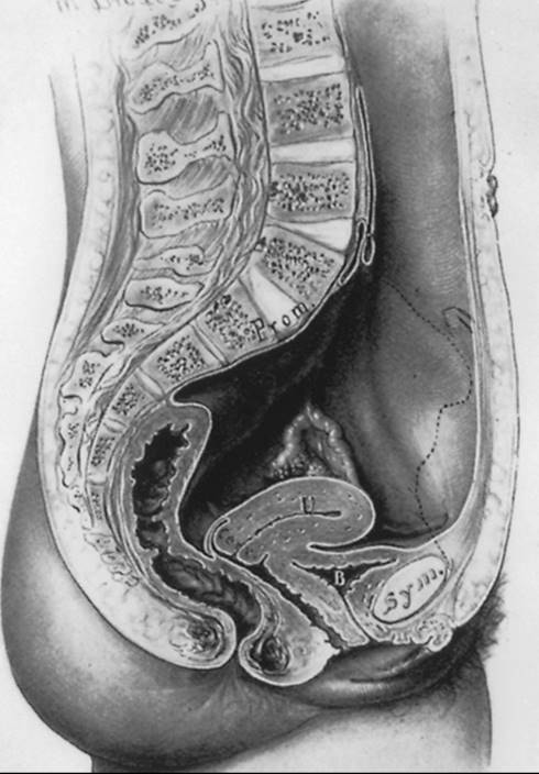
The pelvis lies at the bottom of the abdominopelvic cavity, and the pelvic floor closes the canal within the bony pelvis (Fig. 45.1). The pelvic floor forms a supportive layer that prevents the abdominal and pelvic organs from falling through the opening within the pelvic bones. Its structural role can best be appreciated by considering a surgeon's hand placed through a transabdominal incision that pushes caudally on the pelvic organs. All of the structures that prevent this hand from passing through the pelvic canal constitute the pelvic floor. In addition to this supportive role, the pelvic floor must accommodate conception and parturition, while also controlling storage and evacuation of urine and feces. To understand the pelvic floor and genital prolapse, it is necessary to understand the mechanical strategies that evolution has put in place to prevent downward descent of the pelvic organs, as well as the process by which genital prolapse occurs. As Victory Bonney pointed out, the phenomenon of prolapse is similar to the maneuver that a scrub nurse uses to evert the in-turned finger of a surgical glove (Fig. 45.2). Compressing the air within the glove drives the invaginated finger outward in much the same way that increases in intraabdominal pressure force the vagina and the uterus to prolapse. It is not the weight of the uterus that is important in the development of prolapse, but rather the forces placed on the pelvic floor by increases in intraabdominal pressure.
|
|
|
FIG. 45.1. Sagittal section of the abdomen and pelvis shows the relation of the pelvic floor to the abdominal cavity. (From Kelly HA. Gynecology.Baltimore: Appleton and Co, 1928:64, with permission.) |
|
|
|
FIG. 45.2. A: Diagrammatic representation of the vagina within the abdomen shows how increases in abdominal pressure (arrow) force the vagina to prolapse. B: This prolapse may be prevented by (C) constricting the lower portion of the vagina, (D) suspending the vagina from the pelvic walls, and (E)forming a flap-valve closure, wherein the vagina is pinned against surrounding structures. |
Two mechanical principles explain how the pelvic floor prevents prolapse (see Fig. 45.2). First, the uterus and vagina are attached to the walls of the pelvis by a series of ligaments and fascial structures that suspend the organs from the pelvic sidewalls. Second, the levator ani muscles constrict the lumina of these organs, forming an occlusive layer on which the pelvic organs may rest. It is a combination of these two factors—suspension of the genital tract by the ligaments and fasciae and closure of the pelvic floor by the levator ani—that holds the vagina over the levator ani muscles and forms a flap-valve closure. This flap-valve mechanism is instrumental in keeping the posterior cul-de-sac closed and preventing the development of an enterocele.
Epidemiology of Surgically Managed Pelvic Organ Prolapse and Urinary Incontinence
Prevalence
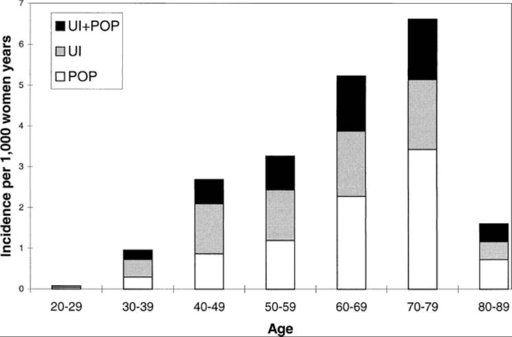
Olsen and colleagues reported the prevalence of surgery for pelvic organ prolapse and urinary incontinence within a defined population. They studied 149,554 women over 25 years of age who were members of the Kaiser Permanente Northwest Health Maintenance Organization. During 1995, they identified 384 women who had surgical treatment for either pelvic organ prolapse or urinary incontinence, or both of these problems. A woman's lifetime risk for needing a single operation by age 80 years was 11.1%. Among this group of women, there was great variety in types and sizes of prolapse (Table 45.1), and the overall incidence of surgery increased with age (Fig. 45.3). The higher incidence was primarily due to the increase in surgery among older women for prolapse rather than for incontinence. Repeated operation was remarkably common, with 29.2% of patients requiring a second surgery. There is increasing risk of pelvic organ prolapse with subsequent parity (Fig. 45.4), and the study of the obstetric factors that cause pelvic floor injury has been neglected.
|
|
|
TABLE 45.1. Preoperative prolapse severity according to operative site |
|
|
|
FIG. 45.3. Age-specific incidence of surgery for pelvic organ prolapse or urinary incontinence. (From Olsen AL, Smith VJ, Bergstrom JO, et al. Epidemiology of surgically managed pelvic organ prolapse and urinary incontinence. Obstet Gynecol 1997;89:501, with permission.) UI, urinary incontinence; POP, pelvic organ prolapse. |
|
|
|
FIG. 45.4. Increase in risk of needing surgery for prolapse with increasing parity. (From Mant J, Painter R, Vessey M. Epidemiology of genital prolapse: observations from the Oxford Family Planning Association study. Br J Obstet Gynaecol 1997;104:579–585, with permission.) |
These data show the remarkable frequency with which women require surgery for incontinence and prolapse but certainly underestimate the prevalence of these problems within the population. One would expect that many women who have prolapse and incontinence choose not to have surgery, so these data represent only those women operated on. Therefore, serious consideration needs to be given these conditions.
Vaginal Delivery and Pelvic Organ Prolapse
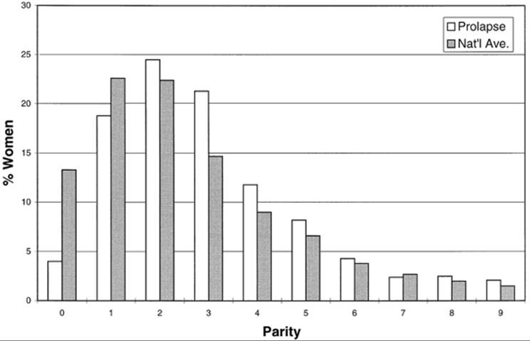
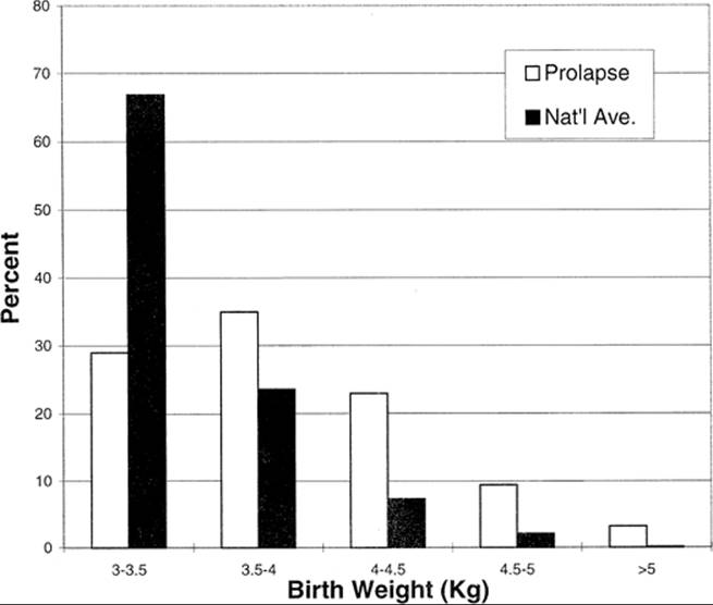
It is commonly accepted that vaginal delivery increases the likelihood that pelvic organ prolapse will develop. Mant, using the Oxford family planning cohort, found that the likelihood of a woman developing prolapse increased approximately eight-fold after two vaginal births and about 12-fold with four or more vaginal deliveries. Timonen and co-workers compared information concerning parity in 1,422 patients operated on for prolapse in their hospital between 1955 and 1965 with national parity data in Finland; these data are displayed in Figure 45.5. Only 4% of women with prolapse are nulliparous, while among the Finnish population 13.3% of women are nulliparous. In addition, they evaluated the size of the largest infant delivered by women in whom prolapse subsequently developed (Fig. 45.6) and compared that with birthweight information for Finland (N = 52,955) during the same period (1957–1958). These data lend some sense of the degree to which vaginal birth influences pelvic organ prolapse.
|
|
|
FIG. 45.5. Parity information for women operated on for pelvic organ prolapse compared with national average. (From Timonen S, Nuoranne E, Meyer B. Genital prolapse: etiological factors. Ann Chir Gynaecol Fenn 1968;57:363, with permission.) |
|
|
|
FIG. 45.6. Birthweight of largest infant delivered vaginally to women with pelvic organ prolapse and normative birthweight data for Finland. (From Timonen S, Nuoranne E, Meyer B. Genital prolapse: etiological factors. Ann Chir Gynaecol Fenn 1968;57:363, with permission.) |
BASIC ANATOMY AND PATHOPHYSIOLOGY OF THE PELVIC FLOOR
Viscerofascial Layer
The topmost layer of the pelvic floor is a combination of the pelvic viscera and their connections to the pelvic walls and will be referred to as the viscerofascial layer. Although it is common to speak of the fasciae and ligaments as separate from the pelvic organs, unless these fibrous structures have something to attach to (e.g., the pelvic organs) they have no structural integrity.
The uterus and vagina are attached to the pelvic walls by the fibrous tissue referred to as the endopelvic fascia. It forms a sheetlike mesentery that is continuous from the uterine artery to the point at which the vagina fuses with the levator ani muscles as it passes through the urogenital hiatus. The tissues that connect the uterus are called the parametria, and those that attach to the vagina are the paracolpium. Although they are given regional names, they are actually one continuous entity. The parametria comprise the cardinal and uterosacral ligaments. These are two different elements of the same tissue (Fig. 45.7). The uterosacral ligaments are the visible and palpable medial margin of the cardinal–uterosacral ligament complex. As is true of the remainder of the parametria, they contain smooth muscle, nerves, and blood vessels and are not the same type of tissue seen in the fascia of the rectus abdominus muscle, which is dense regular connective tissue.
|
|
|
FIG. 45.7. Sagittal section of the pelvis shows the support structures of the genital tract. A: The bladder, urethra, and uterine corpus (above the cervix) have been removed. B: All of the pelvic organs have been removed to show the levator ani muscles. |
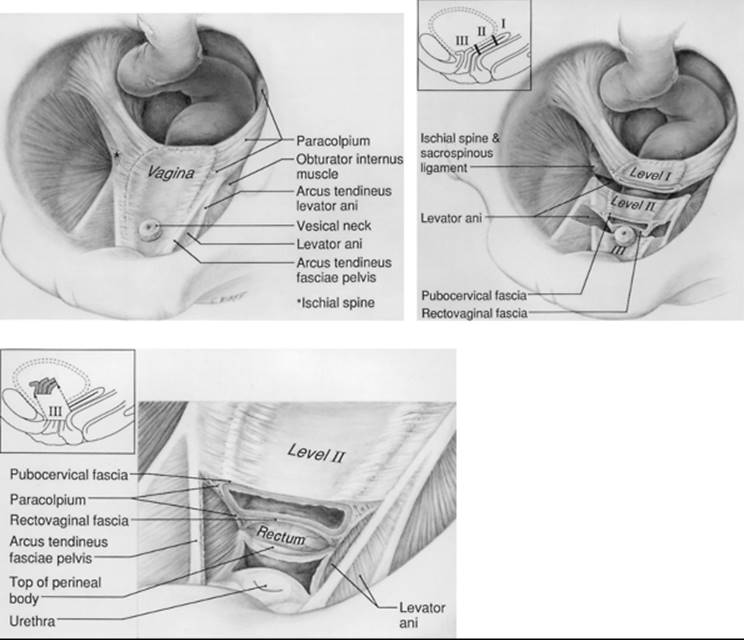
Opposite the external cervical os, the sheet of tissue that attaches the genital tract to the pelvic wall arbitrarily changes name from the parametrium to the paracolpium. The paracolpium has two portions (Fig. 45.8). The upper portion (i.e., level I) consists of a relatively long sheet of tissue that suspends the vagina by attaching it to the pelvic wall in an area similar to that of the cardinal–uterosacral ligament complex. It is this portion that prevents the upper vagina from prolapsing after the uterus has been removed.
|
|
|
FIG. 45.8. Support structures of the vagina after hysterectomy. The bladder has been removed to expose the vagina. A: The paracolpium. B: The different levels of support structures. C: The details of the pubocervical and rectovaginal fasciae after a wedge of vagina and urethra has been removed (inset).(From DeLancey JOL. Anatomic aspects of vaginal eversion after hysterectomy. Am J Obstet Gynecol 1992;166:1717, with permission.) |
In the midportion of the vagina, the paracolpium attaches the vagina laterally and more directly to the pelvic walls (i.e., level II). This stretches the vagina transversely between these two lateral attachments (see Fig. 45.8B). This arrangement has functional significance. The structural layer that supports the bladder (i.e., pubocervical fascia) is composed of the anterior vaginal wall and its attachment through the endopelvic fascia to the pelvic wall. The term fascia is used commonly, but this is not a layer separate from the vagina. A term has been proposed for this layer, the fibromuscular layer of the vagina, that contains both smooth muscle and connective tissue. We use these terms interchangeably in the remainder of the text, because the surgeon generally refers to this layer as fascia. Similarly, the posterior vaginal fibromuscular layer and its connection to the pelvic walls forms the restraining layer that prevents the rectum from protruding forward. In the distal vagina (i.e., level III), the vaginal wall is attached directly to surrounding structures without any intervening paracolpium.
The support that lies under the urethra has special importance for urinary incontinence. The endopelvic fascia in this region is better developed and is tougher than the tissues of the upper vagina, in the area under the bladder. This provides better support for the vesical neck than for the bladder. This layer of suburethral endopelvic fascia attaches laterally to the arcus tendineus fasciae pelvis and also to the medial border of the levator ani muscles. Loss of this normal support of the urethra at the vesical neck is responsible for stress incontinence of urine.
Levator Ani Muscles
Below the uterus, vagina, bladder, and rectum lie the levator ani muscles (see Fig. 45.7B). The medial portion of the levator ani muscles frequently has been called the pubococcygeus, but the pubovisceral muscle is a better term because it extends from the pubic bone and attaches to the vagina and rectum, with only a few insignificant fibers ending in the coccyx (Lawson, 1971). This strong, robust, fatigue-resistant striated muscle starts on the inner surface of the pubic bone near the midline and passes behind the rectum, to return to the pubic bone on the other side. The lateral walls of the vagina are attached to this muscle, which then inserts into the anus in the intersphincteric groove between the internal and external anal sphincter, as well as passing around its dorsal surface in a slinglike loop. The normal resting tone of this muscle squeezes the rectum, vagina, and urethra closed by compressing them against the pubic bone. The pubo-rectalis muscle originates lateral to the pubovisceral muscle and forms a sling behind the rectum at the anorectal angle. Arising from the lateral pelvic walls (at the tendineus arch of the levator ani muscles) is the iliococcygeal muscle that forms a horizontal shelf on which the upper pelvic organs rest.
Interaction Between the Muscles and Fasciae
The interaction between the pelvic floor muscles and ligaments is critical to proper function. As long as the pelvic floor musculature functions normally, the pelvic floor is closed, and the ligaments and fasciae are under no tension. They simply act to stabilize the organs in their position above the levator ani muscles. When the pelvic floor muscles relax or are damaged, the pelvic floor opens. The vagina lies between the high intraabdominal pressure and low atmospheric pressure, where it must be held in place by the ligaments. Although the ligaments can sustain these loads for short periods, if the pelvic floor muscles do not close the pelvic floor, then it is more likely that the connective tissue will become damaged and eventually fail to hold the vagina in place.
ISSUES RELATING TO INCONTINENCE
The mechanisms of urinary and fecal incontinence are more difficult to explain than the mechanical basis of genital tract support, and they are not understood entirely. As is true for genital prolapse, urinary continence depends on both support of the urethra and its ability to remain closed as a result of its innate constriction. Normal support of the urethra allows it to be compressed closed by increases in abdominal pressure (i.e., pressure transmission). Although both support of the urethra and pressure transmission are important for stress urinary continence, they are not the only factors involved. Stress incontinence may occur in women with normal urethral support of the vesical neck and urethra but with inadequate constriction by the muscles within the urethral walls. If the α-adrenergically innervated smooth muscle of the vesical neck does not function normally and the vesical neck is open, this is referred to as type III incontinence. This type of incontinence is differentiated from types I and II incontinence, which involve different degrees of support loss. When the proximal urethra is patent at rest, urine already lies below the area where pressure transmission has its effect and, therefore, bypasses this mechanism of continence.
The support of the urethra is not due exclusively to fascial structures; active muscle contraction plays an important role in urethral support. In addition, the musculature of the urethra contracts during a cough and increases urethral closure. Therefore, neuromuscular mechanisms play an important role in the pathophysiology of stress incontinence and must be considered in understanding this group of diseases.
By the same token, anal continence can be explained only partially by the mechanical structures that support the anorectum and by preservation of the anorectal angle through contraction of the puborectalis. Anal incontinence can occur with normal support of the anorectum and preserved anorectal angle. There are other important factors in maintaining anal continence, including normal integrity of the muscles of the anal sphincter complex and their innervation.
CAUSES OF PELVIC FLOOR DAMAGE
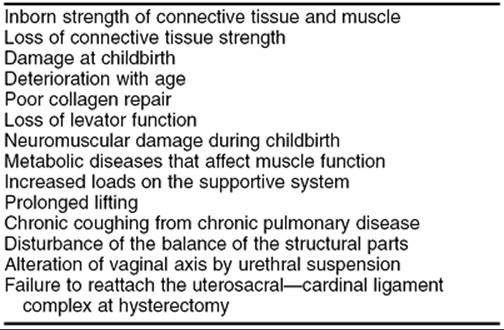
Several factors influence the development of genital prolapse and urinary incontinence (Table 45.2). The inherent strength of the muscles and connective tissue of an individual, damage to these structures that occurs with birth, the rate at which they deteriorate with age, and the loads that they are subjected to during life all play a role in the development of genital prolapse and incontinence. Understanding the interplay among these various factors is critical to appreciating the pathophysiology of genital prolapse.
|
|
|
TABLE 45.2. Factors involved in pelvic organ prolapse |
Although it is obvious that the native strength of the levator ani muscles and endopelvic fascia plays an important role in prolapse, the changes that vaginal birth causes in the pelvic floor deserve specific examination. Distension of the vagina during vaginal birth usually is blamed for prolapse, yet the cervix undergoes a much greater degree of dilation during parturition but recovers sufficiently so that forceful dilation is needed if dilation and curettage is done later in life. Damage to the levator ani muscles and their innervation is common during vaginal birth, and loss of the muscle's ability to support the pelvic organs and unload the ligaments may be partially responsible for the breakage and elongation of the ligaments later in life.
Certainly, some damage to the connective tissue of the parametria and paracolpium occurs during childbirth and can contribute to subsequent prolapse. Although usually thought of as stretching, prolapse also includes instances in which the connective tissue supports rupture. This acute injury mechanism is much more likely than stretching, because usually it is rupture that occurs with overload of other ligaments in the body (e.g., knee). Chronic injury through attenuation of the endopelvic fascia with age contributes and explains why prolapse occurs many years after childbirth. Failure of connective tissue to heal the many minor injuries that occur from day to day may also play a role. Age and chronic disease may cause weakening of the tissues, and heavy lifting or chronic coughing may cause progressive damage. This wear and tear on the connective tissues depends on the magnitude of the stresses placed on them, as well as how well damage is repaired by the body.
DIAGNOSIS AND CLASSIFICATION
Determining the type and severity of prolapse in any given patient is a skill that should be acquired through practice and careful observation. Characterizing the degree of support loss as normal or abnormal depends on comparisons with the findings in normal multiparous women in the examiner's experience. It is, therefore, helpful to perform the same examination on a sufficient number of asymptomatic patients without prolapse to become familiar with the range of normal support. In performing an examination to determine the type and severity of prolapse, the practitioner has two important points to consider:
· Examination must be made with the patient straining forcefully enough that the prolapse is at its greatest.
· The examiner must examine each element of support independently.
If a patient is not able to strain sufficiently in the lithotomy position so that the prolapse is at its largest, examination in the standing position may be necessary. This is a critical point, because it is only when the prolapse can be seen in its fullest extent that all of its various elements can be assessed. If the entire extent of the prolapse is not observed, some element may be overlooked. For example, a large cystocele may be seen initially when the patient strains. It may be only with continued effort by the patient that an enterocele and prolapse of the vaginal apex can be demonstrated. To make sure all aspects of the prolapse can be evaluated, the patient should be asked how large her prolapse is at its largest, and the physician should persist in the examination until that size is achieved. Once the prolapse is visible, the elements of the vagina and pelvic organs that have prolapsed can be evaluated. The examination should then focus on what specific defects in support are present and how severe the prolapse is, and there should be some evaluation of the cause of the prolapse. Once the prolapse is maximally developed, the physician should begin by identifying how much the anterior wall, cervix, and posterior wall have prolapsed. The anterior and posterior walls should be examined separately by retracting the opposite wall with the posterior half of a vaginal speculum. A stepwise, site-specific examination is important because a large cystocele, for example, may hold a potential rectocele in place and, therefore, hide it. If a rectocele is not recognized preoperatively, its repair may be overlooked and the defect can become symptomatic postoperatively. These observations have been confirmed on dynamic colpoproctography imaging studies of the pelvic floor, with contrast placed in the bladder and rectum while the patient strains in the standing position. If the patient has a full bladder when imaging is performed, a rectocele can be obstructed and will not be evident until the patient empties her bladder. Care must also be taken to assess how much of the loss of support is from a defect of the apical (level 1) support. It is not uncommon to correct the apical defects and find that much of what was considered a cystocele and rectocele have been corrected. Examination while under anesthesia is used to evaluate pelvic masses, but it is not the optimal time to identify defects caused by prolapse because of the patient's inability to perform the Valsalva maneuver and because of loss of normal levator tone.
EVALUATING INDIVIDUAL ELEMENTS OF SUPPORT
Anterior Vaginal Wall
Examination of the anterior vaginal wall should establish the status of urethral support, as well as bladder support. The urethra is fused with the lower 3 to 4 cm of the vaginal wall, and abnormal support in this region is properly referred to as a urethrocele (Fig. 45.9). Defective support of the upper portion of the vagina is called a cystocele, because the bladder lies adjacent to this portion of the vaginal wall (Fig. 45.10). The urethrovesical crease, normally visible on examination, forms the line of demarcation between these two areas of support (Fig. 45.11). When support of the entire anterior wall is defective, the term cystourethrocele is used.
|
|
|
FIG. 45.9. Displacement cystourethrocele with intact rugal folds caused by lateral detachment of the pubocervical fascia. (Copyright © DeLancey, 1993.) |
|
|
|
FIG. 45.10. Distension cystourethrocele caused by midline failure of the pubocervical fascia. (Copyright © DeLancey, 1993.) |
|
|
|
FIG. 45.11. Angulation in the anterior vaginal wall, called the urethrovesical crease (arrow), indicates the location of the urethrovesical junction. (Copyright © DeLancey, 1993.) |
The anterior vaginal wall should be above the hymenal ring during straining. Descent of the lower anterior vaginal wall to the level of the hymenal ring during straining is characteristic of a urethrocele and is seen often in patients with stress urinary incontinence. This is due to loss of urethral support and corresponds to the loss of the posterior urethrovesical angle on radiographic studies of patients with stress incontinence. The lower anterior vaginal wall is mobile in all women and may move significantly in continent multiparas. Therefore, motion of this region does not establish stress incontinence but rather indicates the degree to which the support of the urethra has failed. Descent below the hymenal ring is definitely abnormal and indicates a cystourethrocele whether or not stress incontinence is present.
The anterior vaginal wall above the urethrovesical crease usually lies in a flat plane at about a 45-degree angle from the horizontal (see Fig. 45.11). Descent below the level of the hymenal ring is significant. This descent can be caused by one of three entities:
· Separation of the paravaginal attachment of the pubocervical fascia from the white line due to detachment from the ischial spine
· Loss of the vagina's attachment to the cervix
· Tearing in the pubocervical fascia that results in herniation of the bladder through this layer
Uterus and Vaginal Apex
The vagina and cervix are fused with one another, and prolapse of the uterine cervix is associated invariably with prolapse of the upper vagina, as well. When the uterus descends below its normal level, the term uterovaginal prolapse is appropriate, although uterine prolapse commonly is used. In patients in whom the uterus has been removed, descent of the vaginal apex below its normal position in the pelvis is referred to as prolapse of the vaginal apex, and when the vagina turns entirely inside out, the term vaginal eversion is used.
The location of the cervix customarily is used to gauge the severity of uterine prolapse (Fig. 45.12). Its position relative to the hymenal ring should be noted while the prolapse is at its greatest. If the cervix is not visible because of a cystocele or rectocele, then its location may be palpated while having the patient strain. When the cervix descends to within 1 cm of the hymenal ring, there is a significant loss of support. In instances in which the uterus is not necessarily going to be removed, uterine support should be tested before it is assumed that the uterus is well supported. This can be done by grasping the cervix with a tenaculum or ring forceps and applying traction until it stops descending. Occult prolapse, in which the cervix comes below the hymenal ring, can be detected in this way.
|
|
|
FIG. 45.12. Uterine prolapse with the cervix extending 3 cm below the hymen. (Copyright © DeLancey, 1993.) |
In addition to determining how far the cervix descends, its length should be measured. Cervical elongation is frequent in individuals with prolapse, and the uterine corpus often may lie in its normal location. Awareness of cervical elongation preoperatively will allow the surgeon to proceed expeditiously with the hysterectomy, rather than hoping with every pedicle that the uterine arteries will soon appear.
Posterior Vaginal Wall
The posterior vaginal wall is the site of both rectoceles and enteroceles. Evaluation and correction of these two problems challenge even the most experienced gynecologic surgeon, and they are probably the most difficult to understand of all pelvic support defects. Because dyspareunia can follow repair, correction of asymptomatic posterior wall defects is not without risk. On the other hand, having a rectocele or enterocele develop after vaginal hysterectomy and anterior colporrhaphy is an undesirable outcome, and careful consideration of the support of the posterior vaginal wall is important.
Three questions should be asked by the physician when examining the posterior wall.
· Is it supported normally?
· If not, is it a true rectocele or a pseudorectocele?
· Is an enterocele present?
A rectocele is present when the anterior rectal wall and overlying vagina protrude below the hymenal ring. An enterocele exists when the cul-de-sac becomes distended with the intestine and bulges the posterior vaginal wall outward. There are also occasions in which the posterior wall appears to bulge into the vagina, not because of poor support of the rectal wall but because of a deficiency in the perineal body. This has been referred to by Nichols and Randall as a pseudorectocele and can be differentiated easily from a true rectocele because the anterior rectal wall contour is normal on rectal examination. Another type of pseudorectocele may be suspected when there is apical descent of the upper vagina or cervix and apparent loss of posterior support. However, often when the normal apical support is restored (by temporarily supporting it with ring forceps in the office or after surgical repair of the apical descent), a suspected rectocele is not evident. This is important to determine preoperatively, because loss of tone of the levator ani muscle and anal sphincter muscle with muscle paralyzing agents during anesthesia make it harder to establish the existence of a true rectocele.
Enterocele
There is always a cul-de-sac between the upper vagina and the rectum. This allows a culdocentesis to be performed and a colpotomy to be made through the posterior vaginal wall at the beginning of a vaginal hysterectomy. The peritoneal pouch normally extends 3 to 4 cm beyond the junction of the vagina and cervix. Therefore, the absence of an enterocele in normal women must be explained by factors that keep the cul-de-sac closed rather than by the absence of a peritoneal space between the upper vagina and rectum. It is the suspension of the upper vagina near the sacrum in a position where it may rest over the rectum and intact levator plate that keeps this space closed.
There are two types of enteroceles: pulsion enterocele and traction enterocele. A pulsion enterocele exists when the cul-de-sac is distended and appears as a bulging mass that is inflated by increases in abdominal pressure. This may occur with either the vaginal apex or uterus well suspended, in which case the cervix or vaginal apex is at a normal level and the enterocele dissects between the vagina and the rectum. When an enterocele is associated with prolapse of the uterus or vaginal apex, then the prolapse and enterocele occur together.
A traction enterocele represents a situation in which prolapse of the uterus pulls the cul-de-sac peritoneum down with it, but there is no bulging or distension of the cul-de-sac when abdominal pressure rises. This condition usually is found at the time of vaginal hysterectomy when the cervix has prolapsed. It represents a potential enterocele rather than an actual enterocele, because there is no bulging mass separate from the uterus.
Unlike uterine prolapse, which is obvious because of the protrusion of the easily recognized uterine cervix, enteroceles and rectoceles rarely are evident on examination. Therefore, the key to detecting an enterocele lies in actively looking for it whenever a patient who has prolapse is examined. Detection of an enterocele is performed best in the awake, straining patient by noting a mass of small intestine between the rectum and vagina; it may not be suspected in a supine individual at rest.
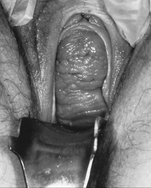
Anatomically, an enterocele extends from the apex of the vagina downward, whereas a rectocele typically begins in the lower portion of the vagina. An enterocele sometimes is evident as a bulge that overrides the more caudal rectocele (Fig. 45.13). Careful inspection of the posterior vaginal wall with a speculum retracting the anterior wall sometimes can suggest that an enterocele is present. The key to detecting a pulsion enterocele lies in palpating the small bowel between the vagina and rectum during rectovaginal examination, with the patient straining so that the prolapse is protruding. To do this, an index finger is placed in the rectum and a thumb is placed in the vagina. Then, with the patient straining, the rectovaginal space may be palpated to detect the bulge of the enterocele and the presence of small bowel, omentum, or large bowel.
|
|
|
FIG. 45.13. “Double hump” sign of an enterocele overriding a rectocele. (Copyright © DeLancey, 1993.) |
Rectocele
The hallmark of a typical rectocele is the formation of a pocket that allows the anterior rectal wall to balloon downward through the introitus. When a rectal examination is performed with the prolapse fully developed, a rectocele exists if there is an extension of the rectal lumen below the axis of the anus (Fig. 45.14). This not only provides the diagnosis but also illustrates the mechanism by which rectoceles create their symptoms. As long as the anterior rectal wall has a smooth contour and no sacculation, even though it may be more mobile than normal, stool will pass through the anus. However, when a pocket develops as the patient strains, stool becomes trapped in it, and difficulty with evacuation can occur.
|
|
|
FIG. 45.14. A: Pelvic examination shows a rectocele. B: Lateral bead chain cystourethrogram with the patient supine and with contrast in the vagina and rectum shows a protruding rectocele. (Copyright © DeLancey, 1993.) |
Prolapse Subsequent to Hysterectomy
Special consideration should be given to patients who have prolapse after hysterectomy to assess whether or not prolapse of the vaginal apex is present. When the uterus is in situ, the cervix calls attention to the poor support of the cervix and upper vagina. In instances of posthysterectomy vaginal prolapse, descent of the vaginal apex is more easily missed. If it is overlooked and an anteroposterior colporrhaphy is not accompanied by suspension of the vaginal apex, the colporrhaphy will fail to cure the apical prolapse, and the problem is not corrected. Overlooking apical support loss also can lead to overly aggressive excision of vaginal tissues during anteroposterior colporrhaphy and a shortened vagina. Examination of patients who have previously had a hysterectomy should include a specific effort to determine the location of the vaginal apex when the prolapse is at its largest. The apex is identified by the vaginal scar at the hysterectomy site (Fig. 45.15). Vaginal prolapse is present when the hysterectomy scar lies below the level of the hymenal ring. If the apex descends to within the lower one third of the vagina with straining, a significant deficit in support of the apex is present, and the vagina should be resuspended during repair.
|
|
|
FIG. 45.15. Eversion of the vagina after hysterectomy. Note that the vaginal apex, indicated by the puckered scar where the cervix had been removed, lies below the hymenal ring. (Copyright © DeLancey, 1993.) |
PELVIC ORGAN PROLAPSE CLASSIFICATION
Several classification systems have been used to describe the sizes and types of pelvic organ prolapse in an individual woman. Because many of these systems use similar words to indicate different degrees of prolapse, confusion has arisen concerning the size of prolapse. For example, grade 2 uterine prolapse in some systems indicates that the cervix descends halfway between its normal position and the introitus. In other classifications, grade 2 can mean that one half of the uterus is outside the introitus. A system that standardizes terminology has been adopted by several groups, including the International Continence Society, the American Urogynecologic Society, and the Society of Gynecologic Surgeons. This standardized terminology (Pelvic Organ Prolapse Quantification, or POP-Q) provides a system that can describe the type of prolapse, as well as quantify the degree of prolapse in each area. Although this standardized system seems somewhat cumbersome when described in writing, in actual practice it is quite simple. The following section first considers the measurements that describe the type and size of prolapse and then discusses the measurements concerned with the changes in the urogenital hiatus in the levator ani muscles through which the prolapse descends.
Measurements Describing Prolapse Type and Size
To describe the nature of a woman's prolapse, it is necessary to do the following: (a) document what part or parts of the genital tract have prolapsed and (b) indicate how far down each part of the vaginal wall or cervix has descended.
Prolapse description must include a consideration of anterior vaginal wall descent, posterior wall descent, and uterine descent (or prolapse of the vaginal apex after hysterectomy). Furthermore, because different parts of the anterior wall might suffer support damage, the system provides for determining the status of each level of vaginal support. For example, the distal anterior vaginal wall adjacent to the urethra may be well supported, while the portion of the vagina under the bladder may prolapse. This system addresses the need to make individual assessments of different parts of the vaginal wall.
The three levels of vaginal support (see Fig. 45.8) must be assessed, corresponding to the different anatomic regions of vaginal support.
· Level I: support of the vaginal apex and uterus
· Level II: support of the bladder and rectum
· Level III: support of the urethra and perineal body
In levels II and III, the anterior and posterior vaginal wall are considered separately, while in level I the cervix (or vaginal apex) and posterior fornix must be assessed.
To understand the POP-Q classification system, refer to Figure 45.16. The size and type of prolapse are measured by determining the location of a series of points on the anterior and posterior vaginal walls relative to the hymenal ring. Points at each of the three levels are measured. Positive numbers reflect measurements of the vaginal points that have prolapsed below the level of the hymen and negative numbers reflect measurements above the hymen. It should be noted that this descriptive scheme does not distinguish between rectocele and enterocele but simply provides a way to quantify the amount of vaginal wall descent in each specific area. Additional examination and written comments concerning these important differences should be made.
|
|
|
FIG. 45.16. Six sites (points Aant, Bant, C, D, Bpost, and Apost), genital hiatus (gh), perineal body (pb), and total vaginal length (tvl) used for pelvic organ support quantitation. |
A summary of the measurements obtained during the POP-Q examination is noted in Table 45.3.
|
|
|
TABLE 45.3. Stages of pelvic organ prolapse based on measurement of specific sites |
I. Vaginal points (vaginal profile): patient straining maximally
A. Level I: apex and cervix: points C and D (D is omitted if patient has had a hysterectomy)
B. Level II: midvaginal points
1. Anterior wall: Ba or Bant
2. Posterior wall: Bp or Bpost
C. Level III: distal vagina (perineum and urethrovesical neck)
1. Anterior wall: Aaor Aant
2. Posterior wall: Ap or Apost
3. External measurements: obtained with patient at rest and again straining
D. Genital hiatus: length gh
E. Perineal body: length: pb
II. Internal digital measurements: obtained with patient at rest with vaginal apex restored to normal position
A. Total vaginal length: tvl
To measure the lower third of vaginal support (level III), the location of a pair of points that normally lies 3 cm above the hymenal ring is assessed. Points measured at level III are called points A. One can imagine marking these vaginal points 3 cm above the hymen with a marker and then recording the position of these points in relation to the hymen with the subject straining maximally. Anteriorly, this corresponds to the approximate location of the urethrovesical junction, and this measurement assesses urethral descent (Aa or Aant). Posteriorly, this region is normally occupied by the tissues of the perineal body (Ap or Apost). By definition, the highest possible position of either point Aant or Apost is 3 cm above the hymen (-3), and the lowest position is 3 cm below the hymen (+3).
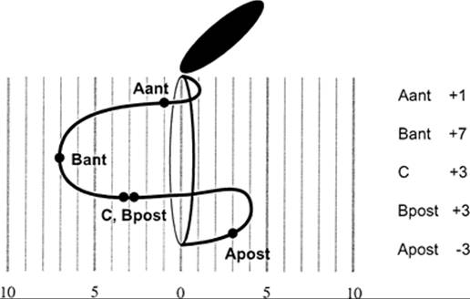
To assess midvaginal support (level II), the most dependent part of the vaginal wall above point Aant is used. This point is called Ba, or Bant, on the anterior wall and Bp, or Bpost, on the posterior wall. This is, therefore, not a fixed point along the surface of the vagina, but rather it is marked at whatever location is the most caudal (distal) portion of that vaginal segment at maximal prolapse protrusion. In a normally supported vagina, this will be the same as point Aant, whereas in a woman with procidentia it will be the same as point C. The same is true for the posterior vaginal wall at point Bpost. Point C corresponds to the most distal portion of the uterine cervix or of the hysterectomy scar in the vagina in those patients who have had the uterus removed. Point D denotes the posterior fornix (that point at which the posterior vaginal wall changes direction). In addition to the positions of these points, the total length of the vagina is noted. Once these data have been gathered, a simple line diagram can be constructed by plotting these points relative to the hymen to provide a graphic representation of the prolapse (Fig. 45.17).
|
|
|
FIG. 45.17. Diagram of prolapse sites. (From Viereck V, Peschers U, Singer M, et al. Metrische Quantifizierung des weiblichen Genitalprolapses: eine sinnvolle Neuerung in der Prolapsdiagnostik? Geburtshilfe Frauenheilk 1997;57:177, with permission.) |
So far, each element of the prolapse has been considered separately. It is also possible to give an overall description to the size of the prolapse by looking at the most dependent part of the protruding vagina or uterus. In this way, different stages—stage 0 through stage IV-can be defined; a description of these is shown in Table 45.3. A briefer version of the original description of the POP-Q staging system has been summarized, also.
The choice of the term stage here is somewhat unfortunate, because most women with stage I and II support are anatomically normal. The implication that they have a stage of prolapse is incorrect. Hopefully this problem with terminology will be corrected in the future.
Pelvic Floor Measurements
In normal women, the levator ani muscles close the pelvic floor. In women with pelvic organ prolapse, the urogenital hiatus within the levator ani muscles is the opening through which the vagina prolapses. This hiatus is enlarged in women with pelvic organ prolapse. The size of the urogenital hiatus and thickness of the perineal body can easily be measured to describe the changes that have occurred in the pelvic floor. The anteroposterior diameter of the genital hiatus extends from the arch of the pubic bone to the front of the perineal body, while the thickness of the perineal body is measured from the anterior margin of the perineal body to the center of the anal verge. The urogenital hiatus is held closed by the constant activity of the levator ani muscles, and the diameter of this opening enlarges in many women with prolapse.
This classification system is detailed and specific. It requires careful examination and assessment. Although at first it seems quite detailed, it is simply the quantitative documentation of the individual defects that experienced surgeons always have found necessary to assess. Some clinicians will not find it expedient to measure each of these sites, but intelligent, detailed analysis of each site of support is important to plan properly any repair for pelvic organ prolapse.
SYMPTOMS
All types of prolapse have several symptoms in common. Once the vagina prolapses below the introitus, it becomes the structural layer between the high pressures in the abdominal space and the relatively low atmospheric pressure. The downward force that this pressure differential creates puts tension on the fasciae and ligaments that support the vagina and uterus. This results in a dragging sensation where the tissues connect to the pelvic wall, usually identified by patients as occurring in the groin, and in sacral backache caused by traction on the uterosacral ligaments. This type of discomfort resolves when the patient lies down and the downward pressure is reduced. In addition, exposure of the moist vaginal walls leads to a sensation of perineal wetness that may be confused with urinary incontinence, and it also can give rise to ulceration of the vaginal wall. Most patients have an underlying sense of insecurity that is difficult for them to describe and is often expressed as a feeling that “something is just not right.” Sometimes patients who feel the cervix or vagina protruding have fears that they have a cancer and may be relieved to find that the condition is related to prolapse. Although patients may find it difficult to put their symptoms into words, the symptoms can cause significant distress and should not be ignored.
Symptoms of Anterior Wall Prolapse
The symptoms of cystourethrocele are varied, and the two primary ones are paradoxical. On the one hand, loss of support of the urethra and the lower vaginal wall is associated with stress urinary incontinence, whereas loss of support of the upper anterior vaginal wall and bladder base can cause difficulty in emptying the bladder. This inability to empty the bladder completely is probably related to voiding by the Valsalva maneuver. If there is a detrusor contraction, there should be no reason for a woman with a cystocele not to empty her bladder, and many women with a significant cystocele have normal postvoid residual urine volumes. When a woman strains to void, however, the cystocele simply gets bigger, and no impulse is provided for urine to flow through the urethra.
In addition to these functional symptoms, many patients with a cystourethrocele complain of urinary urgency and frequency. This probably arises from stretching of the bladder base that accompanies its prolapse through the vaginal introitus; it is often less pronounced at night when patients are supine.
Patients have a varying amount of support loss under the urethra or bladder, and symptoms vary along the spectrum from incontinence to urinary retention. As is true for other forms of prolapse, it is important to correlate a patient's symptoms with the physical findings so these problems can be addressed.
Symptoms Associated With Prolapse of the Uterus, Prolapse of the Vaginal Apex, or Enterocele
Few specific symptoms are related to prolapse of the uterus, prolapse of the vaginal apex, or enterocele. Patients with these conditions usually complain of the generalized symptoms of prolapse mentioned above. Some have urgency and frequency, probably related to pressure of the prolapse on the bladder base, but this is variable. In addition, patients with large, thin enteroceles occasionally have a sense of impending rupture. Although this is an uncommon problem, it should not be overlooked.
Symptoms of Rectocele
The cardinal symptom of a rectocele is difficulty in emptying the rectum. As a woman bears down to evacuate the rectum, stool is pushed into the rectocele, and the harder she strains, the bigger the rectocele becomes. Because constipation is common in older women, it is important to differentiate between infrequent bowel movements due to poor colonic motility or inadequate dietary fiber and difficulty due to a rectocele. Many women have found that if they press between the vagina and rectum to elevate the rectocele, this maneuver helps with defecation. This finding supports the fact that the rectocele is the source of the problem.
WHO SHOULD BE TREATED SURGICALLY
A decision about when an operation for prolapse should be performed is based on the individual woman's situation. It depends on the size of the prolapse, the presence or absence of symptoms, and whether or not physiologic complications have arisen because a prolapse is present. Conservative treatment with a pessary may be considered. There is little data on continuance rates with long-term pessary use. Contrary to prior recommendations that pessaries should be offered only to those women who are not operative candidates or to those awaiting surgery, a survey of urogynecologists found that 77% would offer a pessary for symptomatic patients as first-line treatment.
When the prolapse lies at or above the level of the hymenal ring, surgery should be performed only if definite symptoms are present and can be attributed reliably to the prolapse. Examples of this include:
· A patient with a cystourethrocele at the level of the hymen who has significant stress incontinence
· A woman with the cervix in the lower one third of the vagina, who has the characteristic dragging discomfort of a prolapse that resolves when she lies down or puts a supportive pessary in place
· A patient with a small rectocele in whom a definite pocket can be detected on rectal examination and elevation of the perineum relieves problems with defecation
It is unusual for women in whom the uterus has not yet descended to the level of the hymenal ring to have symptoms caused by descent of the uterus. Symptoms of pressure, back pain, or feelings that something is coming out should be studied to confirm that they are related to the prolapse, and not to other factors, before a decision to operate is made. If the symptoms go away when a pessary is placed or a patient notices prompt relief when she lies down, this helps to confirm that the prolapse is the source of the symptoms. A woman with low back pain whose discomfort persists after she is supine is more likely to have arthritic or musculoskeletal pain than pain caused by uterine prolapse, and the results of surgery are likely to disappoint her.
Among patients whose prolapse descends several centimeters below the hymenal ring, symptoms are usually present, and surgery will relieve this distress. Some women with a large prolapse will deny any symptoms, because they do not wish to undergo surgery or because they simply are not troubled by the prolapse. Their wishes should be respected. On the other hand, potential complications from the prolapse should be considered and discussed frankly.
A prolapse should be repaired, despite a lack of symptoms, if a patient has recurrent urinary tract infections associated with an increased postvoid residual urine volume. Ureteral dilation caused by the prolapse, which leads to an impairment of renal function, occurs in some patients with large prolapses and should be considered an indication for repair. An intravenous pyelogram and renal function testing can detect these abnormalities and indicate the need for treatment. The intravenous pyelogram should include films made with the patient standing and the prolapse present, because the supine position may mask significant dilation.
SURGICAL PROCEDURES
Vaginal Hysterectomy and Anteroposterior Colporrhaphy
Vaginal hysterectomy with appropriate anteroposterior colporrhaphy is the operation most often performed for the treatment of uterovaginal prolapse. Because simple removal of the uterus does nothing to correct the prolapse of the vaginal walls that accompanies descent of the cervix, it is necessary to consider how an operation works to cure these conditions properly.
Basic Principles Underlying the Surgical Repair of Prolapse
Removal of the uterus does not improve support of the genital tract. Successful repair depends on the way in which a surgeon resuspends the vagina and repairs other defects in pelvic organ support when performing a vaginal hysterectomy and anteroposterior repair. Once the uterus has been removed, the vaginal apex should be attached to a point that is higher in the pelvis than the vagina to elevate it to a normal position. During the vaginal hysterectomy, the cardinal and uterosacral ligaments can be shortened and used to resuspend the vagina. In addition, techniques such as McCall culdoplasty anchor a suture to a point on the uterosacral ligaments higher than the preoperative position of the vagina and use this suture to pull the vaginal apex to a point higher in the pelvis. It is the elevation of the vagina rather than removal of the uterus that is critical.
Preoperative Considerations
In addition to the usual thorough history, physical examination, and other medical measures necessary prior to any major operation, preoperative preparation for vaginal hysterectomy should include a pelvic examination, Pap smear, and urinalysis. Special care should be taken, as previously described, to determine the exact type and degree of prolapse and to detect any potential ovarian malignancy. A saline enema (e.g., Fleet enema) the night before surgery empties the rectum in the event that a rectal examination is required during surgery and provides more room posteriorly, improving exposure. Patients should receive a prophylactic antibiotic to minimize the risk of postoperative infections (cefazolin sodium 1 g intramuscularly or intravenously, just before going to the operating room, is appropriate). Cervical cytologic examination results should be documented prior to hysterectomy, and if abnormal uterine bleeding is present, the endometrium should be sampled and tested preoperatively. Finally, special consideration should be given to assessing whether or not concomitant cystocele, stress incontinence, enterocele, or rectocele is present.
Before the hysterectomy is begun, a sterile preparation of the vulva and vagina is performed. An indwelling urinary catheter usually is not placed at this point, to allow the bladder to accumulate enough urine so that an inadvertent cystotomy may be recognized easily. Care should be taken in positioning and draping the patient to avoid compression of the femoral or peroneal nerve, but there should be adequate access to the operative site. An examination should be performed under anesthesia to, once again, assess whether or not any abnormalities exist that would make vaginal hysterectomy impossible and would indicate a switch to an abdominal approach.
Technique of Vaginal Hysterectomy
The elements of vaginal hysterectomy in the United States are derived from the operation described by Heaney (Fig. 45.18). A circumscribing incision is made through the vaginal wall at the cervicovaginal junction (see Fig. 45.18A). Anteriorly, the incision is placed at the lower edge of the bladder to allow entry into the vesicocervical space. The lower edge of the bladder can be palpated against the cervix to determine where this incision should be made. In addition, the junction of the vaginal rugae and the smooth surface of the cervix can be used as landmarks to identify the proper site of incision. Some operators inject saline into the tissues around the cervix to facilitate dissection. This injection should not contain epinephrine, because its use increases the incidence of postoperative pelvic infection. Several studies have documented decreased blood loss after infiltration of a dilute vasopressin solution at the cervical portio, without concomitant increased infection rates. The incision should be made through the full thickness of the vaginal wall. This can be determined by placing a retractor anteriorly while pulling forcefully on the cervix so that the vaginal wall is placed under tension. Once the vagina has been transected it separates, exposing the underlying cleavage plane. On the posterior aspect of the cervix, this exposes the area of the cul-de-sac. Blunt dissection here frees the peritoneum, which can then be grasped and entered (see Fig. 45.18B). Care should be taken not to dissect the peritoneum off the uterus, because that makes entry more difficult. Once the cul-de-sac incision has been extended laterally as far as the medial margins of the uterosacral ligaments, it can be sutured to the posterior vaginal cuff to minimize bleeding. At this point, it is appropriate to palpate the adnexal structures through the colpotomy to detect unsuspected tumors and to make a final decision concerning the feasibility of the vaginal approach, if any doubt existed. This is an easy time to acquire this valuable information, and if unsuspected disease is found, an abdominal approach can be taken.
|
|
|
FIG. 45.18. A: Vaginal hysterectomy is initiated by circumscribing the cervix at the cervicovaginal junction. B: The posterior cul-de-sac is opened. C: The peritoneum of the anterior cul-de-sac is incised. D: The base of the uterosacral and cardinal ligament usually is clamped in two bites. E: The upper cardinal ligament is clamped prior to its transection. F: The uterine fundus is delivered, and the connections between the adnexal structures and uterine corpus are clamped. G: Ligaments and vaginal cuff as they appear after hysterectomy. The posterior cuff is whip-stitched (inset). H: Technique for resuspension of the vaginal cuff and obliteration of the cul-de-sac: 1, placement of suture through exteriorized ligaments and vaginal wall; 2, reefing sutures placed in peritoneum; 3, modified internal McCall suture; 4, high purse-string suture to close the cul-de-sac. (From Mattingly RF, Thompson JD, eds. Te Linde's operative gynecology, sixth ed. Philadelphia: JB Lippincott Co, 1985:554, with permission.) |
Next, the long blade of an Auvrard retractor is placed in the cul-de-sac, and attention is turned to entering the anterior cul-de-sac. Dissection along the vesicocervical plane is best performed by lifting the fascia anteriorly while cutting with Mayo scissors placed tip-down on the cervix. This avoids the problems that sometimes occur when a knife is used, because the scissors held in this orientation will not cut into the dense fibrous tissue of the cervix. Once the cleavage plane of the vesicocervical space is reached, gentle dissection with a gloved finger is all that is needed to elevate the bladder off of the cervix. If the bladder pillars are prominent, they can be clamped next to the cervix, transected, and ligated to provide better lateral exposure. The location of the peritoneum may be detected by moving a finger from side to side and feeling the way in which the two slippery mesothelial surfaces slide on one anther. The peritoneum can then be lifted, transilluminated, and incised (see Fig. 45.18C). After the incision is extended laterally, a suture is placed in its vesical edge and held with a hemostat to facilitate its identification at the time of reperitonealization.
Once the cul-de-sacs have been entered, the ureter should be palpated to determine its position prior to shortening and transecting the cardinal ligaments. This is a critical step, because only if the ligaments are shortened will the vagina be held at a level higher than that of the prolapsed cervix. A retractor is placed in the lateral fornix of the vagina and a finger in the anterior cul-de-sac. Because the ureter must pass under the uterine artery and end in the bladder anterior to the examining finger, it can be palpated against the retractor blade. The characteristic snap of the ureter permits its identification, and it can be followed for some distance by moving the retractor and examining finger anteriorly and posteriorly.
After the ureter is identified the uterosacral and cardinal ligaments are shortened by clamping them at a safe distance from the ureter, somewhat lateral to the cervix (see Fig. 45.18D, Fig. 45.18E). After the ligaments are cut and suture-ligated, the sutures are left long and marked for use in resuspending the vagina at the end of the procedure. The uterine arteries are similarly clamped, transected, and suture-ligated, but they are not tagged, to avoid dislodging the suture from this vascular pedicle.
At this point, the corpus of the uterus usually is brought through the posterior colpotomy by pulling on it with a tenaculum, to expose the connections between the adnexal structures and the uterus (see Fig. 45.18F). When this is not possible because of a small vagina or large uterus, the uterus can be divided along its sagittal plane, and one half can be pushed up into the peritoneal cavity. A long tenaculum is left attached to the elevated half of the uterus to allow its later retrieval, while the contralateral adnexal pedicles are dealt with. Clamps are placed across the adnexal structures, with care taken not to include bowel or adjacent structures, and the pedicles are transected.
Next, consideration should be given to removal of the ovaries. If the ovaries are to be removed, the mesovarium or infundibulopelvic ligament is clamped, the ovary is removed with or without the fallopian tube, and the remaining pedicle is suture-ligated.
Management of the Cul-de-sac and Suspension of the Vaginal Apex
Once the uterus has been removed and the ligaments have been prepared by shortening them, the reconstructive phase of the operation begins. These steps use the ligaments that were tagged during the hysterectomy (see Fig. 45.18G). The goals are to close the peritoneum and suspend the vaginal apex, while also correcting any existing enlargement of the cul-de-sac. The cul-de-sac peritoneum is attached to the uterus. Whenever the uterus prolapses, it pulls the cul-de-sac peritoneum with it. If this extension is not distended with bowel, it is referred to as a traction enterocele, and when the cul-de-sac is not only elongated but also distended with bowel, it is referred to as a pulsion enterocele, a true enterocele.
When little prolapse has been present (i.e., the cervix does not extend below the hymenal ring), simple closure of the cul-de-sac and attachment of the ligaments to the vaginal cuff suffice. An indwelling catheter usually is placed at this time to drain the bladder. This brings the anterior peritoneum closer to the surgeon and, thereby, facilitates its closure.
Closure of the vaginal cuff should accomplish two things: resuspension of the vaginal apex and closure of the cul-de-sac (see Fig. 45.18H). A suture is placed through the vaginal cuff and through the ends of the suspensory ligaments to reattach them to the vagina. Next, the cul-de-sac may be reefed and a suture placed through the uterosacral ligaments to bring them to the midline. Finally, the peritoneum is closed with a purse-string suture of 0 silk placed at the highest level possible, beginning on the vesical peritoneum. A finger is placed in the peritoneum while this suture is tied, to avoid trapping intraabdominal contents in the suture, and is removed as the suture is snugged down.
Anterior Colporrhaphy
After the uterus has been removed and before the ligaments are reattached to the vaginal cuff, an anterior colporrhaphy is performed for patients with cystoceles (Fig. 45.19). Scissors are used to undermine and incise the mucosa of the anterior vaginal wall as far as the midurethra (see Fig. 45.19A). The vaginal fascia is then mobilized widely from the vaginal epithelium, with use of a combination of sharp and blunt dissection, until the superior lateral sulcus of the vagina is reached (see Fig. 45.19B). Care should be taken to remain superficial in this dissection to avoid transecting the fascia that is to be plicated in the midline. Special attention should be given to adequately developing the fascia that lies under the urethrovesical junction, because this is critical to improving urethral support. Once mobilization has been accomplished, a series of plication sutures are taken in the fascia, just medial to the junction of the anterior and lateral vaginal walls (see Fig. 45.19C).
|
|
|
FIG. 45.19. A: The vaginal epithelium is undermined when commencing anterior colporrhaphy. B: The fascia is separated from the mucosa. C: The fascia is sutured together in the midline. Special care should be taken to ensure adequate support at the urethrovesical junction. D: Excess epithelium is trimmed. E: The epithelium is closed. A bite of the underlying fascia is included to minimize dead space. (From Mattingly RF, Thompson JD, eds. Te Linde's operative gynecology, sixth ed. Philadelphia: JB Lippincott Co, 1985:612, with permission.) |
Plication of the suburethral fascia deserves special attention. Once the first suburethral suture has been placed and tied, it pulls some of the more lateral fascia into view, and this offers an opportunity to place a second suture in this same region and improve the elevation that was possible with the first suture. Once the remainder of the fascia has been closed, the cranial edge of the fascia at the vaginal apex should be tagged so that it may be included in the sutures that reattach it to the cardinal and uterosacral ligaments. This step will help elevate the upper margin of the fascia and pull it higher in the pelvis. An appropriate amount of vaginal epithelium is then trimmed (see Fig. 45.19D), and the epithelium is closed with running or interrupted sutures (see Fig. 45.19E).
Posterior Colpoperineorrhaphy
Next, a rectocele, if present, must be corrected. Although often undertaken at the end of a tiring operation, this repair requires the most skill and judgment of all phases of surgery for prolapse. Overcorrection of the posterior wall may leave the patient unable to have satisfying intercourse, yet a failure to reinforce the supportive position of the posterior vaginal wall may increase the likelihood that prolapse will recur. The goal of this portion of the operation is to reinforce the rectovaginal septum and to return the size of the introitus to normal.
The posterior colporrhaphy is begun by establishing the size that the introitus should be at the end of the procedure. This is accomplished by estimating the amount of vaginal and perineal skin that must be removed to return the introitus to a normal diameter. Allis forceps are placed to grasp a solid bite of tissue at the hymenal ring bilaterally at the margins of the mucosa that is to be excised. These forceps are then approximated in the midline to bring the soon-to-be-united parts of the introitus together. This allows a preview of the degree to which the introitus will be narrowed. Adjustments may then be made in the placement of the forceps to obtain an optimal result before an irrevocable incision is made into the perineum. A patient with a normal introitus who has a rectocele above this level need not have any tissue excised, and a vertical incision can be made in the introitus.
After the skin of the introitus is incised, the mucosa is undermined (Fig. 45.20A). The vagina and perineal body are fused, so dissection in the 3 cm inside the hymenal ring must be done sharply. If the rectovaginal septum is thin, placing a finger in the rectum to guide dissection will minimize the likelihood of proctotomy.
|
|
|
FIG. 45.20. A: To begin posterior colporrhaphy, a transverse incision is made in the perineal body, and a vertical incision is extended toward the vaginal apex. B: The perirectal fascia is mobilized from the mucosa. C: The perirectal fascia is sutured, beginning above the site of the rectocele. D: The inner surface of the levator ani muscles are approximated in the perineal body, and redundant mucosa is excised. E: The mucosa and perineal skin are closed. (From Mattingly RF, Thompson JD, eds. Te Linde's operative gynecology, sixth ed. Philadelphia: JB Lippincott Co, 1985:578, with permission.) |
The rectovaginal space begins at the top of the perineal body and lies between the rectovaginal fascia and the rectum. It is a loose areolar plane and dissects easily. Once it is entered, it should be developed laterally (see Fig. 45.20B) until the inferior lateral sulcus of the vagina has been reached and the inner surfaces of the levator ani muscles can be felt. The upper limit of the dissection depends on the location of the rectocele and should extend, at least, to a point several centimeters above the upper limit of the bulge. When a posterior repair is used in a patient who previously had a hysterectomy, the upper vaginal dissection should include a search for an enterocele.
Once the rectovaginal space has been developed adequately, the repair may begin. With a finger depressing the rectum, the connective tissue lateral to the rectum is grasped with a needle and elevated, and the needle is retrieved (see Fig. 45.20C). A similar bite of the endopelvic fascia on the contralateral side is then taken.
Successive sutures are then placed from that point to the introitus. After this is complete, it is prudent to make sure that no ridge has formed. If it has, the offending suture should be removed and replaced to avoid distortion. After this first layer has been placed, the space between the levator ani muscles may be narrowed by suturing their fascial covering, and the perineal body can be reconstructed (see Fig. 45.20D). This requires a second layer of sutures in the distal 3 cm of the vagina. After these sutures are placed, one final stitch usually is placed at the level of the hymenal ring to establish the triangular nature of the perineal body when seen in sagittal section. Finally, an appropriate amount of vaginal mucosa is trimmed, and the vaginal edges are reapproximated with interrupted or running absorbable sutures (see Fig. 45.20E).
When the operation is finished, the adequacy of the vagina for intercourse should be assessed. Despite precautions to avoid vaginal stenosis, it sometimes is noted after the repair is complete. If so, relaxing incisions may be made in the vaginal wall to relieve the stricture, or part of the repair may be taken down and resutured. Time rarely improves a bad initial repair, and correction in the operating room is far superior to taking the patient back to the operating room later for revision.
Le Fort Partial Colpocleisis
In elderly patients who will no longer be engaging in intercourse, a colpocleisis may be used to cure the prolapse. This operation carries less operative and postoperative morbidity and may be performed under local anesthesia in the rare patient whose medical condition puts her at too great a risk to have a regional anesthetic. It consists of denuding a rectangle of anterior and posterior vagina and sewing them together (Fig. 45.21). This prevents the uterus and vagina from prolapsing. An adjunctive high perineorrhaphy at the time of the colpocleisis forms a shelf for the repair to rest on, so as to lessen the likelihood of prolapse recurring. Channels are left below the cervix and lateral to the closure to provide egress of cervical secretions and blood, should postmenopausal bleeding occur. Because the uterus is not removed and access to the cervix is blocked, care should be exercised in the preoperative evaluation to make sure that endometrial carcinoma is not present or likely to develop. Intraoperative dilation and curettage should be considered, as well, to exclude carcinoma.
|
|
|
FIG. 45.21. A–C: In Le Fort colpocleisis, rectangles of vaginal mucosa are removed from the anterior and posterior vaginal walls. D,E: The denuded areas are then sutured together, leaving (F) channels on each side open. (From Mattingly RF, Thompson JD, eds. Te Linde's operative gynecology, sixth ed. Philadelphia: JB Lippincott Co, 1985:562, with permission.) |
Stress incontinence may follow colpocleisis if there is poor urethral support associated with the prolapse. In this instance, steps should be taken during the operation to improve the situation. Preoperatively, occult stress incontinence may be detected by examining the patient with a full bladder and having her cough with the prolapse reduced. If stress incontinence is elicited, steps must be taken to improve urethral support by plicating the endopelvic fascia under the urethra or considering an incontinence procedure such as a postoperative collagen instillation, a pubovaginal sling, or a tension-free vaginal tape procedure.
Vaginal Repair of the Posthysterectomy Enterocele
The description earlier in this chapter concerning care of an enterocele and the cul-de-sac involves management of an enterocele encountered with the uterus in situ during vaginal hysterectomy. Because enterocele after hysterectomy is a significant problem in its own right, it will be considered separately here. Certain features of enterocele, such as its occurrence after high retropubic urethral suspension, warrant special consideration. Repair of an enterocele should not only obliterate the cul-de-sac but also should pull the vagina over the rectum by shortening the ligamentous suspension of the vagina, so that the normal closure of this space may be maintained. In addition, if the vaginal wall has been stretched because of its protrusion below the pelvic floor, the excess vaginal wall must be excised. Although transabdominal enterocele repairs (e.g., Moschcowitz, Halban) are useful as prophylaxis at the time of abdominal repair, an enterocele that appears after hysterectomy can almost always be repaired vaginally. This is the preferred method, because it avoids the morbidity of an abdominal incision and is much easier to perform.
The vaginal wall is opened over the enterocele sac. If a concomitant rectocele is present, this may be done by extending the incision made for the posterior repair up to the apex of the vagina. If a pure enterocele is present, the incision is made directly over the enterocele. In instances where the prolapsed vaginal wall has become stretched, a diamond-shaped incision may be made to excise the excess vaginal skin (Fig. 45.22A). Once identified, the peritoneum is separated from the vaginal wall (see Fig. 45.22B), and the excess peritoneum is excised. Next, the remnants of the uterosacral ligaments are brought together under the peritoneum and attached to the vaginal apex to improve its support (see Fig. 45.22C). At that point, any excess vaginal wall is trimmed, a posterior repair performed if needed, and the vaginal incision closed. Most enterocele repairs should be accompanied by a posterior colpoperineorrhaphy.
|
|
|
FIG. 45.22. A: The enterocele is exposed, and a diamond-shaped piece of vaginal wall is excised to reduce the size of the stretched upper vagina. B: The peritoneum is dissected from the surrounding structures, and it is closed with a purse-string suture. C: The uterosacral ligaments are defined by creating opposing traction with a retractor and suture to complete repair, after which the vaginal wall is closed. (From Segala CJ. New technique for the repair of vaginal vault prolapse following hysterectomy. Int Surg 1969;51:36, with permission.) |
Posthysterectomy Prolapse
Prolapse subsequent to hysterectomy deserves special consideration. One third to one half of women will have had a hysterectomy by the age of 65, so prolapse in older women often will occur in the absence of a uterus. Although it might appear that these individuals would have only a cystocele or rectocele, because there could be no uterine prolapse, prolapse of the vaginal apex is often present. Apical prolapse must be recognized; otherwise, repair of a cystocele or a rectocele associated with prolapse of the vaginal apex is doomed to failure. Additionally, without recognition of the apical prolapse, the surgeon is likely to excise too much vaginal tissue with anterior and posterior colporrhaphy, leaving the patient with a very shortened, narrow vagina.
When the apex of the vagina, marked by the scar at the former location of the cervix, prolapses below the hymenal ring, vaginal prolapse is present. This usually is accompanied by an enterocele and often is seen in combination with a cystocele or a rectocele. To cure these patients, the apex of the vagina must be returned to and held in its normal position in the pelvis, and the cystocele or rectocele must be repaired. This may be accomplished by suturing the vaginal apex to the sacrospinous ligament (Fig. 45.23A) or to the sacrum, using an interposing graft in an operation known as abdominal sacral colpopexy (Fig. 45.23B), or by performing a high uterosacral suspension. Each of these operations is accompanied by repair of the enterocele and whatever other prolapse must be addressed. In women who are no longer interested in preserving coital function, a complete colpocleisis obliterates the vagina and prevents prolapse from occurring, and this provides an easy, safe, and effective technique to relieve the symptoms.
|
|
|
FIG. 45.23. A: The vagina is fixed to the sacrospinous ligament. (From Morely GW, DeLancey JOL. Sacrospinous ligament fixation for eversion of the vagina. Am J Obstet Gynecol 1988;158:872, with permission.) B: The vaginal apex is attached to the sacrum with an intervening graft. (From Addison WA, Timmons MC, Wall LL, et al. Failed abdominal sacral colpopexy: observations and recommendations. Obstet Gynecol 1989;74:480, with permission.) |
Paravaginal Repair
When a cystourethrocele persists or returns soon after anterior colporrhaphy, this often means that the loss of anterior vaginal wall support is due to a paravaginal separation of the pubocervical fascia from its normal lateral attachment to the arcus tendineus rather than to a midline defect in the fascia. In this instance, repeating the anterior colporrhaphy will not be effective, and repair of the separation will be required. Although the paravaginal repair usually is described as an operation to treat stress incontinence associated with cystourethrocele, it also is helpful in treating cystourethrocele in the absence of stress urinary incontinence, especially when an anterior colporrhaphy has failed. This retropubic operation reattaches the lateral margin of the pubocervical fascia to the pelvic sidewall along a line from the undersurface of the pubic symphysis to the ischial spine, along the line of the arcus tendineus. This reestablishes the normal lateral attachments of the pubocervical fascia and, therefore, prevents downward displacement of the bladder. It has not yet been established as a necessary procedure in the primary treatment of cystourethrocele without stress incontinence but, in instances of recurrent cystocele after anterior repair, simply repeating an operation that has not helped the patient is not desirable.
Conservative Measures
Vaginal pessaries are perhaps the oldest effective treatment for prolapse. Named after an oval stone (i.e., a pessos) used in certain Greek games, innumerable natural and man-made materials have been used to make pessaries. These objects are placed in the vagina. Because they are larger than the introitus, they are retained in the vagina above the pelvic floor musculature and thereby prevent the somewhat smaller uterine cervix from passing through the opening. When the pelvic muscles are relatively normal, these devices work well. When the pelvic floor opening is enlarged because of damage to the levator ani, a pessary may not be retained.
There are countless varieties of pessaries. The ones most commonly used for prolapse are the doughnut-shaped pessaries, either permanently inflated or inflatable. The largest pessary that the vagina will accommodate without undue pressure on the vaginal walls is inserted. Often, patients can be taught how to place and remove them. Having the patient remove the pessary at night minimizes the discharge inevitably associated with its use. Adjunctive treatment with topical or systemic estrogen helps the vaginal mucosa tolerate the foreign body. Because pessaries may cause erosion and ulceration, they should be checked periodically. If an ulcer is developing, pessary use should be discontinued or a smaller one fitted, and the ulcer should be monitored until it heals.
SUMMARY
Pelvic organ prolapse affects between 5% and 10% of women. It arises due to damage to the levator ani muscles and endopelvic fascia that occurs at the time of vaginal birth and the deterioration that occurs in these tissues with advancing age. Women who have these conditions express distress from the constant, unrelenting feelings of pressure and incontinence and from fear that this protrusion may lead to other problems.
The term pelvic organ prolapse encompasses a variety of related conditions that arise when the muscles and connective tissues of the pelvic floor can no longer hold the pelvic organs in their normal positions. The location and nature of the muscle injury and fascial ruptures determine which pelvic organ falls downward. The symptoms experienced by a woman come from this mechanical misalignment, along with the functional derangements of the pelvic viscera determined by neurologic injury and these prolapses. Identifying both the location of the abnormality and the symptoms experienced by the individual woman are critical to successful treatment.
Surgical treatment is aimed at resuspending the prolapsed pelvic organs and rearranging the damaged muscles to place them in the most favorable locations for function. This field is characterized by a great diversity of operative procedures used to correct individual elements of defective pelvic organ support. The particular operation that is chosen depends on the anatomic and functional abnormalities that a woman experiences, as well as the training of the surgeon. Individual surgeons become familiar with a subset of these techniques that allows them to address the problems they face. No two surgeons treat these problems in exactly the same way, and a good surgeon rarely will do two operations that are exactly alike.
SUMMARY POINTS
· Pelvic organ prolapse is a remarkably common and distressing condition.
· Vaginal delivery and advancing age are the most common factors causing pelvic organ prolapse.
· Pelvic organ prolapse arises due to a combination of neuromuscular and connective tissue injury and has variable symptoms associated with its development.
· Evaluating the status of each element of pelvic organ support is critical prior to instituting treatment.
· Surgical treatment of pelvic organ prolapse must be tailored to each woman's condition, so that all areas of pelvic support loss are corrected.
SUGGESTED READINGS
Epidemiology of Surgically Managed Pelvic Organ Prolapse and Urinary Incontinence
Mant J, Painter R, Vessey M. Epidemiology of genital prolapse: observations from the Oxford Family Planning Association study. Br J Obstet Gynaecol1997;104:579–585.
Olsen AL, Smith VJ, Bergstrom JO, et al. Epidemiology of surgically managed pelvic organ prolapse and urinary incontinence. Obstet Gynecol 1997;89:501–506.
Timonen S, Nuoranne E, Meyer B. Genital prolapse: etiological factors. Ann Chir Gynaecol Fenn 1968;57:363–370.
Pelvic Floor and the Nature of the Genital Prolapse
Emge LA, Durfee RB. Pelvic organ prolapse: four thousand years of treatment. Clin Obstet Gynecol 1966;9:997.
Richardson AC, Lyons JB, Williams NL. A new look at pelvic relaxation. Am J Obstet Gynecol 1976;126:568–573.
Basic Anatomy and Pathophysiology of the Pelvic Floor
Campbell RM. The anatomy and histology of the sacrouterine ligaments. Am J Obstet Gynecol 1950;59:1.
DeLancey JOL. Structural anatomy of the posterior compartment as it relates to rectocele. Am J Obstet Gynecol 1999;180:815–823.
DeLancey JOL. Structural support of the urethra as it relates to stress urinary incontinence: the hammock hypothesis. Am J Obstet Gynecol1994;170:1713–1720.
DeLancey JOL. Anatomic aspects of vaginal eversion after hysterectomy. Am J Obstet Gynecol 1992;166:1717.
Kuhn RJP, Hollyock VE. Observations on the anatomy of the rectovaginal pouch and septum. Obstet Gynecol 1982;59:445.
Range RL, Woodburne RT. The gross and microscopic anatomy of the transverse cervical ligaments. Am J Obstet Gynecol 1964;90:460.
Issues Relating to Incontinence
DeLancey JOL. Structural aspects of the extrinsic continence mechanism. Obstet Gynecol 1988;72:296.
McGuire EJ. Urodynamic findings in patients after failure of stress incontinence operations. Prog Clin Biol Res 1981;78:351.
Richardson AC, Edmonds PB, Williams NL. Treatment of stress urinary incontinence due to paravaginal fascial defect. Obstet Gynecol 1981;57:357–362.
Smith ARB, Hosker GL, Warrell DW. The role of pudendal nerve damage in the aetiology of genuine stress incontinence in women. Br J Obstet Gynaecol1989;96:29.
Causes of Pelvic Floor Damage
Bump RC, Norton PA. Epidemiology and natural history of pelvic floor dysfunction. Obstet Gynecol Clin North Am 1998;25:723–746.
Diagnosis and Classification, Evaluating Individual Elements of Support and Pelvic Organ Prolapse Classification
Bump RC, Mattiasson A, Bo K, et al. The standardization of terminology of female pelvic organ prolapse and pelvic floor dysfunction. Am J Obstet Gynecol1996;175:10–17.
Weidner AC, Bump RC. Terminology of pelvic organ prolapse. Curr Opin Obstet Gynecol 1997;9:309–312.
Symptoms
Barber MD, Visco AG, Wyman JF, et al. Continence Program for Women Research Group. Sexual function in women with urinary incontinence and pelvic organ prolapse. Obstet Gynecol 2002;99:281–289.
Ellerkmann RM, Cundiff GW, Melick CF, et al. Correlation of symptoms with location and severity of pelvic organ prolapse. Am J Obstet Gynecol2001:185:1332–1337.
Surgical Procedures
Addison WA, Livengood CH, Sutton GP, et al. Abdominal sacral colpopexy with Mersilene mesh in the retroperitoneal position in the management of posthysterectomy vaginal vault prolapse and enterocele. Am J Obstet Gynecol 1985;153:140.
Addison WA, Timmons MC, Wall LL, et al. Failed abdominal sacral colpopexy: observations and recommendations. Obstet Gynecol 1989;74:480.
Burch JC. Cooper's ligament urethrovesical suspension for stress incontinence. Am J Obstet Gynecol 1968;100:764.
Kammerer-Doak DN, Rogers RG, Johnson Maybach J, et al. Vasopressin as an etiologic factor for infection in gynecologic surgery: a randomized double-blind placebo-controlled trial. Am J Obstet Gynecol 2001;185:1344–1347.
Heaney NS. Report of 565 vaginal hysterectomies performed for benign pelvic disease. Am J Obstet Gynecol 1934;28:751.
Heaney NS. Vaginal hysterectomy: its indications and technique. Am J Surg 1940;48:284.
Lee RA. Atlas of gynecologic surgery. Philadelphia: WB Saunders Co, 1992.
Morley GW, DeLancey JOL. Sacrospinous ligament fixation for eversion of the vagina. Am J Obstet Gynecol 1988;158:872.
Nichols DH, Randall CL. Vaginal surgery, fourth ed. Baltimore: Williams & Wilkins, 1996.
Nichols DH. Sacrospinous fixation for massive eversion of the vagina. Am J Obstet Gynecol 1982;142:901.
Sheth SS. The place of oophorectomy at vaginal hysterectomy. Br J Obstet Gynaecol 1991;98:662.
Symmonds RE, Williams TJ, Lee RA, et al. Post-hysterectomy enterocele and vaginal vault prolapse. Am J Obstet Gynecol 1981;140:852.