Andrew J. Li
Beth Y. Karlan
Gestational trophoblastic neoplasia (GTN) encompasses a broad spectrum of benign and malignant tumors derived from the trophoblast of the human placenta. Although they are rare in incidence, they have the potential to become rapidly fatal diseases that afflict young women in their peak reproductive years.
Traditionally, GTN is divided into three histologic categories: hydatidiform mole, invasive mole (chorioadenoma destruens), and choriocarcinoma. Partial hydatidiform moles and placental site trophoblastic tumors (PSTT) are further recognized as histologically and clinically separate entities under the broad classification of GTN.
Despite the apparent diversity of GTN, these diseases are all derived from the human placental trophoblast and the paternal genome, with only occasional maternal genetic contribution. Human chorionic gonadotropin (hCG) is secreted by these neoplasms and serves as a sensitive tumor marker that correlates well with the clinical course for all GTNs except PSTT.
In 1956, metastatic gestational choriocarcinoma, the most malignant form of these diseases, was shown to be curable with chemotherapy. Many studies have now demonstrated the curability for most of these women, although individualization of therapy remains fundamentally important.
Patients may be treated for malignant GTN on the basis of clinical, radiographic, and serologic (hCG level) determinations without a definitive histologic diagnosis. For this reason, the generic term of GTN is useful, especially when treating patients with metastatic disease that is not readily accessible for pathologic evaluation. Except for PSTT, the initial histologic features of any lesion identified as GTN are less important than the clinical data and hCG level.
This chapter will review the wide spectrum of these human neoplasms and discuss important concepts regarding diagnosis, management, and surveillance for women with GTN.
HYDATIDIFORM MOLE
Two distinct types of molar gestations are recognized: partial and complete hydatidiform moles, both of which have distinct cytogenetic origins, pathologic features, and clinical behavior. Although it is not as clear whether partial hydatidiform mole represents a form of GTN or an extreme form of hydropic degeneration of the placenta in a chromosomally abnormal pregnancy, partial moles should be considered a variant of complete hydatidiform moles with risk of malignant sequelae. Most patients with primary molar gestations do not require adjuvant chemotherapy and may be monitored after therapeutic evacuation with serial hCG level determinations until either spontaneous regression occurs or the patient develops criteria for malignant disease.
Complete Hydatidiform Mole
Complete hydatidiform mole is identified macroscopically by edema and swelling of virtually all chorionic villi, without identifiable fetal parts or amniotic membranes. Hydropic villi are usually 1 to 3 centimeters in diameter, giving the gross appearance of grapelike vesicles. Microscopically, the chorionic villi are hydropic with marked interstitial edema. Fetal vessels are absent in the stroma of the villi. Proliferation of cytotrophoblast and syncytiotrophoblast is observed. Regardless of the degree of trophoblastic proliferation, all patients should be followed in similar fashion. All hydatidiform moles secrete hCG, and this marker is used to monitor tumor regression after evacuation.
Complete moles are almost uniformly diploid with completely paternal chromosomal composition. Most are 46,XX, although a minority will demonstrate a 46,XY karyotype. The most common origin of complete hydatidiform mole is fertilization of an empty egg by a haploid sperm followed by reduplication, although some may result from dispermic fertilization of an empty egg.
Unlike women with partial hydatidiform moles, approximately one third to one half of these patients have uterine enlargement greater than expected for gestational dates. Fetal heart tones are absent. Patients often have vaginal bleeding and spontaneous abortion of the atypical hydropic vesicles. Theca lutein cysts are detected clinically in approximately 20% of patients with complete moles. Pulmonary decompensation, pregnancy-induced hypertension, and hyperthyroidism are observed occasionally. The clinical diagnosis of molar gestation is supported by a characteristic mixed echogenic “snowstorm” image filling the uterus on an ultrasonographic scan.
Partial Hydatidiform Mole
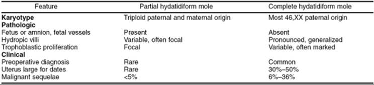
Approximately 1% of pregnancies have a triploid karyotype and resolve following spontaneous abortion; partial hydatidiform moles represent a subset of these pregnancies, with placental histologic features that share features with complete hydatidiform moles. A comparison of karyotypic, pathologic, and clinical features of partial and complete hydatidiform moles is shown in Table 57.1. Partial moles often are associated with identifiable fetal parts or amniotic membranes. Grossly, the placenta demonstrates a mixture of normal and hydropic villi. Microscopic features include normal and hydropic chorionic villi with focal mild hyperplasia of trophoblastic elements. Scalloping of the hydropic villi is common, with trophoblastic inclusions in the stroma. Fetal vessels frequently are observed, with nucleated fetal erythrocytes within the vessels. Normal amniotic membranes are identified often, even if a fetus is not found.
|
|
|
TABLE 57.1. Features of complete and partial hydatidiform moles |
Partial moles almost always are associated with one haploid maternal and two haploid paternal sets of chromosomes. Presumably, this results from dispermic fertilization of a haploid ovum or fertilization of a haploid ovum with a diploid sperm.
Women with partial hydatidiform mole usually have a clinical diagnosis of spontaneous abortion or missed abortion. Often, hydropic villi are not identified on ultrasonography, and the diagnosis is not suspected until after evacuation of the pregnancy. Initial hCG levels are lower than those seen in patients with complete hydatidiform mole, and prompt postevacuation regression of hCG levels occurs in most cases. Unlike patients with complete moles, who have a 10% to 30% incidence of malignant sequelae, fewer than 5% of the patients with partial moles develop criteria requiring chemotherapy. Regardless of this low risk of malignant sequelae, all women with partial hydatidiform moles should undergo hCG surveillance after evacuation, similar to that recommended for patients with complete hydatidiform mole. If there is any doubt that the products of a conception are molar, postevacuation hCG monitoring should be done.
Invasive Mole
Invasive moles (chorioadenoma destruens) are histologically identical to complete moles, but with invasion into the myometrium without intervening endometrial stroma. Invasive moles usually are diagnosed within 6 months of molar evacuation. Untreated invasive moles tend to invade the uterine wall locally, which can result in uterine perforation and hemorrhage. Direct vascular invasion and metastasis may occur rarely. In these cases, biopsies of distant metastases reveal hydropic villi consistent with invasive mole instead of solid sheets of anaplastic cells consistent with choriocarcinoma.
The identification of an invasive mole from uterine curetting may be difficult unless there is sufficient myometrium to document direct myometrial invasion. Most frequently this is a clinical diagnosis made when persistent, nonmetastatic GTN is diagnosed.
Choriocarcinoma
Choriocarcinoma is a highly anaplastic malignancy derived from trophoblastic elements. No chorionic villi are identified. Grossly, the tumor has a red, granular appearance on cut section with focal, often extensive, central necrosis and hemorrhage. Histologically, the lesion consists of mixed syncytiotrophoblastic and cytotrophoblastic elements with numerous abnormal mitoses, multinucleated giant cells, and extensive areas of necrosis and hemorrhage. Choriocarcinoma rapidly invades the myometrium and uterine vessels, and systemic metastasis results from hematogenous embolization. The lung and vagina are the most common sites of metastasis, with secondary dissemination to the central nervous system (CNS), kidney, liver, and gastrointestinal tract.
Choriocarcinomas may develop after any type of pregnancy. Approximately 50% of cases are preceded by hydatidiform mole, and the remaining are distributed equally between a normal antecedent term gestation and abortion or ectopic pregnancy. Gestational choriocarcinoma has been observed several years after the last known pregnancy. Spontaneous regression of the primary uterine site has been well documented from autopsy series of patients before the development of effective chemotherapy.
Placental Site Trophoblastic Tumor
PSTTs are locally invasive neoplasms derived from intermediate cells of the placenta. These rare neoplasms are composed of a monomorphic population of intermediate cytotrophoblast cells that secrete human placental lactogen (hPL) and relatively small amounts of hCG. Typically, there is local myometrial invasion with rare systemic metastasis. PSTTs are significantly more resistant to standard chemotherapy than other forms of GTN, and hysterectomy is the initial therapy of choice.
INCIDENCE AND EPIDEMIOLOGY
Approximately 3,000 cases of hydatidiform mole and 500 to 750 cases of malignant GTN are diagnosed in the United States each year. Hydatidiform mole is identified in approximately 1 in 1,500 to 2,000 pregnancies in the United States. There is a marked geographic variation in the incidence of this disease, with rates 5- to 15-fold higher in the Far East and Southeast Asia than in the Western industrialized nations. Some of this variation may be accounted for by the methodology of studies reporting the incidence of molar gestation, because many data were reported from the experience at referral centers and may overestimate the true incidence of molar pregnancies in the general population. Racial differences also may account for some of the geographic variations; Japanese immigrants to Hawaii have an incidence of molar gestation intermediate between that of native Hawaiians and native Japanese. Nutritional factors may also be important in the development of hydatidiform mole, including deficiencies of protein or animal fat and fat-soluble carotene.
Known risk factors for hydatidiform mole include previous molar pregnancies and maternal age. Women with a history of hydatidiform mole have a 4 to 5 times higher risk for development of a subsequent molar gestation. Women at the extremes of reproductive age are also at increased risk of developing a hydatidiform mole. Several studies have confirmed that risk increases with advanced maternal age, and others have suggested an increased risk for younger women or adolescents. The impact of paternal age on the incidence of hydatidiform mole is difficult to separate from the effect of maternal age.
The incidence of partial hydatidiform mole is unknown. Presumably, many are undiagnosed due to insufficient histologic analysis of tissue from spontaneous and induced abortions. Some pathologists may not be familiar with the diagnosis of partial hydatidiform mole, and karyotyping seldom is performed on material obtained from spontaneous abortions. One report reclassified approximately 10% of all moles in their studies as partial hydatidiform moles on the basis of histologic analysis.
Invasive mole follows approximately 15% to 20% of complete hydatidiform moles. In the United States, choriocarcinoma will follow in approximately 1 in 40 moles, 1 in 5,000 ectopic pregnancies, 1 in 15,000 abortions, and 1 in 150,000 normal pregnancies.
MANAGEMENT
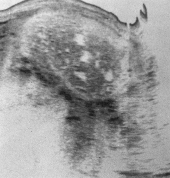
The basic principles of management of the patient with hydatidiform mole include establishment of the diagnosis, evacuation of the molar gestation, and close surveillance of hCG levels after evacuation. Patients with complete hydatidiform moles frequently have spontaneous abortion of hydropic villi, which are pathognomonic for molar pregnancy. Absent fetal heart tones, uterine enlargement different from that expected for the gestational age, hyperemesis, preeclampsia, and a markedly elevated hCG level are clinical indications that may suggest a hydatidiform mole. Ultrasonography is now the diagnostic method of choice for evaluating patients with suspected hydatidiform mole. Ultrasonography demonstrates a characteristic image of multiple echogenic regions within the uterus corresponding to hydropic villi, focal intrauterine hemorrhage, and absence of fetal parts (Fig. 57.1).
|
|
|
FIG. 57.1. Longitudinal ultrasonography reveals a hydatidiform mole. The mixed echoic pattern is caused by hydropic villi and focal intrauterine hemorrhage. |
Evaluation of the patient before evacuation of the hydatidiform mole is directed toward preparing the patient for evacuation, obtaining baseline hCG level information, screening for occult metastatic disease, and screening for associated hyperthyroidism. The following studies are recommended:
· Complete physical and pelvic examinations
· Complete blood count determination
· Blood chemistry levels, including renal, hepatic, and thyroid function tests
· Baseline serum hCG level
· Chest radiograph
· Pelvic ultrasonography
Suction dilation and curettage (D&C) offers a safe, rapid, and effective method of evacuation of hydatidiform mole in most patients. Some patients who do not desire preservation of reproductive function may benefit from primary hysterectomy for evacuation of hydatidiform mole and concurrent sterilization. However, these patients must be followed closely after hysterectomy, because malignant sequelae may still develop. Hysterotomy or induction of labor for molar evacuation is not recommended.
Suction D&C for evacuation of hydatidiform mole has a low complication rate in patients with uterine sizes corresponding to less than 16 weeks of gestation. Oxytocic agents are administered immediately after cervical dilation to aid in postoperative hemostasis. Patients with excessive uterine enlargement have a higher risk of pulmonary complications associated with D&C, which may be related to trophoblastic deportation, preeclampsia, fluid overload, anemia, and hyperthyroidism. In patients with hydatidiform mole complicated by uterine enlargement greater than that of 16 weeks of gestation, baseline arterial blood gas analysis should be obtained preoperatively, with consideration of invasive hemodynamic monitoring (Swan-Ganz catheterization) during the procedure. Evacuation should be performed only after a euvolemic status has been established and blood products readily available for possible transfusion.
Primary hysterectomy is a reasonable alternative for termination of molar gestation in patients with hydatidiform mole who have completed childbearing and desire sterilization. Hysterectomy reduces the incidence of malignant sequelae after evacuation of hydatidiform mole from approximately 20% after suction D&C to less than 5% after hysterectomy. However, this does not eliminate the need for careful follow-up or complete hCG surveillance after termination of hydatidiform mole, because malignant GTN may develop even after hysterectomy. Concurrent surgical extirpation of the adnexae should be considered, as with hysterectomy for benign indications.
Theca lutein cysts are detected clinically in approximately 20% of patients with molar gestations. These cysts are thin walled and highly vascular, and develop as a response to ovarian hyperstimulation from the high hCG levels produced by hydatidiform moles. They typically regress spontaneously over several weeks following molar evacuation. It is preferable to avoid surgery or ovarian manipulation in patients with uncomplicated theca lutein cysts. Rarely, because of abdominal distension and respiratory compromise, they may require aspiration with ultrasonic guidance. Enlarged cysts may undergo torsion, infarction, or rupture, and oophorectomy should be considered in these circumstances.
Some centers prescribe prophylactic short courses of methotrexate or dactinomycin chemotherapy at the time of molar evacuation to decrease the incidence of malignant sequelae in patients with high-risk features. However, prophylactic chemotherapy does not eliminate the chance of subsequent malignancy nor the need for hCG surveillance. Routine prophylactic chemotherapy at the time of molar evacuation for patients with uncomplicated hydatidiform mole is not recommended if reliable hCG surveillance is available.
Surveillance Following Molar Evacuation
The wide availability of sensitive hCG assays provides a backbone for postmolar evacuation triage and therapy. Several sensitive hCG assays are available, measuring the β-subunit of hCG by radioimmunoassay or by radioimmunometric assay. These assays can detect hCG levels elevated above the baseline variations of pituitary gonadotropins. These sensitive hCG assays should be used to monitor patients with GTN after evacuation of hydatidiform mole and during therapy of patients with malignant GTN. Urinary or serum pregnancy screening tests should not be used to follow patients with GTN, because the assays do not have sufficient sensitivity to permit detection of minimal elevations of hCG levels.
The recommendations for postmolar follow-up include determination of serum β-hCG levels every 1 to 2 weeks after evacuation until hCG level is undetectable. Subsequently, hCG levels should be determined for 2 to 4 weeks after the first normal level to confirm spontaneous hCG regression, followed by hCG surveillance every 1 to 2 months for 6 months after the first normal hCG level. In addition, hCG levels should be checked 6 weeks after all subsequent pregnancies due to the patients' elevated risk of subsequent GTN.
Most women with molar pregnancies undergo hCG level regression after evacuation to normal limits and require no further therapy. Strict contraception is recommended during hCG surveillance to avoid an intercurrent pregnancy that would interfere with monitoring. The hCG elevation of an early normal pregnancy may mask the hCG rise associated with postmolar malignant GTN.
Although an early report implicated oral contraceptives as a risk factor for postmolar malignant GTN, subsequent investigators found no significant increase in the risk of malignant GTN associated with the use of oral contraceptives after molar evacuation. After completion of 6 months of hCG level surveillance with normal results, patients may attempt pregnancy if desired. Because the risk of recurrent molar pregnancy is about 1%, they should undergo early screening of all pregnancies with ultrasonography to exclude recurrent molar gestations. Figure 57.2 illustrates an algorithm for diagnosis and follow-up of patients with hydatidiform mole.
|
|
|
FIG. 57.2. Algorithm for diagnosis and treatment of a patient with hydatidiform mole. |
False-positive hCG Test Results
The sensitivity and specificity of serum β-hCG levels makes it an ideal tumor marker in both diagnosis and surveillance of GTN. However, hCG, a glycoprotein comprised of both α- and β-subunits, linked by eight oligosaccharides, has significant heterogeneity in its structure and antigenicity. Free subunits, degraded molecules, molecules with irregular side chains, and fragments of hCG are present in sera of women in pregnancy, with trophoblastic disease, and with nontrophoblastic neoplasms. Furthermore, hCG variants that include hyperglycosylated hCG, nicked hCG, hCG missing the β-subunit C-terminal peptide, free β-subunit, and nicked free β-subunit may also be present. Although all professional laboratory hCG assays use antibodies to different sites on the β-subunit, variations in antibody use may lead to measurements of different hCG-related molecules.
The heterogeneity of hCG and the variability between different hCG assays may result in false-positive test results. The phenomena of “phantom hCG” and “phantom choriocarcinoma” have resulted in erroneous diagnoses of invasive GTN and choriocarcinoma, with unnecessary subsequent chemotherapy and hysterectomy. The U.S.A. hCG Reference Service recommends that when false-positive hCG results are suspected, urine hCG testing for detection of the β-core fragment, the terminal degradation product of hCG and its variants, should be performed. Evaluation also should include serum testing by a laboratory using a different β-hCG assay. In cases with absence of urine hCG and failure to detect serum β-hCG by an alternative laboratory, the initial elevated level likely represents a false-positive test result.
False-positive results also may be due to the presence of heterophilic antibodies. These antibodies are able to complete with hCG in antibody binding used in commercial hCG assays, and may result in persistent positive results. Treatment with a heterophilic antibody blocking reagent has been shown to reduce the incidence of false-positive results, and these reagents have been added to the protocol used by the U.S.A. hCG Reference Service.
MALIGNANT GESTATIONAL TROPHOBLASTIC NEOPLASMS
Malignant Sequelae Following Molar Evacuation
The spectrum of malignant sequelae after evacuation of hydatidiform mole includes intrauterine molar proliferation without invasion (i.e., retained mole), invasive mole, choriocarcinoma, and the clinical identification of metastatic GTN without a histologic diagnosis. The purpose of hCG level surveillance is early detection of trophoblastic neoplasia before the development of complications related to local proliferation, uterine invasion, or distant metastasis.
Before the development of effective chemotherapy for GTN, approximately 9% of women required hysterectomy for malignant sequelae after evacuation of hydatidiform mole. Many series of patients have been reported since the development of chemotherapy, with a wide range in the rate (9% to 36%) of patients requiring therapy after evacuation of hydatidiform mole. These observed differences in the frequency of malignant GTN may reflect inclusion of partial moles in some studies, a different incidence of metastatic disease in patient populations, or different hCG level regression criteria used to define malignant GTN and assign therapy in the various studies.
Histologic and clinical features can be used to define high- and low-risk groups of patients after molar evacuation but are of little value in determining the need for therapy in individual patients. Trophoblastic proliferation, uterine enlargement, theca lutein cysts, respiratory distress syndrome after molar evacuation, and postevacuation uterine hemorrhage all are associated with a higher frequency of postmolar malignant GTN. Prompt uterine involution and regression of theca lutein cysts are favorable prognostic signs. However, the definitive method for predicting development of postmolar malignant GTN is observation of the pattern of hCG regression.
Serial hCG levels are obtained at 1-week intervals with chest radiographs every 2 to 4 weeks as long as the hCG levels are elevated. Patients are treated with chemotherapy according to several criteria as follows: hCG level rise, hCG level plateau for 3 or more consecutive weekly levels, appearance of metastasis, or histologic evidence of invasive mole or choriocarcinoma.
Diagnosis
Malignant GTN is diagnosed in women with rising or paltering hCG levels or identification of metastasis after evacuation of a hydatidiform mole. Histologic diagnosis of invasive mole or choriocarcinoma is a criterion for malignant GTN. Patients who developed malignant GTN after nonmolar gestations often sought treatment for atypical symptoms attributable to distant metastases. Gastrointestinal or urologic hemorrhage, hemoptysis, or neurologic symptoms due to cerebral hemorrhage may be the clinical features. Irregular uterine bleeding or amenorrhea may be observed. Rarely, patients have clinical hyperthyroidism. Under these circumstances, the diagnosis of malignant GTN is facilitated with serum hCG testing and the exclusion of normal pregnancy. The possibility of metastatic GTN should be considered in any woman of the reproductive age group who has metastatic disease involving the lungs or distant sites from an unknown primary site of malignancy.
After the diagnosis has been made, the following clinical, laboratory, and radiographic evaluations are recommended for a patient with malignant GTN:
· Physical and pelvic examinations
· Baseline hCG level
· Complete blood count and baseline chemistry determinations
· Chest radiograph
· Pelvic ultrasonography
· Computed tomography (CT) of brain, chest, and abdomen–pelvis
Before a woman is treated for malignant GTN with chemotherapy, it is essential to exclude an intrauterine pregnancy with a pelvic ultrasonographic scan. Approximately 50% of patients with malignant GTN have pulmonary metastasis detected by routine chest radiographs. The clinical significance of small pulmonary metastases detected only by whole-lung CT scans is unknown. Because CNS and hepatic metastases may develop without clinical or radiographic evidence of pulmonary or vaginal metastases, the remainder of the radiologic studies are recommended strongly, regardless of whether abnormalities are detected by physical examination or chest radiograph. The role of magnetic resonance imaging studies or positron emission tomography in the evaluation of women with GTN is not yet defined.
Occult CNS metastases may be detected using lumbar puncture with simultaneous serum and cerebrospinal fluid (CSF) hCG determinations. The plasma-to-CSF hCG ratio is normally greater than 60:1 in the absence of CNS metastasis and is usually less than 60:1 in patients with CNS metastasis. Some investigators have reported falsely lowered plasma-to-CSF hCG ratios for patients without GTN undergoing first-trimester abortions and for patients with nonmetastatic GTN. CSF hCG determinations are most frequently utilized in evaluation of patients who have developed resistance to chemotherapy, with residual disease documented by elevated serum hCG levels, when the site of disease is obscure. Routine CSF hCG determination, however, is not part of initial GTN staging.
Surgery may be useful for patients with malignant GTN, but it rarely is indicated for staging or diagnosis alone. Histologic evaluation of tissue obtained by D&C may confirm the diagnosis and may be useful in the patient experiencing vaginal bleeding, but the procedure carries the risk of uterine perforation and hemorrhage. Laparoscopy, craniotomy, and thoracotomy rarely are justified to establish the primary diagnosis of malignant GTN, because this diagnosis can be made on the basis of elevated hCG levels with radiographic evidence of metastasis after excluding pregnancy.
All patients with malignant GTN must be thoroughly evaluated for metastatic disease. Selection of the initial therapy and subsequent survival largely depend on identification of poor prognostic factors in patients with metastatic disease. Figure 57.3 demonstrates an algorithm of management of malignant GTN.
|
|
|
FIG. 57.3. Algorithm for treatment of a patient with malignant gestational trophoblastic disease. |
Staging and Classification
Due to considerable overlap in the clinical course of the histologic entities that constitute malignant GTN, and because a complete histologic evaluation of an individual patient with GTN is rarely possible, a variety of classification and staging systems have been used to assess risk and assign initial therapy and prognosis for these patients. Several clinical findings are important in categorizing patients for treatment, and any classification system must take these factors into consideration to be clinically useful.
A simple clinical classification system based on risk factors for malignant sequelae may be used to assign initial therapy for patients with malignant GTN. This system takes into account factors that predict failure of initial single-agent chemotherapy to effect cure and allows identification of patients who would benefit from initial aggressive multidrug chemotherapy (Table 57.2). After radiographic studies have been completed, the patient is considered to have nonmetastatic GTN if there is no evidence of extrauterine spread of disease. This category is not subdivided into good-prognosis and poor-prognosis categories because these patients can achieve approximately 100% remission rates using current chemotherapeutic regimens. The histologic diagnosis of choriocarcinoma mandates treatment but does not change the initial choice of therapy. If there is any clinical or radiographic evidence of extrauterine metastasis, the patient is classified as having metastatic GTN. These patients are divided further into good-prognosis and poor-prognosis categories on the basis of factors that predict the failure of primary single-agent chemotherapy with methotrexate or dactinomycin.
|
|
|
TABLE 57.2. Clinical classification of malignant gestational trophoblastic neoplasia |
The International Federation of Gynecologists and Obstetricians (FIGO) developed a staging system for GTN that is based on the anatomic site of disease, conforming to the FIGO staging systems for other gynecologic malignancies:
· Stage I: Disease confined to the uterine corpus
· Stage II: Metastasis to the vagina or pelvis
· Stage III: Metastasis to the lung
· Stage IV: Other extrapelvic metastasis
Although essentially all patients with stage IV disease are at high risk, this system does not recognize the prognostic importance of other factors, such as the initial hCG level, other direct and indirect measurements of tumor burden, or duration of disease.
The World Health Organization (WHO) devised a prognostic index scoring system (Table 57.3) based on Bagshawe's analysis of the prognostic factors of his patient population. In addition to using a weighted scale for risk factors analyzed by the clinical classification, such as hCG level and duration of disease, the system identifies a graded range of additional risk factors. Ectopic pregnancies and abortions have an intermediate risk between molar and term pregnancies. Blood types and age contribute to the score. The size of the largest tumor, the number of metastatic sites, and site of metastasis are considered indirect approximations of tumor burden. After computation of each risk factor, the patient is considered to be at low risk if the score is 4 or less, at intermediate risk with a score of 5 to 7, and at high risk with a score of 8 or more. The WHO prognostic index scoring system has been found to correlate well with survival after conventional combination chemotherapy protocols. Not all of the WHO factors, however, have been evaluated critically to determine whether the graded scoring system is valid. Figure 57.3 illustrates a plan of evaluation and therapy for patients with malignant GTN.
|
|
|
TABLE 57.3. World Health Organization prognostic scoring system for gestational trophoblastic neoplasia |
Therapy
Before the development of effective chemotherapy against malignant GTN, surgical therapy was not successful in curing most patients, even women with nonmetastatic disease confined to the uterus. Since the introduction of methotrexate in the 1950s, malignant GTN has become the most curable of human solid tumors. Therapy should be conducted by physicians who have considerable experience in the management of these diseases, and a reliable hCG assay should be available so that therapeutic changes can be instituted quickly when necessary. Although chemotherapy has largely supplanted surgery and radiation therapy as the first-line management of malignant GTN, these modalities continue to play an important role in the management of patients with malignant GTN.
Chemotherapy
The initial chemotherapy should be selected on the basis of one of the systems of classification of malignant GTN (Table 57.4). Patients with nonmetastatic and good-prognosis GTN are treated initially with single-agent chemotherapy regimens using methotrexate or dactinomycin. Patients with poor-prognosis metastatic GTN require initial chemotherapy using combinations of active agents. Currently, the most frequently used combination is the EMA-CO regimen (etoposide, methotrexate with folic acid rescue, dactinomycin alternating with cyclophosphamide, and vincristine [Oncovin]).
|
|
|
TABLE 57.4. Chemotherapy for malignant gestational trophoblastic neoplasms |
Chemotherapy for Nonmetastatic Disease
The most widely used initial chemotherapy regimen for patients with nonmetastatic GTN includes courses of alternating doses of methotrexate (1 mg/kg [maximum 50 mg] on days 1, 3, 5, and 7) and folinic acid rescue (0.1 mg/kg on days 2, 4, 6, and 8), both administered intramuscularly. A variety of other agents and schedules, including daily and “pulsed” dactinomycin, also have been used to treat nonmetastatic GTN, with essentially equivalent remission rates. Reports indicate that weekly courses of methotrexate administered intravenously as a single dose of 30 mg/m2 have an advantage over other schedules of methotrexate in reducing cost and toxicity and are equally effective. Close hematologic and renal and liver chemistry monitoring must be done during therapy. Multiple studies show remission rates in this stage of disease to approach 100% when patients are treated appropriately.
Although methotrexate and dactinomycin continue to be the mainstays of treatment for low-risk disease, there is no consensus on a single superior regimen. The Gynecologic Oncology Group, a multicenter collaborative clinical trials association, is accruing patients into a randomized, phase III trial of weekly intravenous methotrexate versus pulsed dactinomycin as primary management for low-risk GTN.
The major goals of therapy for patients with nonmetastatic GTN include administration of active, relatively nontoxic regimens of chemotherapy with close hCG level monitoring. For most of these patients, chemotherapy alone is adequate treatment, and surgery is unnecessary. However, primary hysterectomy may be used for patients who desire sterilization, because this may reduce the number of cycles of chemotherapy necessary to achieve remission and cure.
Chemotherapy for Good-prognosis Metastatic Disease
Initial therapy with repetitive cycles of 5-day courses of single-agent methotrexate or dactinomycin is preferred for patients with good-prognosis metastatic GTN. For these patients without high-risk factors, the ultimate sustained remission rate also approaches 100%. Patients are treated initially with repetitive 5-day cycles of methotrexate administered intravenously at a dose of 0.4 mg/kg per day or dactinomycin administered intravenously daily at a dose of 500 µg. Chemotherapy is repeated every 14 days. Again, hematologic and renal and liver chemistry monitoring must be done, and colony-stimulating factors may be required to keep the treatment on schedule. Remission rates with either agent are approximately 60%, and most patients with drug-resistant disease are salvaged with the alternative single agent. Occasionally, patients may require hysterectomy or other surgical procedures to extirpate loci of persistent disease or else require a change in therapy to a multidrug chemotherapy regimen.
Chemotherapy for Poor-prognosis Disease
Multidrug chemotherapy is used as the initial therapy for patients with poor-prognosis metastatic GTN. Survival is poor for this group if the initial therapy is with only a single agent, and it is important to diagnose poor-prognosis, high-risk features in these patients so that appropriate initial therapy may be instituted in a timely fashion.
The most successful and least toxic multiagent regimen is EMA-CO. Other salvage regimens include MAC (methotrexate, actinomycin D, and cyclophosphamide [Cytoxan]), and the modified Bagshawe regimen of hydroxyurea, methotrexate, vincristine, cyclophosphamide, dactinomycin, and doxorubicin (Adriamycin). Because VP-16, cisplatin, 5-fluorouracil, vinca alkaloids, and bleomycin all demonstrate activity against GTN, other salvage regimens are based on combinations employing some of these agents. Regardless of regimen, another cycle of chemotherapy is administered as soon as toxicity from the previous cycle has cleared. Use of stem cell factor support should be used when needed to keep the treatment on schedule. Although toxicity may be marked with these combinations, aggressive initial multiagent chemotherapy usually is necessary to ensure optimal survival for patients in this category. Patients with poor-prognosis metastatic GTN should be treated at centers that specialize in the therapy of patients with GTN. Physician experience with GTN and these aggressive multimodality therapies appears to improve the outcome for these patients.
Patients with poor-prognosis metastatic GTN who have failed standard chemotherapy regimens are extremely challenging when designing appropriate coordination of chemotherapy and other therapeutic modalities to achieve salvage. Frequently, patients must be treated without regard for toxicity and must be supported through episodes of profound bone marrow suppression, sepsis, and nutritional deprivation to have a chance at salvage.
Surgical and Radiation Therapy
Hysterectomy
Due to improvements in chemotherapy, hysterectomy rarely is indicated as the initial therapy for women with malignant GTN. However, for patients with nonmetastatic or good-prognosis metastatic GTN with uterine disease, hysterectomy appears to decrease the duration of hospital stay and number of courses of chemotherapy required to achieve remission. Delayed or secondary hysterectomy is required as salvage therapy for approximately 10% of patients in these categories, but chemotherapy alone is successful in curing approximately 85% of patients with nonmetastatic and good-prognosis metastatic GTN. Among women with poor-prognosis metastatic GTN, preservation of childbearing capacity must be of secondary importance, but primary or delayed hysterectomy does not appear to offer as many benefits as in the therapy of patients with low-risk disease.
Surgical procedures for the purpose of removing sites of GTN are performed during a cycle of chemotherapy. Theoretically, this prevents dissemination of disease caused by embolization of GTN during the surgical manipulation of tissues. The complications of surgery and wound healing do not appear to be increased using this approach.
Thoracotomy
Thoracotomy with pulmonary segmental resection has been the most frequently performed procedure other than hysterectomy to remove drug-resistant disease. The radiographic regression of pulmonary nodules may lag far behind the response measured by hCG levels, and the persistence of a lung nodule after hCG normalization should not necessarily be interpreted as indicating persistent disease.
There have been reports of success with resection of solitary pulmonary nodules in carefully selected women with drug-resistant disease. Before considering pulmonary resection, it is important to exclude the possibility of disease elsewhere. If the patient has not had a hysterectomy, occult pelvic GTN should be evaluated with arteriography or other imaging techniques, such as magnetic resonance imaging. Prompt hCG remission after pulmonary resection predicts a favorable outcome.
Brain and Liver Metastasis
Whole-brain and whole-liver irradiation are used often as adjuncts to chemotherapy in the treatment of patients with metastasis to these sites. Whole-brain irradiation of 3,000 cGy over 10 days should be instituted immediately in conjunction with chemotherapy after brain metastasis is diagnosed. The rationale for this treatment is to prevent hemorrhage from these highly vascular metastases. With this approach, survival rates of approximately 85% are achieved for patients receiving primary therapy for brain metastasis; survival rates of approximately 50% are achieved for all patients with brain metastasis, including those developing metastasis during chemotherapy or at the time of recurrence. An alternate approach includes the combined use of high-dose systemic methotrexate with intermittent intrathecal methotrexate.
Craniotomy is not often required for the primary therapy or diagnosis of brain metastasis from GTN and usually is not successful when used alone. However, neurosurgical consultation should be obtained early in the course of therapy in the event that hemorrhage into brain lesions occurs and craniotomy is required for stabilization of the patient.
Whole-liver irradiation of approximately 2,000 cGy delivered over 10 days may be used to treat liver metastasis and prevent hepatic hemorrhage. Some investigators have not used hepatic irradiation to avoid exacerbation of the toxic hepatic effects of chemotherapy. Others have advocated selective occlusion of the hepatic artery by means of ligation or embolization when bleeding has occurred. Patients with hepatic metastasis have a poor prognosis and require aggressive chemotherapy, with careful monitoring during therapy to maximize the chances for survival. Whole-organ irradiation does not appear to compromise tolerance of aggressive chemotherapy.
Placental Site Trophoblastic Tumors
PSTT is a rare placental neoplasm that is histologically and clinically distinct from other forms of GTN. PSTTs lack significant volume of syncytiotrophoblast elements and do not secrete high levels of hCG. As such, serum hCG levels are not a reliable tumor marker for PSTT as they are in other forms of GTN. PSTT usually is locally aggressive, with invasion into the myometrium. It typically is resistant to traditional chemotherapeutic agents. Most women with PSTT require hysterectomy, although anecdotal reports suggest a minority may be cured by D&C alone. Rarely, PSTT may pursue a more aggressive course, characterized by distant metastasis and rapidly progressive disease.
Monitoring Therapy
Laboratory Evaluations
During chemotherapy, hematologic, renal, and hepatic indices should be monitored carefully. Toxicity from methotrexate-based and dactinomycin-based chemotherapy is relatively predictable. Unless a patient is receiving salvage chemotherapy for drug-resistant or recurrent GTN, new cycles of therapy should be withheld unless the total leukocyte count is greater than 3,000 cells/mm3, the platelet count is greater than 100,000 cells/mm3, and the renal and hepatic indices are normal. Radiographic studies of metastatic lesions and pelvic examination should be repeated frequently to monitor response to therapy.
More important than radiographic surveillance is closely following the hCG level response during therapy. Sensitive assays of the hCG levels should be performed at 1-week intervals during therapy. Chemotherapy should be changed if the hCG titer has not dropped at least 25% after a treatment cycle or if toxicity does not permit adequate dosage or frequency of administration.
Human Chorionic Gonadotropin Level Remission and Surveillance
Complete remission is defined as three consecutive weekly hCG levels in the normal, undetectable range. After remission has been achieved, hCG levels should be followed every 1 to 2 weeks for the first 3 months after completion of therapy, every 2 to 4 weeks for the subsequent 3 months, and every 1 to 2 months for the completion of the first year of surveillance. Recurrent episodes of GTN usually develop within a few months after completion of therapy, but late recurrences develop in a few cases. The assay of hCG levels should be repeated indefinitely at 6-month intervals. Patients are counseled to avoid pregnancy through the first year of hCG surveillance; most are treated with oral contraceptives for efficiency and to avoid low-level interference with the hCG assays caused by luteinizing hormone.
Prevention of Recurrent Disease
Despite the accuracy of hCG assays, a tumor burden of 104 cells may exist despite normal serum hCG levels. Recurrence rates after therapy for GTN have been 3% to 26%, depending on the patient population being studied. Patients with poor-prognosis metastatic GTN usually have a much higher recurrence rate than patients with low-risk disease. Most investigators agree that consolidation chemotherapy should be administered beyond the first normal hCG level. Typically, patients undergo one additional cycle of chemotherapy beyond the first normal hCG level for nonmetastatic disease, two cycles of consolidation chemotherapy for good-prognosis metastatic GTN, and three to four cycles of maintenance chemotherapy for poor-prognosis metastatic GTN. Toxicity must be considered in continuing therapy for these patients.
Reproduction After Therapy
Most women treated for GTN are cured by chemotherapy without resorting to hysterectomy. Several reports have documented that there is little or no increased risk of congenital malformation of infants during subsequent pregnancies. There may be a slight increase in the incidence of spontaneous abortions in this population, but this increase may be an artifact of increased hCG surveillance and identification of preclinical pregnancies in women who have previously received treatment for GTN. There is also an increased incidence of repeat molar gestation in patients who have had a hydatidiform mole (approximately 1%). Patients who have received intensive therapy for poor-prognosis metastatic GTN often undergo hysterectomy during therapy or may develop ovarian failure as a result of prolonged multidrug chemotherapy. Only a few patients in this category with uterine conservation are able to conceive or desire to attempt pregnancy after therapy.
Although the risk of congenital anomalies is not increased significantly after chemotherapy for malignant GTN, obstetric complication rates may be. Major obstetric complications can be observed in as many as 9% of these pregnancies. The incidence of placenta accreta, in particular, appears to be increased.
Women who have been treated successfully for GTN should be advised that pregnancy should be deferred for at least 1 year after therapy to allow for hCG level surveillance. They should be reassured about the low incidence of congenital malformations and recurrent GTN in subsequent pregnancies. An ultrasonographic scan should be performed early in pregnancy to exclude the possibility of recurrent molar gestation. A chest radiograph and serum hCG level should be obtained 6 to 8 weeks after delivery to screen for the rare case of recurrent choriocarcinoma developing after a subsequent normal pregnancy.
SUMMARY POINTS
· Gestational trophoblastic neoplasia represents a spectrum of human tumors, ranging from benign with potential malignant sequelae to highly malignant.
· Primary hydatidiform mole (complete) usually becomes apparent as a pregnancy with threatened first-trimester vaginal bleeding. Other frequent findings include discordant uterine size, ovarian cystic enlargement, and hyperemesis, but may include hyperthyroidism and preeclampsia.
· Evacuation of hydatidiform mole is best done by suction D&C, with caution used regarding trophoblastic deportation and pulmonary embolization.
· Individualization of therapy is critical for successful treatment of patients with GTN. Although chemotherapy is the primary modality for therapy, surgery and radiotherapy may also have a role.
· The hCG level is a sensitive and reliable marker for GTN and can be used effectively to aid in diagnosis, monitor therapy, and maintain follow-up evaluation.
SUGGESTED READINGS
False-positive hCG Testing
Cole LA, Butler S. Detection of hCG in trophoblastic disease: the USA hCG Reference Service experience. J Reprod Med 2002;47:433–444.
Management of Hydatiform Mole
Berkowitz RS, Goldstein DP, Bernstein MR. Ten years' experience with methotrexate and folinic acid as primary treatment for gestational trophoblastic disease. Gynecol Oncol 1986;23:111–118.
Curry SL, Hammond CB, Tyrey L, et al. Hydatidiform mole: diagnosis, management and long-term follow-up in 347 patients. Obstet Gynecol 1975;45:1–8.
Hancock BW, Tidy JA. Current management of molar pregnancy. J Reprod Med 2002;47:347–354.
Management of Low-risk Disease
Garrett AP, Garner EO, Goldstein DP, et al. Methotrexate infusion and folinic acid as primary therapy for nonmetastatic and low-risk metastatic gestational trophoblastic tumors: 15 years of experience. J Reprod Med 2002;47:355–362.
Management of High-risk Disease
DuBeshter B, Berkowitz RS, Goldstein DP, et al. Metastatic gestational trophoblastic disease: experience at the New England Trophoblastic Disease Center, 1965 to 1985. Obstet Gynecol 1987;69:390–395.
Lurain JR. Advances in management of high-risk gestational trophoblastic tumors. J Reprod Med 2002;47:451–459.
Newlands ES, Holden L, Seckl MJ, et al. Management of brain metastases in patients with high-risk gestational trophoblastic tumors. J Reprod Med2002;47:465–471.
Choriocarcinoma
Athanassiou A, Begent RH, Newlands ES, et al. Central nervous system metastases of choriocarcinoma: 23 years' experience at Charing Cross Hospital. Cancer 1983;52:1728–1735.
Bagshaw KD. Treatment of high-risk choriocarcinoma. J Reprod Med 1984;29:813–820.
Placental Site Trophoblastic Tumors
Feltmate CM, Genest DR, Goldstein DP, et al. Advances in the understanding of placental site trophoblastic tumor. J Reprod Med 2002;47:337–341.
Papadopoulos AJ, Foskett M, Seckl MJ, et al. Twenty-five years' clinical experience with placental site trophoblastic tumors. J Reprod Med 2002;47:460–464.
Chemotherapy in Trophoblastic Disease
Homesley HD, Blessing JA, Rettenmaier M, et al. Weekly methotrexate for nonmetastatic gestational trophoblastic disease. Obstet Gynecol 1988;72:413–418.
Lurain JR, Brewer JI. Treatment of high-risk gestational trophoblastic disease with methotrexate actinomycin-D, and cyclophosphamide chemotherapy. Obstet Gynecol 1985;65:830–836.
Lurain JR, Elfstrand EP. Single agent methotrexate for treatment of nonmetastatic gestational trophoblastic tumors. Am J Obstet Gynecol 1995;172:574–579.
Soto-Wright V, Goldstein DP, Bernstein MR, et al. Management of gestational trophoblastic tumors with etoposide, methotrexate and actinomycin-D. Gynecol Oncol 1997;64:156–159.