Arteries of the Abdomen
Fig. 15.1 Abdominal aorta and major branches
Anterior view. The abdominal aorta enters the abdomen at the T12 level through the aortic aperture of the diaphragm (see p. 54). Before bifurcating at L4 into its terminal branches, the common iliac arteries, the abdominal aorta gives off the renal arteries (see p. 209) and three major trunks that supply the organs of the alimentary canal:
Celiac trunk: Supplies the structures of the foregut, the anterior portion of the alimentary canal. The foregut consists of the esophagus (distal half), stomach, duodenum (proximal half), liver, gallbladder, and pancreas (superior portion).
Superior mesenteric artery: Supplies the structures of the midgut: the duodenum (distal half), jejunum and ileum, cecum and appendix, ascending and transverse colons, and right colic (hepatic) flexure.
Inferior mesenteric artery: Supplies the structures of the hindgut: the transverse colon (distal third), left colic (splenic) flexure, descending and sigmoid colons, rectum, and anal canal (upper part).


Fig. 15.2 Celiac trunk
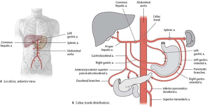
Fig. 15.3 Superior mesenteric artery
Anterior view.

Fig. 15.4 Inferior mesenteric artery
Anterior view.

Fig. 15.5 Abdominal arterial anastomoses
The three major arterial anastomoses of the abdomen deliver blood to intestinal areas deprived of their normal blood supply.

Abdominal Aorta & Renal Arteries
Fig. 15.6 Abdominal aorta
Anterior view of the female abdomen. Removed: Abdominal organs and peritoneum. The abdominal aorta is the distal continuation of the thoracic aorta (see p. 68). It enters the abdomen at the T12 level and bifurcates into the common iliac arteries at L4.
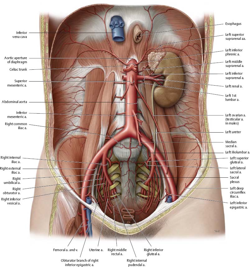
Fig. 15.7 Renal arteries
Left kidney, anterior view. The renal arteries arise at approximately the level of L2. Each renal artery divides into an anterior and a posterior branch. The anterior branch further divides into four segmental arteries (circled).

![]() Clinical
Clinical
Renal hypertension
The kidney is an important blood pressure sensor and regulator. Stenosis of the renal artery reduces blood flow through the kidney and stimulates increased production of renin, a hormone that cleaves angiotensinogen to form angiotensin I. Subsequent cleavage yields angiotensin II, which induces vasoconstriction and an increase in blood pressure. Renal hypertension must be excluded (or confirmed) when diagnosing high blood pressure.
Stenosis of the right renal artery (arrow), visible via arteriography.

Celiac Trunk
![]() The distribution of the celiac trunk is shown on p. 207.
The distribution of the celiac trunk is shown on p. 207.
Fig. 15.8 Celiac trunk: Stomach, liver, and gallbladder
Anterior view. Opened: Lesser omentum. Incised: Greater omentum. The celiac trunk arises from the abdominal aorta at about the level of L1.
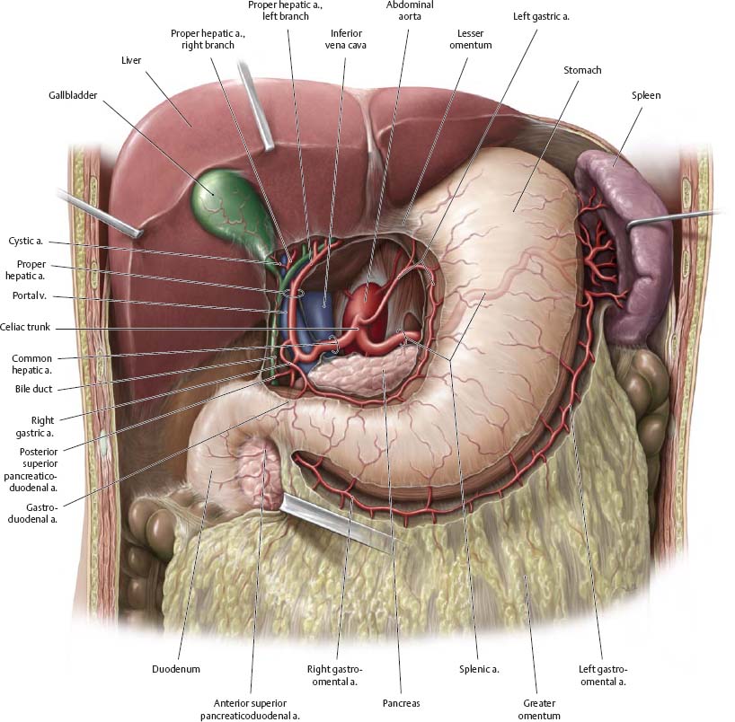
Fig. 15.9 Celiac trunk: Pancreas, duodenum, and spleen
Anterior view. Removed: Stomach (body) and lesser omentum.

Superior & Inferior Mesenteric Arteries
Fig. 15.10 Superior mesenteric artery
Anterior view. Partially removed: Stomach and peritoneum. Note: The middle colic artery has been truncated (see Fig. 15.11). The superior and inferior mesenteric arteries arise from the aorta opposite L2 and L3, respectively.

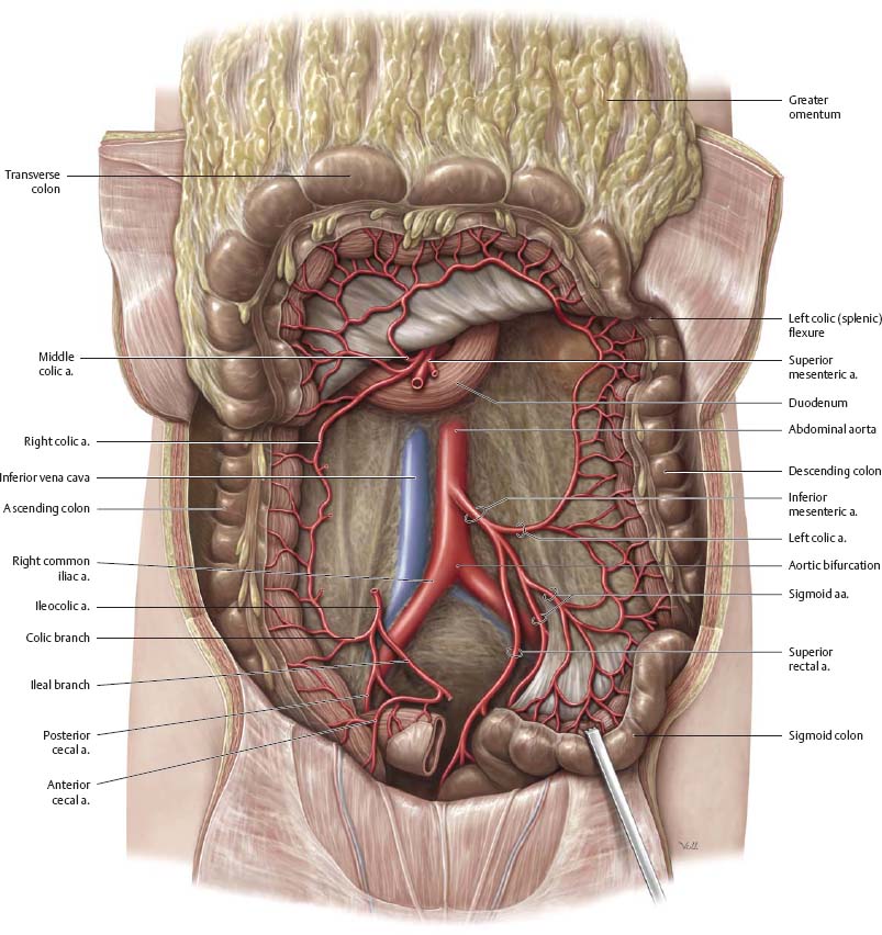
Fig. 15.11 Inferior mesenteric artery
Anterior view. Removed: Jejunum and ileum. Reflected: Transverse colon.
Veins of the Abdomen
Fig. 15.12 Inferior vena cava: Location
Anterior view.
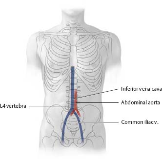
Fig. 15.13 Tributaries of the renal veins
Anterior view.
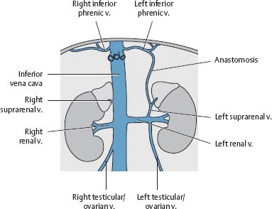

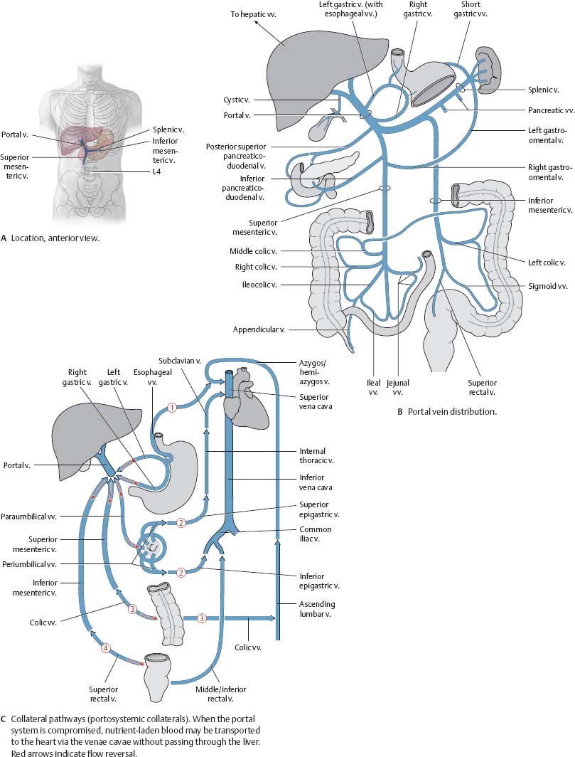
Fig. 15.14 Portal vein
The portal vein (see p. 218) drains venous blood from the abdominopelvic organs supplied by the celiac trunk and superior and inferior mesenteric arteries.
![]() Clinical
Clinical
Cancer metastases
Tumors in the region drained by the superior rectal vein may spread through the portal venous system to the capillary bed of the liver (hepatic metastasis). Tumors drained by the middle or inferior rectal veins may metastasize to the capillary bed of the lung (pulmonary metastasis) via the inferior vena cava and right heart.
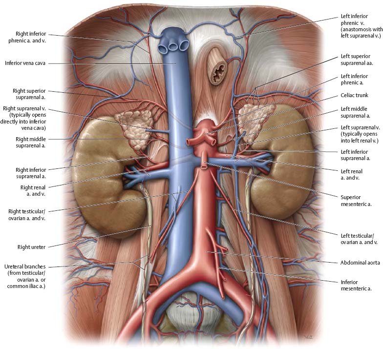
Inferior Vena Cava & Renal Veins
Fig. 15.15 Inferior vena cava
Anterior view of the female abdomen. Removed: All organs (except the left kidney and suprarenal gland).
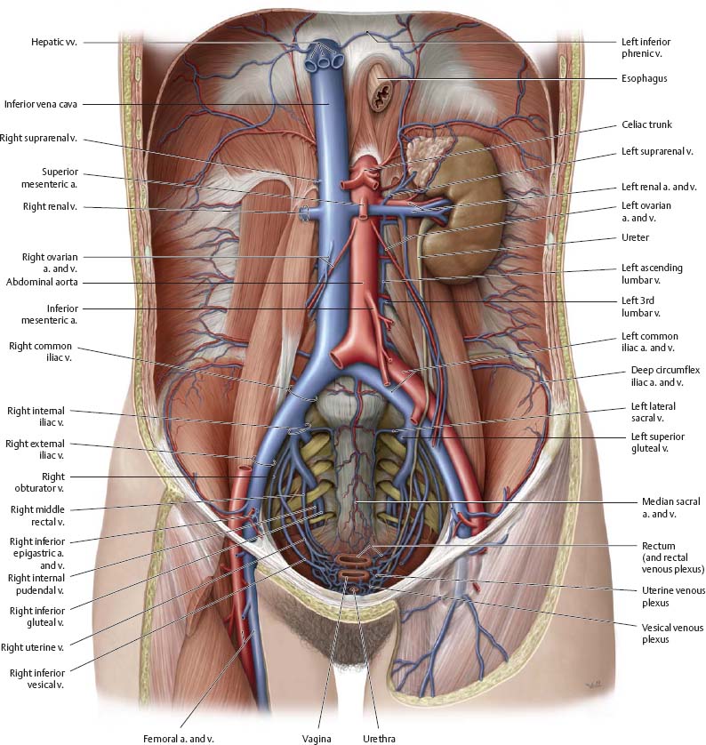
Fig. 15.16 Renal veins
Anterior view. See p. 209 for the renal arteries in isolation.
Portal Vein
![]() The portal vein is typically formed by the union of the superior mesenteric and the splenic veins posterior to the neck of the pancreas. The distribution of the portal vein is shown on p. 215.
The portal vein is typically formed by the union of the superior mesenteric and the splenic veins posterior to the neck of the pancreas. The distribution of the portal vein is shown on p. 215.
Fig. 15.17 Portal vein: Stomach and duodenum
Anterior view. Removed: Liver, lesser omentum, and peritoneum. Opened: Greater omentum.

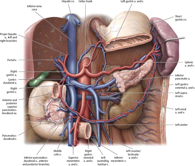
Fig. 15.18 Portal vein: Pancreas and spleen
Anterior view. Partially removed: Stomach, pancreas, and peritoneum.
Superior & Inferior Mesenteric Veins
Fig. 15.19 Superior mesenteric vein
Anterior view. Partially removed: Stomach, pancreas, peritoneum, mesentery, and transverse colon. Displaced: Small intestine.
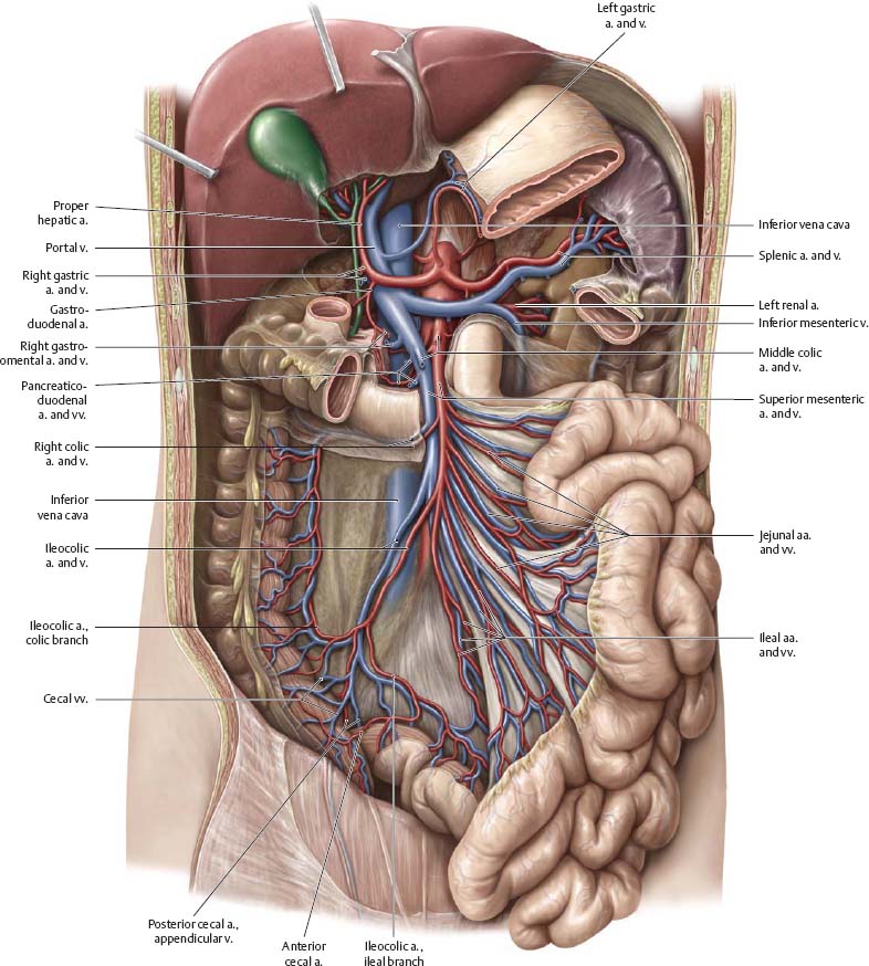
Fig. 15.20 Inferior mesenteric vein
Anterior view. Removed: Stomach, pancreas, small intestine, and peritoneum.

Arteries & Veins of the Pelvis


Fig. 15.21 Blood vessels of the pelvis
Idealized right hemipelvis, left lateral view.

Arteries & Veins of the Rectum & Genitalia
Fig. 15.22 Blood vessels of the rectum
Posterior view. The main blood supply to the rectum is from the superior rectal arteries; the middle rectal arteries serve as an anastomosis between the superior and inferior rectal arteries.

Fig. 15.23 Blood vessels of the genitalia
Anterior view.
