Nonviral infectious agents can affect the liver, contributing to a hepatitis-like picture clinically and morphologically. These conditions are rarely encountered by a pathologist not practicing in respective endemic areas. However, in the era of modern travel as well as changing responses to therapy, they can occasionally be seen anywhere (42). Many infectious disorders occur as complications of acquired immunodeficiency syndrome (AIDS) or in other immunocompromised conditions, causd by chemotherapy or in the setting of organ transplantation.
BACTERIAL CAUSES OF HEPATITIS
Typhoid Fever
Typhoid fever, caused by Salmonella typhi, is rare in Western countries, except when acquired abroad (42). Early, the liver shows nonspecific reactive changes including lymphocytosis, focal hepatocyte necrosis, and Kupffer cell hypertrophy. Erythrophagocytosis can sometimes be seen, as can small nonnecrotizing epithelioid granulomas and microvesicular steatosis. In the symptomatic stage (fastigium) hepatosplenomegaly is present, sometimes with jaundice. Granulomas, when present, can enlarge and become necrotic (e-Figs. 22.1, 22.2). Bacilli are generally not demonstrable with Gram's stain, although they are readily recoverable from the bile and gallbladder. Cholangitis is rare.
Brucellosis
Humans can be infected with Brucella through occupational exposure or ingestion of milk products (17). Brucellosis mimics many clinical conditions. The infective agent is difficult to culture, and serologic tests establish the diagnosis. A nonspecific hepatitis, with nonnecrotizing granulomas, is seen (13). Cirrhosis is unusual.
Melioidosis
Burkholderia pseudomallei, the cause of melioidosis, occurs in tropical zones and only rarely in the Western hemisphere (7). Approximately 10% of Americans who served in the Vietnam War had antibodies, but there were only 300 cases and 50 deaths documented (41). Hepatosplenomegaly and jaundice are common. In acute stages abscesses of varying sizes are seen with bacilli demonstrable with Gram's or Giemsa stain. Necrotic granulomas then form; they either resemble typical tuberculosis granulomas or show stellate appearance, similar to those seen in Yersinia infections or cat-scratch disease.
Listeriosis
Listeria monocytogenes affects pregnant women and can be transmitted transplacentally (11). It also occurs in immunosuppressed individuals and only rarely in immunocompetent adults (12). Microabscesses are seen in the liver, with abundant Gram-negative rods. Granulomas can also be seen. In some cases, however, the biopsy can show features resembling viral hepatitis.
Cat-Scratch Disease
The liver is rarely involved in cat-scratch disease without clinically demonstrable lymphadenopathy (20). Characteristic granulomas of cat-scratch disease are described as stellate, usually with central necrosis. Bacilli are generally not demonstrable with Gram's stain but can be seen with silver impregnation methods. Bile ducts can be involved.
Actinomycosis
Actinomycosis of the liver is usually secondary to intra-abdominal infection, with transmission via the portal vein or thoracic infection. There are often multiple hepatic abscesses, which contain colonies of Actinomyces.
SPIROCHETAL CAUSES OF HEPATITIS
Syphilis
Hepatic syphilis is exceedingly rare. The liver is typically not involved in primary syphilis.
Secondary Syphilis
In secondary syphilis, spirochetes become widely disseminated. Hepatitis can clinically be documented (6,37). There is often focal necrosis of hepatocytes, with portal inflammation and periductal inflammatory infiltrate rich in polymorphonuclear leukocytes and sometimes granulomas. Portal vessels can show vasculitis (51,53).
Tertiary Syphilis
The gumma is the characteristic lesion of tertiary syphilis. It can be quite large and clinically mistaken for a tumor. Gummas are giant cell granulomas with central necrosis, similar to those of tuberculosis. In contrast to caseation, the outlines of cellular architecture are not completely effaced. Plasma cells are usually prominent and an associated obliterative endarteritis may be present. Healing by fibrosis distorts the liver into nodule-like divisions of varying size (hepar lobatum) (53) (e-Figs. 22.3, 22.4). There is portal hypertension.
Congenital Syphilis
Congenital syphilis can manifest early during pregnancy or can be neonatal. Multiple, tiny, miliary-like foci of necrosis with giant cell response and associated portal inflammation are seen (e-Figs. 22.5, 22.6). Silver impregnation demonstrates numerous spirochetes when necrosis is prominent. Progressive pericellular fibrosis with liver plate atrophy and, ultimately, hepar lobatum may be seen.
Leptospirosis (Weil Disease)
Leptospira icterohemorrhagica is acquired from animals, but severe hepatorenal disease can be caused by various Leptospira serotypes. Biopsy shows severe cholestasis, most prominent in zones 3 and 2, with varying degrees of regenerative activity, including binucleate forms and mitoses (10). Extensive hepatocyte apoptosis is seen, with viral replication within hepatocytes (54), but balloon cells are relatively uncommon. Portal and lobular inflammation are not prominent. There is little or no steatosis.
Borreliosis (Relapsing Fever)
Liver biopsy, which is rarely performed in borreliosis because of the typical coagulopathy, shows multifocal liver cell necrosis with hemorrhage. Lymphocytes and polymorphonuclear leukocytes infiltrate sinusoids, with prominent Kupffer cells and erythrophagocytosis (34).
Lyme Disease
Lyme disease is a multisystem infection, with both acute and chronic phases. Liver involvement is usually not dominant (25,29). Acutely, mild portal tract inflammation is seen. In recurrent disease, hepatitis is more prominent, with prominent sinusoidal infiltration by lymphocytes and polymorphonuclear leukocytes. Hepatocytes are ballooned, with mitoses, and there is microvesicular steatosis and Kupffer cell hyperplasia.
MYCOBACTERIAL CAUSES OF HEPATITIS
Tuberculosis
Tuberculosis remains a disease of major importance worldwide, with frequent involvement of the liver (16). Typically, caseating granulomas can be seen in the lobule or portal tracts. Bacilli can be few or even undetectable, although molecular techniques, such as polymerase chain reaction, can establish the diagnosis (3,56).
Early, the lesion resembles a typical bacterial microabscess; this stage is exceedingly transient and rarely seen in the biopsy material. Granulomas sometimes have a relatively modest epithelioid and giant cell reaction, and the dominant feature may be central fibrinoid necrosis in which acid-fast bacilli are generally easily demonstrated (immature granuloma) (e-Figs. 22.7, 22.8).
In immunosuppressed individuals, innumerable acid-fast bacilli may be seen, with a picture resembling that of Mycobacterium avium-intracellulare (MAI) complex infection.
Leprosy
Liver involvement is uncommon (15). In lepromatous patients, abundant foamy macrophage clusters are distributed randomly throughout the liver, with acid-fast bacilli easily demonstrable. A polymorphonuclear neutrophilic reaction is seen at this stage (36). In the tuberculoid stage, granulomas are seen but acid-fast bacilli are scanty or absent. Amyloidosis can also be seen in the liver biopsy in patients with long-standing disease.
RICKETTSIAL AND CHLAMYDIAL CAUSES OF HEPATITIS
Q Fever
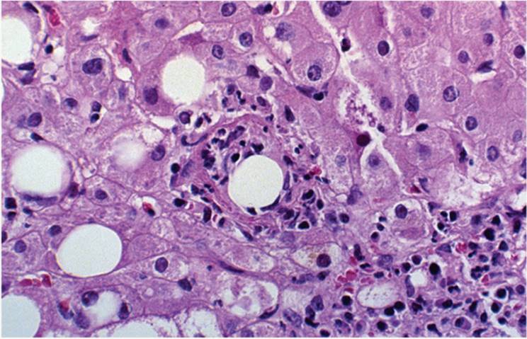
Q fever, caused by Coxiella burnetti, is transmitted by inhalation. Next to fever, hepatic involvement is the most common clinical feature. The typical lesion is a fibrin-ring granuloma, with a peripheral rim of epithelioid histiocytes with lymphocytes and occasional Langhans giant cells, within which there is a fibrin layer (Fig. 22.1) (5,32,47). The fibrin-ring granuloma characteristically surrounds an empty space that is usually about the size of a large fat vesicle (e-Figs. 22.9-22.13). Tuberculoid granulomas with central necrosis are also seen. The fibrin-ring granuloma is not pathognomonic for Q fever and can be seen in other infections (39,44,45).
Boutonneuse Fever
In boutonneuse fever, liver inflammation occurs without clinical evidence of hepatitis. Usually there is focal liver cell necrosis without granulomas, often accompanied by a lymphocytic infiltrate but with unremarkable portal tracts (26,31,58). Immunohistochemical reactions demonstrate rickettsial antigen in sinusoidal endothelial cells.
Chlamydial Infection
The liver can be involved in psittacosis and in genital infection. In psittacosis, there may be jaundice and hepatomegaly. The liver shows focal hepatocyte necrosis and Kupffer cell hypertrophy (60). In genital chlamydial infection (Chlamydia trachomatis) there may be an associated perihepatitis (Fitz-Hugh and Curtis syndrome) (60).
|
|
|
FIGURE 22.1 Fibrin-ring granuloma in Q fever (hematoxylin-eosin, original magnification ×200). |
PROTOZOAL CAUSES OF HEPATITIS
Malaria
The characteristic feature of malaria in the liver is malarial pigment (a molecule consisting of iron and a protein moiety) in erythrocytes and Kupffer cells during the stage of parasitemia and later in portal macrophages (Fig. 22.2, e-Figs. 22.13, 22.14). Sinusoids are distended by red blood cells in which parasites are often visible. Lobular or portal inflammation is generally mild (19,21,33). Liver cells may show glycogen depletion. With shock there may be zone 3 necrosis and cholestasis. Malarial antigen can be demonstrated within liver cells (23).
Amebiasis
The hallmark of liver involvement is the amebic abscess, usually in the right lobe and often multiple. The diagnosis should be considered in any case of liver abscess (30). Prior to the development of an abscess, the amebic trophozoites can be found in the sinusoids, with associated edema and focal coagulative necrosis and only a mild polymorphonuclear leukocyte response (2) (e-Fig. 22.15). The trophozoites resemble macrophages but tend to have a somewhat bluer cytoplasm. Erythrophagocytosis is common. The organisms can be highlighted with trichrome stain and periodic acid-Schiff reaction, and demonstrated immunohistochemically. With increasing lysis, the central, necrotic portion of the abscess enlarges. Older lesions become fibrotic. The surrounding liver is often edematous, showing the changes typical of space-occupying lesions. There may be marked cholestasis (46).
|
|
|
FIGURE 22.2 Malarial pigment in Kupffer cells (hematoxylin-eosin, original magnification ×400). |
Leishmaniasis
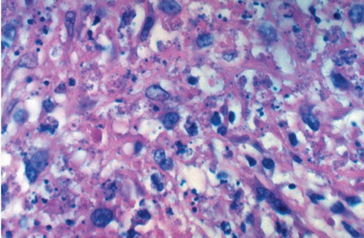
Visceral leishmaniasis (kala azar) is caused by several species of Leishmania and is seen in AIDS (9). Kupffer cells are prominent, and there is a diffuse sinusoidal and portal chronic inflammation with many plasma cells (Fig. 22.3). Parasites are generally demonstrable in both portal macrophages and Kupffer cells and, rarely, in hepatocytes (22). In another form of hepatic leishmaniasis, macrophage clusters are seen within the parenchyma containing only rare parasites. Epithelioid granulomas, with varying degrees of central necrosis, may be seen, and fibrin-ring granulomas, similar to those of Q fever, have been described (44). Steatosis is common, and there may be jaundice. Sometimes there may be hemorrhagic necrosis, perhaps a result of shock.
|
|
|
FIGURE 22.3 Leishmaniasis in a patient with acquired immune deficiency syndrome (hematoxylin-eosin, original magnification ×400). |
Balantidiasis
Balantidium coli is a rare cause of dysentery and, as with amebiasis, can cause liver abscess.
Toxoplasmosis
Congenital toxoplasmosis may present as giant cell hepatitis. More often the liver biopsy shows multiple foci of necrosis, with enlarged sinusoidal cells in which the parasites can be demonstrated. In acquired toxoplasmosis there can be a severe hepatitis with foci of necrosis and cholestasis (56). Nonnecrotizing giant and epithelioid cell granulomas can also be seen (59).
Babesiosis
In babesiosis, transmitted by ticks, the liver shows parasite-containing erythrocytes distending sinusoids, with Kupffer cell hyperplasia and zone 3 ischemic necrosis.
HELMINTHIC CAUSES OF HEPATITIS
Ascariasis
The Ascaris worms fill and distend the grossly visible bile ducts, causing liver biopsy changes similar to those of any large duct obstruction (28,35). (e-Figs. 22.16-2.18)
Schistosomiasis
In the early stages of schistosomiasis, eggs become trapped in portal vein radicles where they elicit an eosinophilic reaction; sometimes this results in an eosinophilic abscess with abundant fibrin deposition (24,48). Subsequently, an epithelioid and giant cell granuloma forms (12). Schistosomal pigment, indistinguishable from malarial pigment, is present in portal macrophages and Kupffer cells. Portal fibrosis eventually develops, leading to portal hypertension and the characteristic pipe stem pattern of fibrosis seen microscopically (24) (e-Fig. 22.19).
Echinococcosis
The larval forms of Echinococcus granulosus and Echinococcus multilocularis cause hydatid disease of the liver. Liver biopsy can be dangerous, potentially leading to dissemination through the abdominal cavity.
E. granulosus forms a spherical cyst with a complex wall consisting of an outer acellular laminated membrane and an inner transparent germinal membrane to which the budding protoscolices attach. Host reaction is modest with some granulation tissue. Eosinophils are usually not prominent.
In E. multilocularis there is no fibrous rim and the cyst wall is relatively thin. The wall is laminated, but there is no germinal layer and protoscolices are not seen. The membranes invade necrotic liver tissue, and there is a variable, generally mild, host response (e-Fig. 22.20).
Clonorchiasis
The liver fluke Clonorchis sinensis, found principally in the Far East, is acquired by the ingestion of uncooked freshwater fish (38). The worms thrive in the distal bile ducts, but in severe cases they can be found in the proximal bile ducts and the gallbladder. The liver biopsy initially shows dilated interlobular bile ducts and ascending cholangitis, and sometimes abscess formation, with subsequent chronic inflammation, including many eosinophils, and fibrosis. Worms are generally not seen in the interlobular bile ducts. The larger bile ducts show epithelial hyperplasia. Multicentric, mucin-secreting cholangiocarcinoma may develop (8,27).
Opisthorchiasis
Infection with Opisthorchis species flukes occurs less frequently than with Clonorchis, but the manifestations are essentially the same, including the development of cholangiocarcinoma.
Enterobiasis
Rarely Enterobius vermicularis can manifest as nodules of the liver (enterobioma) (18,43). A necrotic center is surrounded by a granulomatous reaction, which becomes fibrotic.
Strongyloidiasis
The larvae of Strongyloides vermicularis are present in small portal vessels and in sinusoids, often without significant inflammation. There may be a chronic inflammatory cell infiltrate, often with multinucleated giant cells (49).
Capillariasis
Adult worms and eggs of Capillaria hepatica can be seen in the liver, with marked infiltration by eosinophils and a granulomatous reaction.
Toxocariasis (Visceral Larva Migrans)
Visceral larva migrans is primarily a disease of children (52). The liver shows small foci of necrosis, with many eosinophils and variable numbers of histiocytes and giant cells (55). The granulomas are often somewhat cylindrical and elongated, representing long tracts of tissue destruction.
Fascioliasis
Humans are only occasionally infected as a result of eating contaminated watercress. The fluke migrates to the large bile ducts and the gallbladder (38). As the fluke migrates through the liver, an inflammatory tract develops marked necrosis and eosinophilia, with eventual fibrosis. Bile duct dilatation occurs, as in clonorchiasis, with inflammatory and proliferative changes of the bile duct epithelium (1,17,51). Cholangiocarcinoma has not been reported.
REFERENCES
1. Acosta-Ferreira W, Vercelli-Hetta J, Falconi LM. Fasciola hepatica human infection. Virchows Arch A 1979;383:319-327.
2. Adams EB, MacLeod IN. Invasive amebiasis. II. Amebic liver abscess and its complications. Medicine 1977;56:325-334.
3. Akcan Y, Tuncer S, Hayran M, et al. PCR on disseminated tuberculosis in bone marrow and liver biopsy specimens: correlation to histopathological and clinical diagnosis. Scand J Infect Dis 1997;29:271-274.
4. Andrade ZA, Peixoto E, Guerret S, et al. Hepatic connective tissue changes in hepatosplenic schistosomiasis. Hum Pathol 1992;23:566-673.
5. Atienza P, Raymond M-J, Degott C, et al. Chronic Q fever hepatitis complicated by extensive fibrosis. Gastroenterology 1988;95:478-481.
6. Baker AL, Kaplan MM, Wolfe HJ, et al. Liver disease associated with early syphilis. N Engl J Med 1971;284:1422-1423.
7. Barnes OF, Appleman MD, Cosgrove MM. A case of melioidosis originating in North America. Am Rev Respir Dis 1986;134:170-171.
8. Belamaric J. Intrahepatic bile duct carcinoma and C. sinensis infection in Hong Kong. Cancer 1973;31:468-473.
9. Berenguer J, Moreno S, Cercenado E, et al. Visceral leishmaniasis in patients infected with human immunodeficiency virus (HIV). Ann Intern Med 1989;111:129-132.
10. Bhamarapravati N, Boonyapaknavig V, Viarnuvatti V, et al. Liver changes in leptospirosis: a study of needle biopsies in 22 cases. Am J Proctol 1966;17:480-487.
11. Bourgeois N, Jacobs F, Tavares ML, et al. Listeria monocytogenes in a liver transplant recipient: case report and review of the literature. J Hepatol 1993;18:284-289.
12. Brito JM, Borojevic R. Liver granulomas in schistosomiasis: mast cell-dependent induction of SCF expression in hepatic stellate cells is mediated by TNF-alpha. J Leukoc Biol 1997;62:389-396.
13. Cervantes F, Carbonell J, Bruguera M, et al. Liver disease in brucellosis. A clinical and pathologic study of 40 cases. Postgrad Med J 1982;58:346-350.
14. Chen MG, Mott KE. Progress in assessment of morbidity due to Fasciola hepatica infection. Trop Dis Bull 1990;87:R1-R37.
15. Chen TSN, Drutz DJ, Whelan GE. Hepatic granulomas in leprosy. Arch Pathol Lab Med 1976;100:182-185.
16. Cook GC. Liver involvement in systemic infection. Eur J Gastroenterol Hepatol 1997;9: 1239-1247.
17. Corbel MJ. Brucellosis: an overview. Emerg Infect Dis 1997;3:213-221.
18. Daly JJ, Baker GF. Pinworm granuloma of the liver. Am J Trop Med Hyg 1984;33:62-64.
19. de Brito T, Barone AA, Faria RM. Human liver biopsy in P. falciparum and P. vivax malaria: a light and electron microscopy study. Virchows Arch A 1969;348:220-229.
20. Delbeke D, Sandler MP, Shaff MI, et al. Cat-scratch disease: report of a case with liver lesions and no lymphadenopathy. J Nucl Med 1988;29:1454-1456.
21. Deller JJ, Cifarelli PS, Berque S, et al. Malaria hepatitis. Milit Med 1967;132:614-620.
22. Duarte MIS, Mariano ON, Corbett CEP. Liver parenchymal cell parasitism in human visceral leishmaniasis. Virchows Arch A 1989;415:1-6.
23. Duarte MI, Boulos M, Segurado AA, et al. Hyperreactive malarious splenomegaly: immunohistochemical demonstration of Plasmodium falciparum antigen in liver cells. Trans R Soc Trop Med Hyg 1997;91:429-430.
24. Dunn MW, Berkowitz FE, Miller JJ, et al. Hepatosplenic cat-scratch diseases and abdominal pain. Pediatr Infect Dis J 1997;16:269-72.
25. Duray PH. Clinical pathologic correlations of Lyme disease. Rev Infect Dis 1989;11 (Suppl 6):S1487-S1493.
26. Ferreira VA, Vianna MR, Yasuda PH, et al. Detection of leptospiral antigen in the human liver and kidney using an immunoperoxidase staining procedure. J Pathol 1987;151:125-131.
27. Flavell DJ. Liver-fluke infection as an aetiological factor in bile-duct carcinoma of man. Trans R Soc Trop Med Hyg 1981;75:814-824.
28. Gayotto LCDC, Muszkar RML, Souza IV. Hepatobiliary alterations in massive miliary ascariasis. Histopathological aspects of an autopsy case. Rev Inst Med Trop Sao Paulo 1990;32:91-95.
29. Goellner MH, Agger WA, Burgess JH, et al. Hepatitis due to recurrent Lyme disease. Ann Intern Med 1988;108:707-708.
30. Greenstein AJ, Lowenthal BA, Hammer GF, et al. Continuing patterns of disease in pyogenic abscess: a study of 38 cases. Am J Gastroenterol 1984;79:217-226.
31. Guardia J, Martinez-Vazquez JM, Moragas A. The liver in boutonneuse fever. Gut 1974; 15:549-551.
32. Hofmann CE, Heaton JW Jr. Q fever hepatitis: clinical manifestations and pathological findings. Gastroenterology 1982;83:474-479.
33. Joshi YK, Tandon SK, Acharya SK, et al. Acute hepatic failure due to Plasmodium falciparum liver injury. Liver 1986;6:357-360.
34. Judge DM, Samuel I, Perine PL, et al. Louse-born relapsing fever in man. Arch Pathol Lab Med 1974;97:136-140.
35. Khuroo M, Zarger SA, Mahajan R. Hepatobiliary and pancreatic ascariasis in India. Lancet 1990;1:1503-1503.
36. Kramarsky B, Edmondson HA, Peters RL, et al. Lepromatous leprosy in reaction. Arch Pathol Lab Med 1968;85:516-531.
37. Lee RV, Thornton GF, Conn HO. Liver disease associated with secondary syphilis. N Engl J Med 1972;284:1423-1425.
38. Liu LX, Harinasuta KT. Liver and intestinal flukes. Gastroenterol Clin North Am 1996; 25:627-636.
39. Lobdell DH. “Ring” granulomas in cytomegalovirus hepatitis. Arch Pathol Lab Med 1987;11:880-882.
40. Maincent G, Labadie H, Fabre M, et al. Tertiary hepatic syphilis. A treatable cause of multilobular liver. Dig Dis Sci 1997;42:447-450.
41. McCormick JB, Sexton DJ, McMurray JG, et al. Human-to-human transmission of Pseudomonas pseudomallei. Ann Intern Med 1973;83:512-513.
42. Mermin JH, Townes JM, Gerber M, et al. Typhoid fever in the United States, 1985-1994: changing risks of international travel and increasing antimicrobial resistance. Arch Intern Med 1998;158:633-638.
43. Mondou EN, Gnepp DR. Hepatic granuloma resulting from Enterobius vermicularis. Am J Clin Pathol 1989;91:97-100.
44. Moreno A, Marazuela M, Yerba M, et al. Hepatic fibrin-ring granulomas in visceral leishmaniasis. Gastroenterology 1988;95:1123-1126.
45. Murphy E, Griffiths MR, Hunter JA, et al. Fibrin-ring granulomas: a non-specific reaction to liver injury? Histopathology 1991;19:91-93.
46. Nigam P, Gupta AK, Kapoor KK, et al. Cholestasis in amoebic liver abscess. Gut 1985;26: 140-145.
47. Pellegrin M, Delsol G, Aubergant JC, et al. Granulomatous hepatitis in Q fever. Hum Pathol 1980;11:51-57.
48. Pereira LM, McFarlane BM, Massarolo P, et al. Specific liver autoreactivity in schistosomiasis mansoni. Trans R Soc Trop Med Hyg 1997;91:310-314.
49. Poltera AA, Katsimbura N. Granulomatous hepatitis due to Strongyloides stercoralis. J Pathol 1974;113:241-246.
50. Rizkallah MF, Meyer L, Ayoug EM. Hepatic and splenic abscesses in cat-scratch disease. Pediatr Infect Dis J 1988;7:191-195.
51. Romeu J, Rybak B, Dave P, et al. Spirochetal vasculitis and bile ductular damage in early hepatic syphilis. Am J Gastroenterol 1980;74:352-354.
52. Schantz PM. Toxocara larva migrans now. Am J Trop Med Hyg 1989;41(Suppl):21-34.
53. Sobel HJ, Wolf EH. Liver involvement in early syphilis. Arch Pathol Lab Med 1972;93: 565-568.
54. Sun HC, Deng PF, Wang CJ, et al. Liver involvement in epidemic haemorrhagic fever: in-situ hybridization, immunohistochemical and pathological studies. J Gastroenterol Hepatol 1997;12:540-546.
55. Taylor MRH, Keane CT, O'Connor P, et al. The expanded spectrum of toxocaral disease. Lancet 1988;1:692-695.
56. Tiwari I, Rolland CF, Popple AW. Cholestatic jaundice due to toxoplasma hepatitis. Postgrad Med J 1982;58:299-300.
57. Vago L, Barberis M, Gori A, et al. Nested polymerase chain reaction for Mycobacterium tuberculosis IS6110 sequence on formalin-fixed paraffin-embedded tissues with granulomatous diseases for rapid diagnosis of tuberculosis. Am J Clin Pathol 1998;109: 411-415.
58. Walker DH, Staiti A, Mansueto G, et al. Frequent occurrence of hepatic lesions in boutonneuse fever. Acta Trop 1986;43:175-181.
59. Weitberg AB, Alper JC, Diamond I, et al. Acute granulomatous hepatitis in the course of acquired toxoplasmosis. N Engl J Med 1979;300:1093-1096.
60. Wollner-Hanssen P, Weström L, Märdh P-A. Perihepatitis and chlamydial salpingitis. Lancet 1980;1:901-904.
61. Yow EM, Brennan JC, Preston J, et al. The pathology of psittacosis: a report of two cases with hepatitis. Am J Med 1959;27:739-747.