Benign proliferative and neoplastic processes arise from hepatocytes, biliary epithelium, stromal (mesenchymal) elements, or combinations of those elements (Tables 23.1 and 23.2). These tumors are relatively rare. Mesenchymal tumors, such as lipoma and leiomyoma, resemble those at other sites and will not be discussed in great detail.
BENIGN HEPATOCELLULAR TUMORS AND TUMOR-LIKE CONDITIONS
Benign hepatocellular tumors are uncommon. In the past, most were diagnosed incidentally, when they reached significant dimensions, or at autopsy. Liver cell adenomas became more common with the widespread use of oral contraceptives. Increasing awareness of benign liver tumors and sophisticated imaging techniques allow for detection of small tumors. However, it is still difficult for the pathologist to establish the correct diagnosis for these processes, especially when only a core biopsy is available.
Hepatocellular Adenoma
Prior to the introduction of contraceptive pills, hepatocellular adenoma was one of the rarest of human tumors (54,55,86). The estimated incidence is 3 to 4 cases per 100,000 (41,54,63,68). Anabolic steroids (9,54) and other medications (59) have been associated, as have metabolic diseases, including glycogenosis I (18,50,84), glycogenosis IV (3), tyrosinemia (115), diabetes mellitus (34), galactosemia (27), and thalassemia (16). Rarely, adenomas develop spontaneously (39,117). Adenomas developing in the setting of a metabolic disease have an increased incidence of hepatocellular carcinoma (HCC) (9,39,41,54,56,66).
Patients may be asymptomatic or can present with right upper quadrant abdominal pain. If the tumor is large, it may be palpable. Subcapsular adenomas can rupture with intraperitoneal hemorrhage and acute abdominal manifestations (45,55,84). Adenomas can regress spontaneously (13,28) and after withdrawal of medications (13,28,54,82,86,93,102). Adenomas are usually single and most often right lobe, but can be multiple (6,16,52,71,89,90). They vary considerably in size and, with advanced imaging techniques, are detected when quite small (75,84,88).
|
TABLE 23.1 Benign Hepatocellular Tumors and Tumor-Like Conditions |
|||||||||||||||||||||||||||||||||||||||||||||||||||||||||||||||||||||
|
|||||||||||||||||||||||||||||||||||||||||||||||||||||||||||||||||||||
|
TABLE 23.2 Mesenchymal Tumors and Tumor-Like Conditions of the Liver |
|||||||||||||||||||||||||||||||||||||||||||||||||||||||||||||||||||||
|
|||||||||||||||||||||||||||||||||||||||||||||||||||||||||||||||||||||
|
|
|
FIGURE 23.1 Liver cell adenoma. Bland hepatocytes arranged in two- or three-cell-thick liver plates (hematoxylin-eosin, original magnification ×100). |
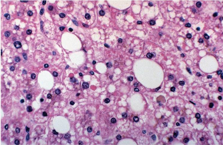
PATHOLOGY. Liver cell adenomas consist of bland-appearing hepatocytes, arranged in two- and three-cell-thick liver plates (Figs. 23.1, 23.2, e-Figs. 23.1-23.8). Bile-containing acinar structures can be seen. Liver cells can be either larger or smaller than the surrounding nonneoplastic hepatocytes and may have a relatively increased nuclear/cytoplasmic ratio, resembling well-differentiated hepatocellular carcinoma. Some adenomas express hormone receptors (70). Adenoma cells can also contain more glycogen than the surrounding liver and appear relatively pale. Various cytoplasmic materials and inclusions can be seen, including Mallory hyalin, lipofuscin, giant mitochondria, and oncocytic change (55,83,89) (Fig. 23.3). Mitotic figures are rare. Focal necrosis, with or without apoptosis, microgranulomas, and extramedullary hematopoiesis, may be seen (63,68,84). Some adenomas show focally atypical, pleomorphic cells, often with hyperchromatic nuclei, and as many as 10% have giant cell transformation (9,39,43,55,58,73). Fibrosis is not prominent. Vascular elements, particularly thick-walled arteries and arterioles, are seen at the periphery of the tumor. Thick-walled veins are seen as well as small-caliber, thin-walled vascular channels randomly distributed within the tumor. Sinusoidal endothelial cells and Kupffer cells are present (38,55).
|
|
|
FIGURE 23.2 Liver cell adenoma. Reticulin silver preparation highlighting thickened liver cell plates (reticulin silver preparation, original magnification ×100). |
|
|
|
FIGURE 23.3 Liver cell adenoma. Giant mitochondria are present in some hepatocytes (hematoxylin-eosin, original magnification ×400). |
DIFFERENTIAL DIAGNOSIS. Hepatic adenoma must be differentiated from well-differentiated HCC, focal nodular hyperplasia (FNH) (79), macroregenerative nodules (MRNs), nodular regenerative hyperplasia (NRH), multiple hepatocellular adenomatosis, focal steatosis, and sometimes even normal liver.
In HCC, the nuclear/cytoplasmic ratio is high and cellular atypia is usually more pronounced. There is usually only a scanty reticulin network in HCC in contrast with an adenoma in which the reticulin network is usually maintained. Adenoma is distinguished from FNH by the absence of portal tract structures and terminal hepatic venules (central veins), as well as lack of the central fibrotic zone characteristic of FNH. In biopsy material, however, differentiation can be exceedingly difficult. Recent studies have shown that based on molecular criteria, HNF1α and β-catenin mutations and the presence or absence of inflammation, liver cell adenomas can be distinguished from FNH (Table 23.3) (7).
|
TABLE 23.3 Histopathologic, Clinical, and Molecular Features of Benign Liver Lesions |
||||||||||||||||||||||||||||||||||||||||
|
||||||||||||||||||||||||||||||||||||||||
MRNs in cirrhosis or after massive necrosis can be indistinguishable from liver cell adenoma. Cellular atypia is generally not seen. Adenomas rarely occur in cirrhosis, except in the metabolic disorders. NRH can involve the whole liver, and the nodules are quite small, representing exaggerated lobules, usually with surrounding compressed atrophic-appearing hepatocytes.
Multiple Hepatocellular Adenomatosis
Multiple hepatocellular adenomatosis is exceedingly rare (6,17,33,55,63,67,84). Adenomas may be clustered in one lobe but can be diffuse. The diagnosis is applied with four or more adenomas with unremarkable intervening liver parenchyma (5,6,88). Progressive hepatocellular proliferation develops as a response to chronic low-grade ischemia, similar to NRH and possibly partial nodular transformation (110,111). There is no proven association with contraceptive pills or anabolic steroids.
PATHOLOGY. Nodules consist of bland, benign hepatocytes, arranged in one- or two-cell-thick liver plates. Macrovesicular steatosis may be focally present. Areas of hemorrhage can also be present.
Macroregenerative Nodule
MRNs, defined as at least 0.8 cm in size, have been also called adenomatous hyperplasia, adenomatous regeneration, and large regenerative nodules (29,31,32,36). Some are most likely preneoplastic (29,36,61,76,80,88,103), evidenced by their occurrence adjacent to HCC (29,76,88). MRNs usually occur in cirrhosis but also with submassive liver necrosis. MRNs can be multiple (103,109).
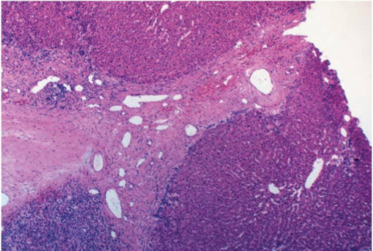
PATHOLOGY. MRN type I (ordinary or typical) is composed of liver cell plates that are two or three cells thick, similar to those in cirrhosis. Portal tracts are usually present. The liver cells are bland, with no significant cytologic atypia, and may contain various intracytoplasmic materials, including fat, hemosiderin, bile, and/or Mallory material (98,101,103,109) (Fig. 23.4, e-Fig. 23.9). In contrast, MRN type II (atypical MRN, atypical adenomatous hyperplasia, borderline lesions) has focal or multifocal small or large cell dysplasia, with altered architectural pattern and irregular liver cell plates, and there may be nodule-in-nodule formation and focal pseudoacinar formation (Fig. 23.5).
DIFFERENTIAL DIAGNOSIS. The distinction between MRN type II and HCC can be very difficult; MRN II nodules may represent true hepatocellular carcinoma in situ (32,61,88). Extensive loss of the underlying reticulin network, irregular thickness of the liver cell plates, and significant cytologic nuclear atypia are features favoring HCC.
|
|
|
FIGURE 23.4 Macroregenerative nodule, type I. Note the relatively bland hepatocytes arranged in two- to three-cell-thick liver plates. Focal macrovesicular steatosis is present (hematoxylin-eosin, original magnification ×40). |
Focal Nodular Hyperplasia
A benign pseudotumor, FNH is one of the most common benign liver lesions, occurring predominantly in young women (15,55,78,81,84,87). Although usually solitary, multiple lesions have been described (42,111). FNH can also be associated with other benign tumors, including adenomas and hemangiomas (20,39,46,71). Most cases are found incidentally, but a small proportion of patients present with abdominal pain or a palpable mass. FNH can be associated with oral contraceptive use (39,54,110,112).
|
|
|
FIGURE 23.5 Macroregenerative nodule, type II (adenomatous hyperplasia). Note the nodule-in-nodule formation and liver cells with large cell dysplasia. This would qualify for so-called borderline lesion (hematoxylin-eosin, original magnification ×100). |
|
|
|
FIGURE 23.6 Focal nodular hyperplasia with central fibrovascular area (hematoxylin-eosin, original magnification ×40). |
The most characteristic feature is the central, stellate fibrovascular zone, which has the historically entrenched name of “fibrous scar” or “scarlike fibrosis (Figs. 23.6 and 23.7, e-Figs. 23.10-23.15). Pathogenesis of FNH can, in many cases, be attributed to a hyperplastic response of the liver to a pre-existing arterial abnormality (110). Some recent studies, however, demonstrated overexpression of glutamine synthase in the periportal areas in very early stages of FNH development, suggesting that the primary event is portal tract and portal vein injury, leading to formation of arteriovenous shunts, ductular reaction, and finally, scar formation (8).
|
|
|
FIGURE 23.7 Focal nodular hyperplasia. Marginal bile ductular proliferation is present (hematoxylin-eosin, original magnification ×40). |
PATHOLOGY. The diagnosis of FNH can be difficult in biopsy material. Hepatocytes surrounding the fibrovascular zone are cytologically bland but may show mild degenerative changes, focal steatosis, or increased glycogen. They are arranged in incomplete nodules or pseudonodules and are partially surrounded by slender fibrous septa extending from the central fibrotic zone. Focal thickening of liver cell plates is common. Portal structures or complete portal tracts are seen. Cholestasis and chronic cholestasis features can be seen, and zone 1 (periportal) hepatocytes can contain copper-binding protein (15,55,84).
The central fibrovascular zone contains mature collagen and numerous vascular channels, most of them medium and large thick-walled arteries. Arteries often show fibromuscular hyperplasia, myointimal proliferation, and myxomatous change, very often with significant lumen narrowing (55,84). Veins are unremarkable. True portal tracts, native interlobular or larger bile ducts are not seen within the central fibrovascular zone, but marginal bile ductular proliferation is common. A mild chronic inflammatory infiltrate, with occasional polymorphonuclear leukocytes, is common but nonspecific. Recently, new histologic forms of FNH have been recognized and a histologic scoring system proposed (30,78). Several recent findings pertinent to the pathogenesis have prompted an updated classification of FNH and liver cell adenoma and those findings are incorporated in Table 23.3 (7).
DIFFERENTIAL DIAGNOSIS. Differential diagnosis includes hepatocellular adenoma, cirrhosis, mixed hamartoma, bile duct adenoma (BDA), well-differentiated HCC, and the fibrolamellar variant of HCC (81,107).
Nodular Regenerative Hyperplasia
Nodular regenerative hyperplasia (NRH) is a benign regenerative lesion composed of hyperplastic hepatocytes forming small nodules not surrounded by fibrous tissue, eventually diffusely distributed. Relatively uncommon, it presents with noncirrhotic portal hypertension or its complications (e.g., ascites, variceal bleeding) (19,55,85,96,99). NRH can sometimes clinically mimic venoocclusive disease (74).
NRH is associated with various hematologic and immunologic diseases (55,74,75,84,85). Immunosuppressive drugs and toxins have also been implicated (74). NRH can be seen following liver transplantation. Recently, NRH has been recognized in patients with inflammatory bowel diseases treated with 6-thioguanine (91). Spontaneous regression of NRH may be seen. The pathogenesis is not entirely clear, but it is postulated that inadequate vascular supply and even local ischemic events play a role (96,111,113).
|
|
|
FIGURE 23.8 Nodular regenerative hyperplasia. Note the well-defined regenerative nodules of liver parenchyma without significant fibrosis (hematoxylin-eosin, original magnification ×100). |
PATHOLOGY. NRH is best recognized at low magnification. Distinct contours of regenerative nodules, composed of unremarkable hepatocytes, are centered on generally unchanged portal tracts, rimmed by slightly or markedly atrophic hepatocytes at the periphery (Figs. 23.8 and 23.9, e-Figs. 23.16-23.22). Liver cell dysplasia is rare (92). Unremarkable sinusoidal endothelial and Kupffer cells are present. Vascular alterations have been described, including inconspicuous portal vein branches and hepatic artery inflammation (55,84). Reticulin silver preparation is helpful, particularly for the earliest detection of NRH (e-Fig. 23.23). A preserved reticulin network with one- and two-cell-thick liver plates is seen with a peripheral rim of compressed hepatocytes (Fig. 23.9).
|
|
|
FIGURE 23.9 Nodular regenerative hyperplasia. Note the compression of underlying reticulin network at the periphery of the nodules (reticulin silver preparation, original magnification ×100). |
Partial Nodular Transformation
Partial nodular transformation (PNT) is an exceptionally rare, benign, tumor-like solitary nodule, 3 to 40 mm in diameter, almost always arising in the hilum. Patients present with portal hypertension, portal vein thrombosis, ascites, and esophageal varices (25,55,92). Fewer than 20 cases have been reported. The pathogenesis is thought to be similar to that of NRH as a response to insufficient blood supply, as with portal vein thrombosis. The extrahepatic portal vein frequently shows evidence of old thrombosis. A hypercoagulable state may contribute to portal vein thrombosis.
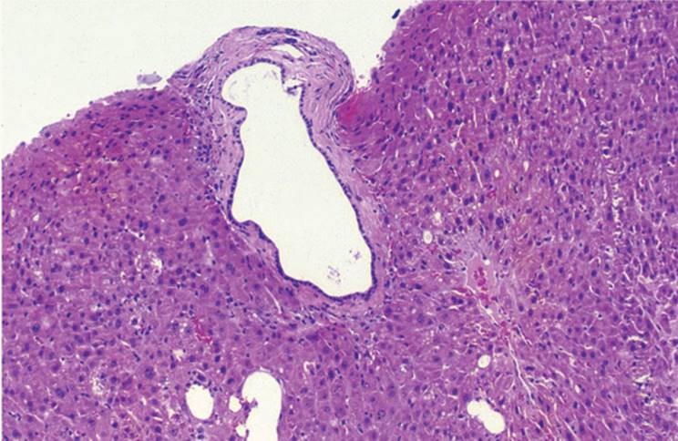
PATHOLOGY. The features of PNT are subtle, with regenerative and incomplete liver nodules and slender fibrous septa that partially circumscribe the nodules. Portal tracts are at the periphery (Fig. 23.10). Bile ductule proliferation is not seen. Portal vein branches are inconspicuous, and occlusion or thrombosis is not usually evident. Sinusoidal dilatation may be seen. Liver plates are focally two to three cells thick, best seen with reticulin silver stain. The hepatocytes are generally bland.
DIFFERENTIAL DIAGNOSIS. Differential diagnosis includes NRH and FNH (Fig. 23.11).
|
|
|
FIGURE 23.10 Partial nodular transformation. Paucicellular portal tract with a slender septum extending into the parenchyma (hematoxylin-eosin, original magnification ×100). |
|
|
|
FIGURE 23.11 Proliferative activity in focal nodular hyperplasia, nodular regenerative hyperplasia, and liver cell adenoma (immunoperoxidase, avidin-streptavidin, antibody to proliferating cell nuclear antigen, original magnification ×200). |
BENIGN BILIARY EPITHELIAL TUMORS
Bile Duct Adenoma
True bile duct adenoma (BDA) is exceedingly rare and usually incidentally discovered. A few millimeters to 2 cm, tumors are well-circumscribed, pale tan, sometimes easily seen on the capsular surface or in the immediate subcapsular region (2). They may be multiple (55).
PATHOLOGY. BDA consists of numerous small, relatively uniform bile duct-like structures that are not cystically dilated. A small amount of bland, paucicellular collagen is between ducts, which are sharply demarcated from the surrounding liver parenchyma (Fig. 23.12, e-figs. 23.24, 23.25). The epithelium is benign but can show hyperchromatic, enlarged nuclei and less cytoplasm than the biliary epithelium of interlobular bile ducts. Mitoses are not seen. A few inflammatory cells, including lymphocytes and polymorphonuclear leukocytes, may be present.
DIFFERENTIAL DIAGNOSIS. Differential diagnosis includes benign biliary (microbiliary) hamartoma (von Meyenburg complex, microhamartoma), biliary adenofibroma, cholangiocarcinoma, and metastatic adenocarcinoma. Bile duct hamartoma is a developmental malformation, part of the spectrum of fibropolycystic diseases of the liver, and may be single or multiple (14,25,55) (see Chapter 6). Distinctly tortuous, or angulated, focally cystically dilated ductlike structures, often filled with inspissated bile or dense eosinophilic proteinaceous acellular debris, are seen, and the surrounding stroma is fibrotic (Fig. 23.13). Bile duct hamartoma is often adjacent to or arises from existing portal tract structures. The epithelial cells in BDA show reactivity for both low and high molecular weight keratins (CAM5.2 and AE1/3), indicating that these cells may originate from either hepatocytes or biliary epithelium, or from a common stem cell. In contrast, bile duct hamartoma reacts strongly with high molecular weight keratin (AE1/3 or CK19). Biliary adenofibroma is similar to BDA but usually larger, with bile duct structures embedded in fibrous stroma. The epithelium may show focal apocrine metaplasia (104).
|
|
|
FIGURE 23.12 Bile duct adenoma showing numerous somewhat irregular bile duct structures embedded in a paucicellular fibrous stroma. Note that the epithelial cells lack any degree of atypia. The whole lesion is sharply demarcated from the surrounding liver parenchyma (hematoxylin-eosin, original magnification ×20). |
|
|
|
FIGURE 23.13 Von Meyenburg complex (microhamartoma) with cystically dilated ductlike structures, arising from or adjacent to the portal tract (hematoxylin-eosin, original magnification ×100). |
Cholangiocarcinoma can develop in the background of BDA (14), as well as with multiple bile duct hamartomas (14,23,48). Differentiation of BDA from metastatic adenocarcinoma can be difficult in frozen section material.
Solitary Unilocular Bile Duct Cyst
Solitary unilocular bile duct cysts are lined by benign columnar or cuboidal biliary-type epithelium, surrounded by a variable amount of indistinct fibrous tissue (55). Mononuclear inflammatory cells can be seen (Fig. 23.14).
DIFFERENTIAL DIAGNOSIS. Diagnoses to consider are parasitic cysts, posttraumatic cysts, and healing abscess wall. Posttraumatic cysts and healing abscess do not have true epithelial lining.
Ciliated Foregut Cyst
This develops from embryonic foregut with differentiation to respiratory-type (bronchial) structures, often with four distinct layers including pseudostratified respiratory-type epithelium, subepithelial tissue, smooth muscle layer, and fibrous capsule (55,100). The ciliated foregut cyst is almost invariably unilocular.
Biliary Cystadenoma
Biliary cystadenoma is a relatively rare lesion. It is usually single and multiloculated with a predilection for the right lobe of the liver (1,53,114). The tumor can be as large as 25 to 30 cm and may replace almost the entire liver lobe. A smooth inner epithelial lining is present.
|
|
|
FIGURE 23.14 Solitary, simple cyst lined by flattened cuboidal epithelium (hematoxylin-eosin, original magnification ×100). |
|
|
|
FIGURE 23.15 Biliary cystadenoma with mesenchymal ovarian-like stroma. The epithelium is nonciliated, columnar, and mucin producing with no cytologic atypia (hematoxylin-eosin, original magnification ×100). |
PATHOLOGY. The cysts are lined by mucin-producing, nonciliated columnar or cuboidal focally flattened epithelium (Fig. 23.15, e-Fig. 23.26). In women, a mesenchymal ovarian-like stroma is usually seen, suggesting that these tumors arise from intrahepatic ectopic ovarian tissue (53,114). Cholesterol crystals, as well as hemosiderin-laden and ceroidladen macrophages, may be prominent. When the fluid is blood tinged, malignancy should be suspected. Three types have been recognized: (a) hepatobiliary cystadenoma with mesenchymal stroma, (b) hepatobiliary cystadenoma without mesenchymal stroma, and (c) intraductal polypoid hepatobiliary cystadenoma. In men there is no mesenchymal stroma. If not completely excised, the tumor may recur. Malignant transformation is relatively common (53,116,116,118).
DIFFERENTIAL DIAGNOSIS. Diagnoses to consider are ciliated foregut cyst, intrahepatic choledochocyst, and parasitic cysts. Intrahepatic choledochocyst occurs in the hilum and does not exhibit mesenchymal stroma. Parasitic cysts usually have fibrous capsule, often with calcifications and without identifiable lining epithelium, and often with identifiable parasites.
Biliary Papillomatosis
Biliary papillomatosis is an unusual, multifocal, histopathologically benign lesion that is relentlessly progressive in growth and ultimately fatal (72,108). Patients are usually male adults who present with symptoms and signs of biliary obstruction.
PATHOLOGY. Intrahepatic and extrahepatic bile ducts are dilated and contain exaggerated papillary growth of the biliary epithelium with supporting fibrovascular stroma. The epithelial cells are in most cases bland, columnar, with mucin-containing cytoplasm and basally located nuclei. Focal inflammation and ulceration may also be seen, and necrotic cellular debris admixed with inflammatory cells may fill the lumen. Complications include bacterial cholangitis, sepsis, and liver failure. Malignant transformation has been reported (77).
BENIGN MESENCHYMAL TUMORS
Benign mesenchymal tumors can be divided into several groups according to their origin. These relatively rare tumors are listed in Table 23.2.
Mixed Epithelial and Mesenchymal Tumors
MESENCHYMAL HAMARTOMA. This uncommon childhood tumor has a male predominance and comprises approximately 5% of pediatric liver tumors. The tumor results from malformation of primitive hepatic mesenchyme. Children present with a painless abdominal mass, with a characteristic imaging appearance. The tumor is usually solitary and large, with a right lobe predilection (22,24,35,62,94).
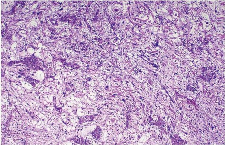
Pathology. Solid tumor areas have a mixture of hepatic and mesenchymal cells, often with vascular proliferation, bile ducts or duct-like structures, and cystically dilated spaces (Fig. 23.16, e-Figs. 23.27-23.30). Scattered stellate mesenchymal cells are embedded in edematous collagenous or myxoid stroma. Cystically dilated spaces represent distended bile duct-like structures. Bile ducts often are at the periphery, with atrophic epithelium and focal acute inflammation. Entrapped hepatocytes in abundant mesenchymal stroma show reactive and regenerative changes. Foci of hematopoiesis are occasionally seen. Thick-walled blood vessels may be at the periphery (26,66).
|
|
|
FIGURE 23.16 Mesenchymal hamartoma composed of a mixture of epithelial (liver and bile duct) cells and embedded in the edematous myxoid stroma (hematoxylin-eosin, original magnification ×100). |
Differential Diagnosis. Differential diagnosis includes embryonal (undifferentiated) sarcoma and infantile hemangioendothelioma. Differentiation from embryonal sarcoma is particularly important, since both lesions may exhibit loose, edematous, myxoid stroma. In most embryonal sarcomas, the cellularity is readily evident, and neoplastic cells show distinctive cytologic features of highly malignant cells.
MIXED HAMARTOMA. Mixed hamartoma is an exceedingly rare solitary tumor usually diagnosed in infants to teenagers (55). Also called mixed adenoma because of the epithelial components, it has bile ductular structures and hepatocytes embedded in dense fibrous stroma lacking vascular structures and portal tracts.
Pathology. The tumor is pseudonodular, with benign hepatocyte nests surrounded by dense fibrous tissue Proliferated bile ductules and thick-walled arterial vessels are at the interface with the fibrous tissue.
Differential Diagnosis. The principal diagnosis to consider is FNH. The central fibrovascular scar of FNH is virtually diagnostic. The diffuse, uniform nodularity of mixed hamartoma, easily recognized in a resected specimen, may not be obvious in biopsy material. Mixed hamartoma has a cirrhosis-like pattern but is focal.
Benign Vascular Tumors
INFANTILE HEMANGIOENDOTHELIOMA. Infantile hemangioendothelioma (IH) can be single, most commonly in the right lobe, or multifocal. Size may vary from a few millimeters to more than 20 cm. Multiple tumors impart a spongiform appearance to the liver. Larger tumors are well circumscribed but not encapsulated and are variegated, often with scattered calcifications (52,54,55,57).
Pathology. The tumor consists of small vessels with plump endothelial cells. Type I IH contains stroma composed of unremarkable collagen and reticulin fibers (20) (Fig. 23.17, e-Figs. 23.31, 23.32). Foci of hematopoiesis are frequently seen. Type II IH is an aggressive variant with multilayered endothelial cells and occasional tufting (119). Type II IH has variable degrees of nuclear pleomorphism, hyperchromasia, and mitoses and may metastasize. Typical angiosarcoma may develop (60,95).
CAVERNOUS HEMANGIOMA. This most common benign tumor, uncommon in children, can be solitary or multiple, often only a few millimeters in size. Hemangiomas can also be extremely large, replacing considerable portions of liver parenchyma (52,55,90). Hemangioma can be multiple and can occur with FNH.
Pathology. Cavernous spaces are lined by a single layer of benign endothelial cells (Fig. 23.18). Antibodies, including factor VIII, CD31, and CD34, can be used but are generally not necessary. Fibrosis and calcifications are common. Sclerosis is seen with larger lesions, with extensive fibrosis, hyalinization, and narrowing; sometimes complete obliteration of vascular spaces occurs.
|
|
|
FIGURE 23.17 Hemangioma with cavernous spaces lined by a single layer of endothelial cells (hematoxylin-eosin, original magnification ×40). |
LYMPHANGIOMA AND HEPATIC LYMPHANGIOMATOSIS. Multiple hepatic lymphangiomatosis is exceedingly rare (106). Solitary lymphangiomas in the liver are even rarer, and their existence is controversial. A systemic form of lymphangiomatosis occurs in children, affecting multiple organs, the skeleton, lymph nodes, soft tissue, and the retroperitoneum, with potentially poor prognosis. Multiple hepatic lymphangiomatosis is seen as clustered multiple soft white-tan tumors with cystically dilated spaces that expel milky fluid when punctured (106). Benign flattened endothelial cells line irregular spaces. The surrounding fibrous stroma shows focal calcifications, and adjacent hepatocytes may be compressed and atrophic.
|
|
|
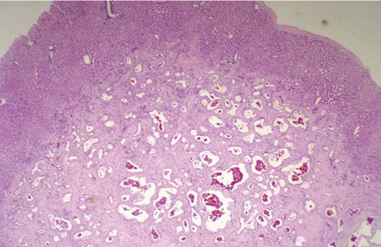
FIGURE 23.18 Angiomyolipoma. The tumor is composed of a mixture of epithelioid cells, with smooth muscle differentiation, variable amount of fat, and thick-walled blood vessels (hematoxylin-eosin, original magnification ×100). |
HEREDITARY HEMORRHAGIC TELANGIECTASIA (OSLER-WEBER-RENDU DISEASE). Telangiectasias can be seen in any part of the liver, including lobules and portal tracts. The primary abnormality is thought to be a dilatation of intrahepatic arteries. Dilated vascular spaces lined by unremarkable endothelial cells are surrounded by fibrous stroma (70,111) (Fig. 23.19). NH can also be seen (111).
Benign Lipomatous Tumors
LIPOMA. Lipoma is one of the rarest benign liver tumors and usually incidental. Usually single and well demarcated, they range from a few millimeters to 2 cm (52,55). The cell of origin has not been identified. Portal tract structures are preserved, and many hepatocytes contain large fat droplets. Differential diagnosis includes pseudolipoma, liver cell adenoma with abundant fat, and so-called focal fatty change.
PSEUDOLIPOMA. Pseudolipoma is another rare, small, encapsulated lesion. It consists of mature lipocytes on the capsular surface or in the immediate subcapsular region, presumed to be from an adherent appendix epiploica, with varying degrees of degeneration, calcification, and occasionally true osseous metaplasia (52,55).
|
|
|
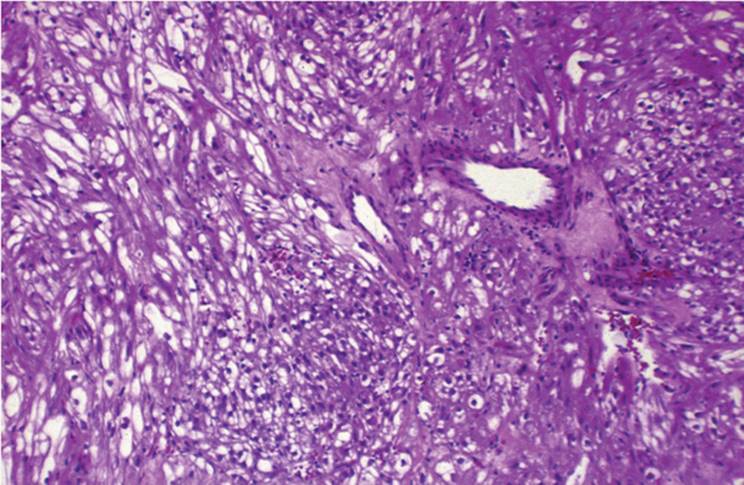
FIGURE 23.19 Angiomyolipoma. The smooth muscle component consists of bundles of epithelioid cells, strongly positive for smooth muscle actin and focal positivity for HMB45 (immunoperoxidase, avidin-streptavidin, original magnification ×100). |
|
|
|
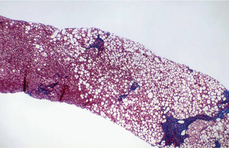
FIGURE 23.20 Focal fatty change. Localized macrovesicular steatosis involving only a portion of liver parenchyma. Surrounding liver is without significant histopathologic findings (hematoxylin-eosin, original magnification ×100). |
ANGIOMYOLIPOMA. Angiomyolipoma is a rare benign lipomatous tumor resembling the more common renal angiomyolipomas. Patients are between 30 and 72 years of age (37). Tumors are usually solitary and may be as large as 20 cm.
Pathology. Three components occur in variable proportions: smooth muscle, blood vessels, and fat (Fig. 23.20, e-Figs. 23.33-23.36). The smooth muscle component is seen as sheets and bundles of spindle and epithelioid cells. Tortuous vascular channels with thick-walled vessels are particularly prominent at the periphery. Lipomatous tissue varies from as little as 5% to more than 90%. Hibernoma-like cells can be seen. Epithelioid cells have granular and eosinophilic cytoplasm, but many have clear glycogen-rich cytoplasm. Tumors with extensive extramedullary hematopoiesis have been termed “myelolipoma” or “angiomyelolipoma.” Immunohistochemical analysis demonstrates diffuse reactivity for smooth muscle actin, focal reactivity for vimentin, and widespread reactivity for S-100 and HMB45. The expression of HMB45 is characteristic (e-Fig. 23.37) (37,47,105). The tumor is benign with a diploid DNA pattern (47). Malignant transformation has not been reported.
Differential Diagnosis. Differential diagnosis includes other benign lipomatous lesions, such as lipoma, pseudolipoma, and focal steatosis. The epithelioid smooth muscle cells may mimic hepatocytes and can be misinterpreted as HCC. Metastatic renal cell carcinoma also has to be excluded.
FOCAL FATTY CHANGE. Focal fatty change, or localized steatosis of the liver, can sometimes be misinterpreted as neoplastic growth on computed tomography scan (11,52,97). The lesion is not encapsulated, and the surrounding liver may be entirely normal.
Differential Diagnosis. In a biopsy, focal fatty change is indistinguishable from other forms of steatosis. Diabetics particularly show this change. The differential diagnosis includes steatotic liver cell adenoma, as well as subcapsular lipoma or pseudolipoma (57).
Benign Fibrous Tumors
FIBROMA. This exceedingly rare benign tumor arises from submesothelial connective tissue. It has also been called localized (solitary) mesothelioma, localized fibrous mesothelioma, and extraovarian fibrothecoma. No association has been made with asbestos exposure.
Pathology. The tumor is composed of bland bundles of fibrocytes, with varying degrees of cellularity. Fibrocytes alternate with dense collagen bands. There is strong immunohistochemical staining for vimentin (52,55).
Differential Diagnosis. The principal diagnoses to consider are fibrosarcoma and leiomyosarcoma when the cells are highly atypical with pleomorphic nuclei and increased numbers of mitoses.
INFLAMMATORY PSEUDOTUMOR. This rare inflammatory lesion presents as a localized parenchymal mass or at the hepatic hilum. Patients may be febrile but usually are asymptomatic (4,5,49,59,65).
Pathology. An abundant and mixed inflammatory infiltrate is seen, including spindle and foamy histiocytes, lymphocytes, and numerous plasma cells (e-Fig. 23.38) Hepatocytes may be entrapped in this infiltrate. The central portion of the lesion is fibroblastic/myofibroblastic, and the tumor is also called inflammatory myofibroblastic tumor. Vascular invasion can sometimes be seen (12).
Differential Diagnosis. Diagnosis is usually difficult. The differential includes both benign and malignant tumors, including lymphoproliferative disorders, histiocytosis X, and healing abscesses. Careful clinical history, imaging studies, and cultures for microorganisms can be helpful.
REFERENCES
1. Akwari OE, Tucker A, Seigler HF, et al. Hepatobiliary cystadenoma with mesenchymal stroma. Ann Surg 1990;211:18-27.
2. Allaire GS, Rabin L, Ishak KG, et al. Bile duct adenoma. A study of 152 cases. Am J Surg Pathol 1988;12:708-715.
3. Alshak NS, Cocjin J, Podesta L, et al. Hepatocellular adenoma in glycogen storage disease type IV. Arch Pathol Lab Med 1994;118:88-91.
4. Amankonah TD, Strom CB, Vierling JM, et al. Inflammatory pseudotumor of the liver as the first manifestation of Crohn's disease. Am J Gastroenterol 2001;96:2520-2522.
5. Anthony PP, Telesinghe PU. Inflammatory pseudotumor of the liver. J Clin Pathol 1986; 39:761-768.
6. Arsenault TM, Johnson CD, Gorman B, et al. Hepatic adenomatosis. Mayo Clin Proc 1996;71:478-480.
7. Bioulac-Sage P, Balaboud C, Bedossa P, et al. Pathological diagnosis of liver cell adenoma and focal nodular hyperplasia: Bordeaux update. J Hepatol 2007;46:521-527.
8. Bioulac-Sage P, Laumonier H, Cubel G, et al. Overexpression of glutamine synthase in focal nodular hyperplasia: early stages in the formation support the hypothesis of a focal hyperarterialization with venous (portal and hepatic) and biliary damage. Comparative Hepatology 2009; 7:2-12.
9. Boyd PR, Mark GJ. Multiple hepatic adenomas and a hepatocellular carcinoma in a man on oral methyl testosterone for eleven years. Cancer 1977;40:1765-1770.
10. Bravo R, Macdonald-Bravo H. Changes in the nuclear distribution of cyclin (PCNA) but not its synthesis depend on DNA replication. EMBO J 1985;4:655-661.
11. Brawer MK, Austin GE, Lewin KJ. Focal fatty changes of the liver, a hitherto poorly recognized entity. Gastroenterology 1980;78:247-252.
12. Broughan TA, Fischer WL, Tuthill RJ. Vascular invasion by hepatic inflammatory pseudotumor. Cancer 1993;71:2934-2940.
13. Buckler H, Provino M, Akovbiantz A, et al. Regression of liver cell adenoma: a follow-up study of three consecutive patients after discontinuation of oral contraceptive use. Gastroenterology 1982;82:775-782.
14. Burns CD, Kuhns JG, Wieman J. Cholangiocarcinoma in association with multiple biliary microhamartomas. Arch Pathol Lab Med 1990;114:1287-1289.
15. Butron Villa MM, Haot J, Desmet VJ. Cholestatic features in focal nodular hyperplasia of the liver. Liver 1984;4:387-395.
16. Cannon RO III, Dusheiko GM, Long JA Jr, et al. Hepatocellular adenoma in a young woman with beta-thalassemia and secondary iron overload. Gastroenterology 1981;81:534-536.
17. Chen KT, Bocian JJ. Multiple hepatic adenomas. Arch Pathol Lab Med 1983;107:274-275.
18. Coire CI, Qizilbash AH, Castelli MF. Hepatic adenomata in type Ia glycogen storage disease. Arch Pathol Lab Med 1987;111:166-169.
19. Colina F, Alberti N, Solis JA, et al. Diffuse nodular regenerative hyperplasia of the liver (DNRH): a clinicopathologic study of 24 cases. Liver 1989;9:253-265.
20. De Carlis L, Pirotta V, Rondinara G, et al. Hepatic adenoma and focal nodular hyperplasia: diagnosis and criteria for treatment. Liver Transplant Surg 1997;3:160-165.
21. Dehner LP, Ishak KG. Vascular tumors of the liver in infants and children: a study of 30 cases and review of the literature. Arch Pathol 1971;92:101-111.
22. Dehner LP, Ewing SL, Sumner HW. Infantile mesenchymal hamartoma of the liver. Histologic and ultrastructural observations. Arch Pathol 1975;99:379-381.
23. Dekker A, Ten Kate FJW, Terpstra OT. Cholangiocarcinoma associated with multiple biliary microhamartomas. Arch Pathol Lab Med 1990;114:1287-1289.
24. De Maioribus CA, Lally KP, Sim K, et al. Mesenchymal hamartoma of the liver: a 35 year review. Arch Surg 1990;125:598-600.
25. Desmet VJ. Congenital diseases of intrahepatic bile ducts: variations on the theme “ductal plate malformation.” Hepatology 1992;16:1069-1083.
26. Dick AP, Gresham GA. Partial nodular transformation of the liver presenting with ascites. Gut 1972;13:289-292.
27. Edmonds AM, Hennigar GR, Crook R. Galactosemia: report of a case with autopsy. Pediatrics 1952;10:40-47.
28. Edmondson HA, Reynolds TB, Henderson B, et al. Regression of liver cell adenomas associated with oral contraceptives. Ann Intern Med 1977;86:180-182.
29. Eguchi A, Nakashima O, Okudaira S, et al. Adenomatous hyperplasia in the vicinity of small hepatocellular carcinoma. Hepatology 1992;15:843-848.
30. Fabre A, Audet P, Vilgrain V, et al. Histologic scoring of liver biopsy in focal nodular hyperplasia with atypical presentation. Hepatology 2002;35:414-420.
31. Ferrell L, Crawford J, Dhillon A, et al. Proposal for standardized criteria for the diagnosis of benign, borderline and malignant hepatocellular lesions arising in chronic advanced liver disease. Am J Surg Pathol 1993;17:1113-1123.
32. Ferrell L, Wright T, Lake J, et al. Incidence and diagnostic features of macroregenerative nodules vs. small hepatocellular carcinoma in cirrhotic livers. Hepatology 1992;16:1372-1381.
33. Flejou J-F, Barge J, Menu Y, et al. Liver adenomatosis: an entity distinct from liver cell adenomas? Gastroenterology 1985;89:1132-1138.
34. Foster JH, Donohue TA, Berman MM. Familial liver-cell adenomas and diabetes mellitus. N Engl J Med 1978;299:239-241.
35. Fried RH, Wardzala A, Wilson RA, et al. Benign cartilaginous tumor (chondroma) in the liver. Arch Pathol Lab Med 1986;110:203-206.
36. Furuya K, Nakamura M, Yamamoto Y, et al. Macroregenerative nodule of the liver: a clinicopathologic study of 345 autopsy cases of chronic liver disease. Cancer 1988;61:99-105.
37. Goodman ZD, Ishak KG. Angiomyolipomas of the liver. Am J Surg Pathol 1984;8:745-750.
38. Goodman ZD, Mikel UV, Lubbers PR, et al. Kupffer cells in hepatocellular adenoma. Am J Surg Pathol 1986;11:191-196.
39. Grange J-D, Guechot J, Legendre C, et al. Liver adenoma and focal nodular hyperplasia in a man with high endogenous sex steroids. Gastroenterology 1987;93:1409-1413.
40. Grigioni WF, D'Errico A, Bacci F, et al. Primary liver neoplasms: evaluation of proliferative index using MoAb Ki67. J Pathol 1989;158:23-29.
41. Gyorffy EJ, Bredfeldt JE, Black WC. Transformation of hepatic cell adenoma to hepatocellular carcinoma due to oral contraceptive use. Ann Intern Med 1989;110:489-490.
42. Haber M, Reuben A, Burrell M, et al. Multiple focal nodular hyperplasia of the liver associated with hemihypertrophy and vascular malformations. Gastroenterology 1995;108:1256-1262.
43. Hall PA, Levison DA, Woods AL, et al. Proliferating cell nuclear antigen (PCNA) immunolocalization in paraffin sections: an index of cell proliferation with evidence of deregulated expression in some neoplasms. J Pathol 1990;162:285-294.
44. Hawkins EP, Jordan GL, McGavran MH. Primary leiomyoma of the liver. Am J Surg Pathol 1980;4:301-304.
45. Hayes D, Lamki H, Hunter IW. Hepatic-cell adenoma presenting with intraperitoneal hemorrhage in the puerperium. BMJ 1977;2:1394.
46. Herman P, Pugliese V, Machado MA, et al. Hepatic adenoma and focal nodular hyperplasia: differential diagnosis and treatment. World J Surg 2000;24:372-376.
47. Hoffman AL, Emre S, Verham RP, et al. Hepatic angiomyolipoma: two case reports of caudate-based lesions and review of the literature. Liver Transplant Surg 1997;3:46-54.
48. Honda N, Cobb C, Lechago J. Bile duct carcinoma associated with multiple von Meyenburg complexes in the liver. Hum Pathol 1986;17:1287-1290.
49. Horiuchi R, Uchida T, Kojima T, et al. Inflammatory pseudotumor of the liver. Clinicopathologic study and review of the literature. Cancer 1990;65:1583-1590.
50. Howell RR, Stevenson RE, Ben-Menchem Y, et al. Hepatic adenomata with type 1 glycogen storage disease. JAMA 1976;236:1481-1484.
51. Hytiroglou P, Linton P, Klion F, et al. Benign schwannoma of the liver. Arch Pathol Lab Med 1993;117:216-218.
52. Ishak KG. Mesenchymal tumors of the liver. In: Okuda K, Peters RL, eds. Hepatocellular carcinoma. New York: John Wiley and Sons, 1976:247-307.
53. Ishak KG, Willis GW, Cummins SD, et al. Biliary cystadenoma and cystadenocarcinoma. Report of 14 cases and review of the literature. Cancer 1977;29:322-338.
54. Ishak KG. Hepatic neoplasms associated with contraceptive and anabolic steroids. In: Lingeman C, ed. Recent advances in cancer research, vol 66. Berlin: Springer-Verlag, 1979:79-128.
55. Ishak KG. Benign tumors and pseudotumors of the liver. Appl Pathol 1988;6:82-104.
56. Janes CH, McGill DB, Ludwig J, et al. Liver cell adenoma at the age of 3 years and transplantation 19 years later after development of carcinoma: a case report. Hepatology 1993;17:583-585.
57. Karhunen PJ. Hepatic pseudolipoma. J Clin Pathol 1985;38:877-879.
58. Kawakita N, Seki S, Sakaguchi H, et al. Analysis of proliferating hepatocytes using a monoclonal antibody against proliferating cell nuclear antigen/cyclin in embedded tissues from various liver diseases fixed in formaldehyde. Am J Pathol 1992;140:513-520.
59. Kessler E, Turani H, Kayser S, et al. Inflammatory pseudotumor of the liver. Liver 1988;8:17-23.
60. Kirchner SG, Hellar RM, Kasselberg AG, et al. Infantile hemangioendothelioma with subsequent malignant degeneration. Pediatr Radiol 1981;11:42-45.
61. Kondo F, Hirooka N, Wada K, et al. Histological features and clinical course of large regenerative nodules: evaluation of their precancerous potentiality. Hepatology 1990;12:592-598.
62. Lack E. Mesenchymal hamartoma of the liver: a clinical and pathologic study of nine cases. Am J Pediatr Hematol Oncol 1986;8:91-98.
63. Le Bail B, Jouhanole H, Deugneier Y, et al. Liver adenomatosis with granulomas in two patients on long-term oral contraceptives. Am J Surg Pathol 1992;16:982-987.
64. Lederman SM, Martin EC, Laffey KT, et al. Hepatic neurofibromatosis, malignant schwannoma and angiosarcoma in von Recklinghausen's disease. Gastroenterology 1989;92:234-239.
65. Li GH, Li JQ, Lin YZ. Inflammatory pseudotumor of the liver. J Surg Oncol 1989;42:244-248.
66. Limmer J, Fleig WE, Leupold D, et al. Hepatocellular carcinoma in type I glycogen storage disease. Hepatology 1988;8:531-537.
67. Lui AFK, Hiratzka LF, Hirose FM. Multiple adenomas of the liver. Cancer 1980;45:1001-1004.
68. Maltjalian DA, Graham CH. Liver adenoma with granulomas. The appearance of granulomas in oral contraceptive-related hepatocellular adenoma and in the surrounding nontumorous liver. Arch Pathol Lab Med 1982;106:244-246.
69. Martini GA. The liver in hereditary hemorrhagic telangiectasia—an inborn error of vascular structure with multiple manifestations: a reappraisal. Gut 1978;19:531-537.
70. Masood S, West AB, Barwick KW. Expression of steroid hormone receptors in benign hepatic tumors. An immunohistochemical study. Arch Pathol Lab Med 1992;116:1355-1359.
71. Mathieu D, Bruneton JN, Droulliard J, et al. Hepatic adenomas and focal nodular hyperplasia: dynamic CT study. Radiology 1986;160:53-58.
72. Mercadier M, Bodard M, Fingerhut A, et al. Papillomatosis of the intrahepatic bile ducts. World J Surg 1984;8:30-35.
73. Miyachi K, Fritzler MJ, Tan EM. Autoantibody to a nuclear antigen in proliferating cells. J Immunol 1978;121:2228-2234.
74. Morales JM, Prieto C, Colina F, et al. Nodular regenerative hyperplasia of the liver in renal transplantation. Transplant Proc 1987;19:3694-3696.
75. Nakanuma Y, Ohta G, Sasaki K. Nodular regenerative hyperplasia of the liver associated with polyarteritis nodosa. Arch Pathol Lab Med 1984;108:133-135.
76. Nakanuma Y, Terada T, Terasaki S, et al. “Atypical adenomatous hyperplasia” in liver cirrhosis: low grade hepatocellular carcinoma or borderline lesion? Histopathology 1990;17:27-35.
77. Neuman RD, LiVolsi VA, Rosenthal NS, et al. Adenocarcinoma in biliary papillomatosis. Gastroenterology 1976;70:779-782.
78. Nhuyen BN, Flejou JF, Terris B, et al. Focal nodular hyperplasia of the liver. A comprehensive pathologic study of 305 lesions and recognition of new histologic forms. Am J Surg Pathol 1999;23:1441-1445.
79. Nokes SR, Baker ME, Spritzer CE, et al. Hepatic adenoma: MR appearance mimicking focal nodular hyperplasia. J Comput Assist Tomogr 1988;12:885-887.
80. Orsatti G, Theise ND, Thung SN, et al. DNA image cytometric analysis of macroregenerative nodules (adenomatous hyperplasia) of the liver: evidence in support of their preneoplastic nature. Hepatology 1993;17:621-627.
81. Pain JA, Gimson AES, Williams R, et al. Focal nodular hyperplasia of the liver: results of treatment and options in management. Gut 1991;32:524-527.
82. Parker P, Burr I, Slonim A, et al. Regression of hepatic adenoma in type Ia glycogen storage diseases with dietary therapy. Gastroenterology 1981;81:534-536.
83. Paulson EK, McClellan JS, Washington K, et al. Hepatic adenoma: MR characteristics and correlation with pathologic findings. AJR Am J Roentgenol 1994;163:113-116.
84. Petrovic LM. Benign hepatocellular tumors and tumor-like lesions. In: Ferrell L, ed. Diagnostic problems in liver pathology. Philadelphia: Hanley & Belfus, 1994:119-141.
85. Reynolds WJ, Wanless IR. Nodular regenerative hyperplasia of the liver in a patient with rheumatoid vasculitis: a morphometric study suggesting a role for hepatic arteritis in the pathogenesis. J Rheumatol 1984;11:838-842.
86. Rooks JB, Ory HW, Ishak KG, et al. Epidemiology of hepatocellular adenoma: the role of oral contraceptive use. JAMA 1979;242:644-648.
87. Sadowski DC, Lee SS, Wanless IR, et al. Progressive type of focal nodular hyperplasia characterized by multiple tumors and recurrence. Hepatology 1995;21:970-975.
88. Sakamota M, Hirohashi S, Shimosato Y. Early stages of multistep hepatocarcinogenesis: adenomatous hyperplasia and early hepatocellular carcinoma. Hum Pathol 1991;22:172-178.
89. Salisbury JR, Portmann BC. Oncocytic cell adenoma. Histopathology 1987;11:191-196.
90. Schwartz SI, Husser WC. Cavernous hemangioma of the liver: a single institution report of 16 resections. Ann Surg 1987;205:456-465.
91. Shastri S, Dubinsky M, Poordad F, et al. Nodular hyperplasia of the liver (Sabourin lesion) occurring with inflammatory bowel diseases in association with 6-thioguanine therapy. Am J Clin Pathol 2002;118:631.
92. Sherlock S, Feldman CA, Moran B, et al. Partial nodular transformation of the liver with portal hypertension. Am J Med 1966;40:195-203.
93. Steibrecher UP, Lsibona R, Huang SN, et al. Complete regression of hepatocellular adenomas after withdrawal of oral contraceptives. Dig Dis Sci 1981;26:1045-1050.
94. Stocker JT, Ishak KG. Mesenchymal hamartoma of the liver. Report of 30 cases and review of the literature. Pediatr Pathol 1983;1:245-267.
95. Strate SM, Rutledge JC, Weinberg AG. Delayed development of angiosarcoma in multinodular infantile hepatic hemangioendothelioma. Arch Pathol Lab Med 1984;108:943-944.
96. Stromeyer FW, Ishak KG. Nodular transformation (nodular “regenerative” hyperplasia) of the liver. A clinicopathologic study of 30 cases. Hum Pathol 1981;12:60-71.
97. Taylor CR, Taylor KJ, Belleza N. Focal fatty changes in the liver: academic or epidemic? J Clin Gastroenterol 1982;4:475-478.
98. Terada T, Hosos M, Nakanuma Y. Mallory body clustering in adenomatous hyperplasia in human cirrhotic livers: report of four cases. Hum Pathol 1989;20:886-890.
99. Terada T, Nakanuma Y. Cell proliferative activity in adenomatous hyperplasia of the liver and small hepatocellular carcinoma. An immunohistochemical study demonstrating proliferating cell nuclear antigen. Cancer 1981;70:591-598.
100. Terada T, Nakanuma Y, Kono N, et al. Ciliated hepatic foregut cyst. A mucus histochemical, immunohistochemical, and ultrastructural study in three cases in comparison with normal bronchi and intrahepatic bile ducts. Am J Surg Pathol 1990;14:356-363.
101. Terada T, Nakanuma Y. Iron-negative foci in siderotic macroregenerative nodules in human cirrhotic liver. A marker of incipient neoplastic lesions. Arch Pathol Lab Med 1989;113:916-920.
102. Tesluk H, Laurie J. Hepatocellular adenoma: its transformation to carcinoma in a user of oral contraceptives. Arch Pathol Lab Med 1981;105:296-299.
103. Theise ND, Schwartz M, Miller C, et al. Macroregenerative nodules and hepatocellular carcinoma in forty four sequential adult liver explants with cirrhosis. Hepatology 1992;16:949-955.
104. Tsui WMS, Loo KT, Chow LTC, et al. Biliary adenofibroma. A heretofore unrecognized benign biliary tumor of the liver. Am J Surg Pathol 1993;17:186-192.
105. Tsui WMS, Yuen AKT, Tse CCH. Hepatic angiomyolipoma with a deceptive trabecular pattern and HMB-45 reactivity. Histopathology 1992;21:569-571.
106. Van Steenbergen W, Joosten E, Marchal G, et al. Hepatic lymphangiomatosis. Report of a case and review of the literature. Gastroenterology 1985;88:1968-1972.
107. Vecchio FM, Fabrano A, Ghirlanda G, et al. Fibrolamellar carcinoma of the liver. The malignant counterpart of focal nodular hyperplasia with oncocytic change. Am J Clin Pathol 1984;81:521-526.
108. Veloso FT, Ribeiro AT, Teixeira A, et al. Biliary papillomatosis: report of a case with 5-year follow-up. Am J Gastroenterol 1983;78:645-648.
109. Wada K, Kondo F, Kondo Y. Large regenerative nodules and dysplastic nodules in cirrhotic livers: a histopathologic study. Hepatology 1988;8:1684-1688.
110. Wanless IR, Mawdsley C, Adams R. On the pathogenesis of focal nodular hyperplasia. Hepatology 1985;5:1194-1200.
111. Wanless IR, Gryfe A. Nodular transformation of the liver in hereditary hemorrhagic telangiectasia. Arch Pathol Lab Med 1986;110:331-335.
112. Wanless IR, Albrecht S, Bilbao J, et al. Multiple focal nodular hyperplasia associated with vascular malformations of various organs and neoplasia of the brain: a new syndrome. Mod Pathol 1989;2:456-462.
113. Washington K, Lane KL, Meyers WC. Nodular regenerative hyperplasia in partial hepatectomy specimens. Am J Surg Pathol 1993;17:1151-1158.
114. Weihing RR, Shintaku IP, Geller SA, et al. Hepatobiliary and pancreatic mucinous cystadenocarcinomas with mesenchymal stroma: analysis of estrogen and progesterone receptors and expression of tumor associated antigens. Mod Pathol 1997;10:372-379.
115. Weinberg AG, Mize CE, Worthem HG. The occurrence of hepatoma in the chronic form of tyrosinemia. J Pediatr 1976;88:434-438.
116. Wheeler DA, Edmondson HA. Cystadenoma with mesenchymal stroma (CMS) in the liver and bile ducts: a clinicopathologic study of 17 cases, 4 with malignant change. Cancer 1985;56:1434-1445.
117. Wheeler DA, Edmondson HA, Reynolds TB. Spontaneous liver cell adenoma in children. Am J Clin Pathol 1986;85:6-12.
118. Woods GL. Biliary cystadenocarcinoma. Case report of hepatic malignancy originating in benign cystadenoma. Cancer 1981;1:2936-2940.
119. Yasunaga C, Sueishi K, Ohgami H, et al. Heterogeneous expression of endothelial cell markers in infantile hemangioendothelioma. Immunohistochemical study of two solitary cases and one multiple one. Am J Clin Pathol 1989;91:673-681.