Malignant neoplasms, both secondary and primary, are common in the liver. Metastases are far more common than primary liver malignancies.
PRIMARY EPITHELIAL TUMORS OF THE LIVER
Hepatocellular Carcinoma
Hepatocellular carcinoma (HCC), a common neoplasm worldwide with as many as one million new cases per year, is most common in Southeast Asia and Central Africa. In North America and Europe the incidence is rising. The most important risk factors are hepatitis B (HBV) and C (HCV) viral infection, hemochromatosis, cirrhosis of any cause including alcoholic liver disease, and many drugs and toxins (2,15,16,25,28,44,48,50,52,60,93,98,100,103,158,169,181,189,199). Less common associations include metabolic and other disorders (1,27,31,39,71,77,90,95,99,108,113,118,130,143,149,158,160,182,191).
HBV DNA is integrated into the host genome and likely promotes carcinogenesis (25,28,93,98,100). Repeated and persistent injury followed by inflammation and regeneration triggers the carcinogenic cascade of events leading to carcinogenesis with many factors contributing (42,64,119,122,152,169,172,180,184,188). Chronic HCV is also associated with a high incidence of HCC. HCV is an RNA virus with a mode of replication different from that of HBV; it is not integrated into the host genome, and the mechanism of hepatocarcinogenesis is still poorly understood (37,58,93,116,145,169,175,188,196).
CLINICAL FEATURES. Varied and nonspecific manifestations include abdominal pain, weight loss, and, with progression, hepatomegaly, jaundice, and signs of bile duct obstruction. Liver tests are abnormal but relatively nonspecific, often reflecting underlying chronic liver disease. Elevated serum α-fetoprotein (AFP) values, although not specific, can indicate development and growth of HCC. Imaging techniques can detect relatively early lesions, but small HCCs (less than 1.5 cm) are often not seen. The prognosis of larger (greater than 6 cm) tumors is poor. The fibrolamellar variant of HCC (FL-HCC) may have a somewhat better prognosis (33,80,81,135,138). HCC can be solitary or multinodular (79,80). In multinodular tumors, the question of multicentric origin versus intrahepatic metastases remains unresolved. As many as 90% of HCCs develop in the background of a cirrhotic liver, especially in high-incidence regions, but HCC also occurs in noncirrhotic liver (33,54,136,180).
|
TABLE 24.1 Primary Epithelial Tumors of the Liver (Based on a Revised World Health Organization Classification) |
||||||||||||
|
||||||||||||
HCC has a propensity for intravascular spread, most often to the portal vein system. Hepatic veins can also be involved, leading to venous out-flow obstruction (Budd-Chiari syndrome). Tumor can extend to the inferior vena cava, the right atrium, and there may be gastric and esophageal varices (33). Direct extension into bile ducts can lead to hemobilia or symptoms of large bile duct obstruction. Spread to adjacent organs, such as stomach and duodenum, also occurs (33,80). Rarely, HCC undergoes spontaneous regression (59,69).
CLASSIFICATION. Numerous classifications and criteria have been proposed for the diagnosis of HCC (38,90). The World Health Organization (WHO) classification (Table 24.1), which emphasizes the importance of both architectural and cytologic features in establishing the diagnosis, is widely accepted (80).
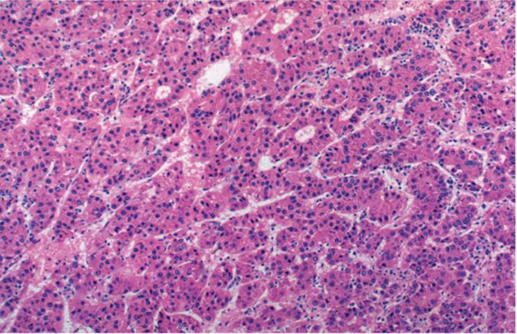
PATHOLOGY: ARCHITECTURAL PATTERNS. Several patterns are seen. The most common is the trabecular or sinusoidal pattern, with exaggerated liver plates, sometimes 15 to 20 cells thick or more, separated by sinusoids that maintain endothelial lining cells confirmed with immunostains for factor VIII, CD31, and CD34 (Fig. 24.1, e-Figs, 24.1,24.2,24.3) (33,81). Kupffer cells are often present, in reduced numbers. Solid areas may be seen, and necrosis may be prominent.
The second common pattern is acinar or pseudoglandular. Glandlike structures, formed by hepatocytes, may contain fibrin, bile, and even histiocytes (Fig. 24.2, e-Figs. 24.4-24.6). Bile can be seen in the cytoplasm of tumor cells, most often the acinar variant, and is pathognomonic for HCC (e-Fig. 24.7). Many HCCs have mixed architecture, with both trabecular and acinar areas (Fig. 24.3, e-Figs. 24.8, 24.9).
|
|
|
FIGURE 24.1 Hepatocellular carcinoma, trabecular pattern (hematoxylin-eosin, original magnification ×100). |
A third pattern is solid, compact, or pelioid with thickened liver cell trabecula. Immunostains can demonstrate sinusoids, but they are compressed by the expanding liver plates, giving the impression of a solid neoplasm (e-Fig. 24.10). Large pseudovascular blood-filled spaces (vacular mimicry) (e-Fig. 24.11) similar to those of peliosis hepatis are often seen and may signify poorer prognosis. These tumors can rupture and cause hemoperitoneum.
|
|
|
FIGURE 24.2 Hepatocellular carcinoma, predominantly acinar pattern. Note the presence of bile within the acinar lumina (hematoxylin-eosin, original magnification ×100). |
|
|
|
FIGURE 24.3 Hepatocellular carcinoma, mixed pattern, trabecular and acinar (hematoxylin-eosin, original magnification ×200). |
Histologic Variants of HCC (Table 24.2)
In many well-differentiated HCCs, hepatocytes are quite bland with minimal cytologic atypia. Cells retain their polygonal shape and eosinophilic finely granular cytoplasm. Nuclei are usually round, and the early sign of atypia is nuclear enlargement with hyperchromasia, irregular nuclear contour, and irregular nuclear chromatin. Nuclei of dysplastic cells often show more cytologic atypia than the nuclei of well-differentiated HCC.
Eosinophilic nucleoli may be prominent. Individual cells may not be enlarged.
|
TABLE 24.2 Histologic Variants of Hepatocellular Carcinoma (HCC) |
||||||
|
||||||
|
|
||||||
|
FIGURE 24.4 Hepatocellular carcinoma, intracytoplasmic inclusions (hematoxylin-eosin, original magnification ×400). |
HEPATOCELLULAR CARCINOMA OF THE USUAL TYPE. HCC cells can be strikingly similar to benign, nonneoplastic hepatocytes, with somewhat more basophilic cytoplasm, and variable degrees of pleomorphism, arranged in several-cells-thick trabeculae.
Various intracytoplasmic inclusions can be seen including Mallory-like material, albumin, fibrinogen, pale bodies, and megamitochondria (33,74,80,140,142,164) (Fig. 24.4). Intranuclear eosinophilic pseudoinclusions, representing focal invaginations of the cytoplasm, are also seen. Mucin is not an HCC product, and its presence should suggest either metastatic adenocarcinoma or cholangiocarcinoma (CCa) (87,106,120,148,151). In HBV associated HCC, viral antigens are only rarely demonstrated in tumor cells (98).
Differential Diagnosis. Conditions to be distinguished in usual type HCC include macroregenerative nodules (MRNs), liver cell adenoma (LCA), CCa, and hepatoblastoma (HB). In some cases, liver cell dysplasia may be quite similar to HCC.
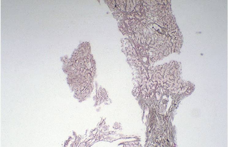
Distinguishing between HCC and MRNs with liver cell dysplasia can be problematic. Liver cells in a well-differentiated HCC can be very bland and resemble benign hepatocytes. In contrast, dysplastic liver cells often show prominent cytologic atypia. The architectural pattern may be similar in both conditions, although liver cell plates are usually thicker in HCC. The diagnosis of well-differentiated HCC can be exceedingly difficult, especially in biopsy samples, and reticulin silver preparation is usually helpful. In most HCCs, including well-differentiated ones, the reticulin network is significantly attenuated or even absent (Fig. 24.5, e-Fig. 24.11). Glypican-3 immunostain is often positive in HCC (e-Figs. 24.13, 24.14) and can be helpful. However, although sensitive, it is not completely specific. It can also be useful in aspirate samples (e-Fig. 24.15).
|
|
|
FIGURE 24.5 Hepatocellular carcinoma, trabecular pattern. Note attenuation and almost complete absence of underlying reticulin pattern (reticulin silver preparation, original magnification ×100). |
Differentiation of HCC from LCA can also be difficult. LCA arises in the noncirrhotic liver, whereas most HCCs are seen with cirrhosis. LCA is usually solid, although focal acinar pattern may also be seen. LCA does not have significant cytologic atypia, and there is no significant attenuation of reticulin network.
Differentiation between acinar HCC and CCa can also be problematic, particularly in biopsies (110). Intracytoplasmic mucin is strong evidence for CCa. In contrast, intracytoplasmic bile in tumor cells is diagnostic of HCC. Desmoplastic stromal reaction is more prominent in CCa but is not always helpful because it is also seen in sclerosing HCC.
Imunohistochemical markers are helpful in differentiating HCC from CCa (e-Figs. 24.16-24.18) (Table 24.3).
SCLEROSING HEPATOCELLULAR CARCINOMA. The sclerosing variant of HCC is associated with hypercalcemia (79,150). The tumor shows trabecular, acinar, or mixed trabecular-acinar architectural pattern. Malignant hepatocytes are embedded in abundant, relatively dense, hypocellular fibrous stroma (33,79). FL-HCC, CCa, and other metastatic adenocarcinomas can be similar.
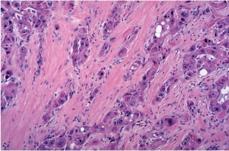
The FL-HCC fibrous stroma is distinctly lamellar (Fig. 24.6, e-Figs. 24.19, 24.20). The hepatocytes are large, polygonal, and eosinophilic, with abundant intracytoplasmic pale bodies. Characteristically, FL-HCC occurs in younger patients in contrast with the sclerosing variant of HCC, which usually occurs in older patients.
|
TABLE 24.3 Immunohistochemical Markers Helpful in Differentiating HCC from Cholangiocarcinoma (Compiled Data) |
|||||||||||||||||||||||||||||||||||||||||||||||||||
|
|||||||||||||||||||||||||||||||||||||||||||||||||||
|
|
|||||||||||||||||||||||||||||||||||||||||||||||||||
|
FIGURE 24.6 Fibrolamellar hepatocellular carcinoma (hematoxylin-eosin, original magnification ×200). |
|
|
|
FIGURE 24.7 Hepatocellular carcinoma, clear cell variant (hematoxylin-eosin, original magnification ×200). |
Metastatic adenocarcinomas, particularly those arising in the biliary tree and pancreas, can be quite desmoplastic, but the stroma is usually not as dense as in sclerosing HCC. Immunohistochemical studies can be helpful (109,159,190,197).
CLEAR CELL HEPATOCELLULAR CARCINOMA. In clear cell HCC, tumor cytoplasm is clear because of abundant glycogen. Clear cells alternate with tumor cells resembling nonneoplastic hepatocytes (Fig. 24.7, e-Fig. 24.21). Clear cell HCC can be associated with hypoglycemia, sometimes with hypercholesterolemia, and may have a somewhat better prognosis than usual HCC (22). Differentiation from other clear cell tumors, particularly metastatic renal cell carcinoma (RCC) or adrenocortical carcinoma (AdCC), can be difficult. Cirrhosis strongly favors the diagnosis of HCC. Vimentin and LeuM-1 immunostaining are characteristic of RCC, and AdCC shows synaptophysin, inhibin, and Melan-A, rare in HCC. Intracellular bile is diagnostic of HCC as is a canalicular pattern with polyclonal CEA (e-Fig. 24.) (33).
SPINDLE CELL (SARCOMATOID) HEPATOCELLULAR CARCINOMA. Spindle cell or sarcomatoid variant of HCC is rare. The tumor consists of spindle cells arranged in fascicles. An organoid pattern can also be seen. Multinucleated giant cells are common (92). Differentiation from true sarcomas, including leiomyosarcoma and fibrosarcoma, may be difficult, but true mesenchymal tumors can express vimentin.
PLEOMORPHIC (GIANT CELL, ANAPLASTIC) HEPATOCELLULAR CARCINOMA. The least common histologic variant of HCC is pleomorphic or giant cell variant, in which the tumor cells are arranged in solid sheets and most tumor cells exhibit bizarre nuclear features. In addition, many cells may be multinucleated (33,76) (Fig. 24.8, e-figs 24.22-24.31).
|
|
|
FIGURE 24.8 Pleomorphic (giant cell) variant of hepatocellular carcinoma (hematoxylin-eosin, original magnification ×200). |
FIBROLAMELLAR HEPATOCELLULAR CARCINOMA. FL-HCC is more often seen in younger adults, in the background of a noncirrhotic liver. Both sexes are equally affected. A generally better prognosis is attributed to patient's age, resectability of the tumor, and absence of cirrhosis (33,46,113,140). Tumor cells show abundant eosinophilic granular cytoplasm and are distinctly oncocytic (Fig. 24.6, e-Figs. 24.32-24.41). Sheets of oncocytic cells are separated by pale, paucicellular, lamellated fibrous stroma imparting the characteristic microscopic appearance. Tumor cells can produce bile. Pale bodies show reactivity with antibody for fibrinogen, demonstrable in frozen sections. Eosinophilic intracytoplasmic globules composed of Creactive protein, fibrinogen or α1-antitrypsin are also seen (17,18,24). Most cases have copper and copper-binding protein.
Histologic Grading of Hepatocellular Carcinoma
In contrast with most other malignant tumors, histologic grading has not proven particularly useful in determining HCC prognosis. Edmondson and Steiner proposed four grades of HCC (47). Most HCCs are grade II or III. Well-differentiated HCC, grade I, may be difficult to distinguish from LCA or MRNs with dysplasia. Grade IV tumors may be indistinguishable from undifferentiated adenocarcinomas from other primary sites (33).
IMMUNOHISTOCHEMISTRY. There is no single reliable and specific immunohistochemical marker for HCC (110). Hepatocytes, both benign and malignant, generally show strong reactivity with low molecular weight keratin (CAM 5.2, CK8) but not with a high molecular weight keratin (CK7, CK19). CCa, in contrast, can be positive for both. AFP is demonstrable in 10% to 70% of HCCs (Table 24.3) (78,91,109,120,151).
Antibodies helpful in establishing the diagnosis of HCC include hepatocyte paraffin 1 (hepar-1), α1-antitrypsin, demonstration of canalicular pattern with polyclonal CEA, and highlighting of the sinusoidal pattern with CD10, CD31, or CD34 (29,109,110,122,151,153). Neuroendocrine differentiation can sometimes be shown with chromogranin and synaptophysin (14). Human chorionic gonadotropin (HCG) and estrogen and progesterone receptors can also be demonstrated in some cases (137).
PROLIFERATION MARKERS. Various antibodies, including those for proliferating cell nuclear antigen and Ki-67 (Mib-1), may be useful in demonstrating the high level of cell proliferation within the tumor but are not useful in establishing the diagnosis (97,148).
FLOW CYTOMETRY AND IMAGE ANALYSIS. Flow cytometry and morphometric studies of HCC have shown conflicting results (40,57,138,139,152,180,188). Image analysis may be useful as an adjunct tool in differentiating between dysplasia, well-differentiated HCC, and poorly differentiated HCC (9).
ELECTRON MICROSCOPY. Electron microscopy may be useful in some cases of HCC, but it is rarely used in routine practice (33).
MOLECULAR STUDIES. Molecular studies have shown significant differences in the expression of various oncogenes in nonneoplastic liver and HCC, including hepatocyte growth factor (HGF) and its receptors c-met and c-myc (122,131,154,156,172,175,183,189). For example, HGF RNA is not expressed in normal or cirrhotic liver tissue but is present in LCA and HCC. Its receptor c-met is also significantly up-regulated in LCA and HCC, as opposed to normal livers, and the expression of HGF and c-met is also associated with higher expression of c-myc protooncogene (122,183). The role of a putative stem cell in liver regeneration and carcinogenesis has been extensively studied in the last several years (178). A computer-assisted imaging neural network expert system can be a helpful adjunct tool in differentiating hepatocellular large cell dysplasia from HCC (9).
Premalignant Conditions
LIVER CELL DYSPLASIA. Liver cell dysplasia was described more than 20 years ago, but its nature and significance remains controversial (11,12). In large cell dysplasia (LCD), cells are large, with prominent hyperchromatic nuclei, often with intranuclear inclusions (10,11,12) (Fig. 24.9, e-Fig. 24.42). The number of affected cells in a regenerative nodule may be variable with single cells or groups, or LCD can occupy the entire nodule. LCD is more commonly seen in cirrhosis resulting from HBV or HCV but can be present in chronic hepatitis and cirrhoses of other causes (34,53,115,127,144,152). LCD usually has an aneuploid DNA pattern and was considered a true preneoplastic condition (9). However, it has been suggested that LCD is not truly preneoplastic, instead reflecting a response to cholestasis in the failing liver (115). In our experience, LCD is only rarely associated with cholestasis. In an experimental model, LCD was a clear precursor to the development of HCC (62).
|
|
|
FIGURE 24.9 Liver cell dysplasia, large cell type (hematoxylin-eosin, original magnification ×200). |
Small cell dysplasia (SCD) is less common than LCD. The liver cell nuclei are enlarged, but the hepatocytes remain relatively small, imparting the histologic appearance of an increased number of cells in a defined histologic field (e-Figs. 24.43. 24.44). There is general agreement that SCD is a reliable indicator of malignancy and, in biopsy, may not be distinguishable from well-differentiated HCC.
MRNs are also discussed in the chapters on benign liver lesions/tumors (Chapter 23) and cirrhosis (Chapter 20) (3,4,49,53,54,58,144,152,172).
Hepatoblastoma
Hepatoblastoma (HB) is a primary liver cell tumor mostly affecting males younger than 3 years of age, only rarely seen in adults (33,80,104). There is failure to thrive, a growing abdominal mass, and high serum AFP values (33). The tumor may be associated with several congenital anomalies, including Down syndrome, Beckwith-Wiedemann syndrome, nephroblastoma, absence of right adrenal gland, fetal hydrops, Meckel diverticulum, and umbilical hernia. Familial cases have been described (33,81,102,161).
PATHOLOGY. Irregular tumor nodules composed of epithelial and mesenchymal components are usually separated by delicate fibrous strands and may initially resemble cirrhotic liver. Epithelial hepatoblastoma can show fetal and embryonal cells. Fetal cells resemble hepatocytes of the fetus at 6 to 8 weeks of gestation, arranged in two- to three-cell-thick liver cell cords. The fetal cells contain variable amounts of glycogen and neutral fat, giving a characteristic alternating light and dark histologic pattern of the tumor. Extramedullary hematopoiesis is often present (33,38,66). Embryonal cells are usually elongated or spindle cells, arranged in cords or rosette-like formations. Some epithelial HBs exhibit striking similarity to HCC, with so-called macrotrabecular growth pattern. These have a poorer prognosis (Fig. 24.10, e-Figs. 24.44-24.51) (38,66).
|
|
|
FIGURE 24.10 Hepatoblastoma, epithelial type with embryonal and fetal cells (hematoxylin-eosin, original magnification ×100). |
Mixed epithelial-mesenchymal HB has, in addition to its usual epithelial component, mesenchymal tissue composed of fibroblasts, collagen, and osteoid (Fig. 24.11). Sometimes these tumors are described as teratoid hepatoblastomas (125). Anaplastic hepatoblastoma is composed of small, undifferentiated cells with hyperchromatic nuclei and scant cytoplasm. The prognosis of this type is worse than that of other types (65,66). Combined hepatoblastoma and yolk sac tumor have also been described (35).
|
|
|
FIGURE 24.11 Hepatoblastoma, mixed type with osteoid-like formation (hematoxylin-eosin, original magnification ×100). |
|
|
|
FIGURE 24.12 Hepatoblastoma, focal positivity for α-fetoprotein (AFP) (immunoperoxidase, antibody for AFP, original magnification ×200). |
Immunohistochemical findings include positivity for low and high molecular weight keratins, CAM 5.2 and AE 1/3, and for AFP, S-100, and vimentin, particularly in embryonal spindle cells (Fig. 24.12). Endocrine differentiation within the tumor has been reported, and chromogranin and neuron-specific enolase may be positive (166). Some HBs secrete human chorionic gonadotropin demonstrable with immunostain (134). Melanincontaining HB has also been reported (167).
Untreated, the prognosis is dismal. Fetal pattern HB may respond to therapy. HB grows rapidly, invading adjacent tissues and organs with distant metastases often in lungs, brain, or bone marrow at diagnosis. Survival generally is better for HB after liver transplantation than for HCC (104).
Cholangiocarcinoma
This adenocarcinoma of bile duct origin can be intrahepatic (peripheral), hilar (Klatskin tumor), or extrahepatic (33). CCa is usually solitary and, unlike HCC, usually arises in a noncirrhotic liver. Factors associated with CCa include primary sclerosing cholangitis, infestation with various oriental flukes (77,169), and congenital biliary cysts, including Caroli disease and choledochal cyst (13,19,23,26,33,36,75), as well as after exposure to thorium dioxide (Thorotrast) (87,165,192). CCa usually develops between 50 and 70 years of age with both sexes equally affected (141). Peripheral CCa is more common, involving the smaller intrahepatic bile ducts. By the time symptoms manifest, CCa is generally significantly large and unresectable. In contrast, hilar CCa presents relatively early with jaundice and other signs and symptoms of bile duct obstruction. Extrahepatic CCa arises anywhere between the hepatic duct and ampulla of Vater. Both hilar and extrahepatic variants of CCa affect mostly men in the sixth and seventh decades (141).
|
|
|
FIGURE 24.13 Cholangiocarcinoma in a patient with known primary sclerosing cholangitis (hematoxylin-eosin, original magnification ×200). |
PATHOLOGY. CCa is a glandular tumor with abundant desmoplastic stromal reaction (Fig. 24.13, e-Figs. 24.52-24.55). There can be papillary or solid growth patterns. Tumor cells are columnar or cuboidal with mildly basophilic cytoplasm, round or ovoid nuclei, and indistinct nucleoli. Intracytoplasmic mucin is often seen.
There are several grades of differentiation. A signet ring component can be present in poorly differentiated, usually papillary CCa variants. Focal squamous differentiation can also occur, with the tumor resembling mucoepidermoid or adenosquamous carcinoma (105,141,157). The tumor locally infiltrates portal tracts and insidiously invades periportal sinusoids. Metastases are usually in regional lymph nodes at the time of diagnosis, and distant metastases to lungs and peritoneal surface may be seen. It may be impossible to distinguish this tumor from adenocar - cinoma originating from pancreas or the extrahepatic portion of the biliary tree.
Differentiating between HCC with predominantly acinar architectural configuration and CCa is often difficult. Reactivity with antibodies for epithelial membrane antigen, high molecular weight keratins, carcinoembryonic antigen, blood group antigens, tissue polypeptide antigens, and carbohydrate antigen CA 19-9 can help establish the diagnosis of CCa (Table 24.3) (20,41,78,91,110,120).
Mixed Cholangiohepatocellular Carcinoma
Mixed cholangiohepatocellular carcinoma is an uncommon tumor with features of HCC and CCa. Patients present with elevated serum AFP values (67) (e-Figs. 24.56, 24.61).
HEPATOID TUMOR. Rare HCC-like malignancies, so-called hepatoid tumors, are histologically indistinguishable from usual HCC. They produce albumin, bile, fibrinogen, AFP, and other hepatocytes products. They have been described in the ovary, stomach, gallbladder, pancreas, lung, kidney, and endometrium (e-Fig. 24.62) (61,73,84,85,86,128,147).
Hepatobiliary Cystadenocarcinoma
Hepatobiliary mucinous cystadenocarcinoma with mesenchymal stroma is a rare malignant tumor, occurring almost exclusive in women, and is indistinguishable from that of the ovary (33,43,68). It can develop in congenital hepatic fibrosis, choledochal cyst, or in the setting of a pre-existing hepatobiliary cystadenoma (5,33) and also arises from ectopic gallbladder rests and bile duct epithelium. This concept is supported by ultrastructural similarities between stroma and epithelium.
PATHOLOGY. Hepatobiliary cystadenocarcinoma is a multilocular cystic neoplasm lined with either simple cuboidal or columnar epithelium, with or without mucin production. A distinctive stroma composed of densely packed spindle cells, resembling either ovarian stroma or fetal mesenchyme, is subjacent to the epithelium (Fig. 24.14, e-figs. 24.63-24.65).
In both hepatobiliary and pancreatic mucinous cystadenocarcinoma, stromal cells express estrogen and progesterone receptors (190) (Fig. 24.15).
|
|
|
FIGURE 24.14 Hepatobiliary cystadenocarcinoma with mesenchymal stroma arising in cystadenoma (hematoxylin-eosin, original magnification ×100). |
|
|
|
FIGURE 24.15 Hepatobiliary cystadenocarcinoma with mesenchymal stroma that expresses estrogen/progesterone receptors (immunoperoxidase, specific antibodies for estrogen (A) and progesterone (B) receptors, original magnification ×200). |
Differential diagnosis includes CCa and CCa with hepatic cysts, both of which have invariably dismal prognosis. Hepatobiliary cystadenocarcinoma, in contrast, is a less aggressive neoplasm, and timely hepatic resection may be curative.
|
TABLE 24.4 Nonepithelial Primary Malignant Tumors of the Liver |
||||||||||||
|
NONEPITHELIAL PRIMARY MALIGNANT TUMORS OF THE LIVER (TABLE 24.4)
Angiosarcoma
Angiosarcoma is the commonest of the primary malignant mesenchymal neoplasms of the liver. Males are most often affected, and increasing age is a significant risk factor. Environmental risk factors include long-term exposure to monomeric vinyl chloride (56,159), thorium dioxide (Thorotrast) (82,87,126,159), arsenic (33), and steroids (49,74,133). Alcoholic cirrhosis and hemochromatosis may also be associated (33).
The biopsy diagnosis may be difficult, and differentiation from epithelioid hemangioendothelioma may be virtually impossible. Differentiation is important, however, since prognosis and treatment differ significantly. Epithelioid hemangioendothelioma can be cured by transplantation, whereas the prognosis of angiosarcoma is invariably dismal (82). There are four distinct gross patterns: diffuse micronodular, diffuse multinodular, massive, and mixed (82,159).
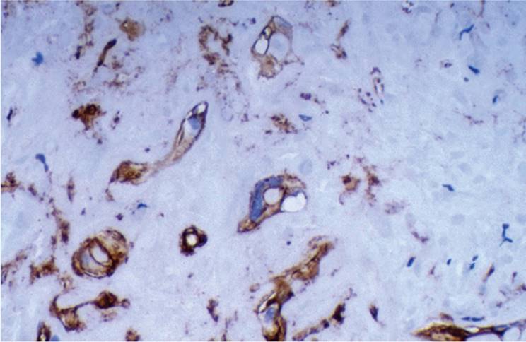
PATHOLOGY. Several patterns occur, often in combination. The sinusoidal or cavernous pattern has dilated sinusoid-like or vascular spaces lined by malignant spindle endothelial cells. The endothelial cells are enlarged, plump, with hyperchromatic, irregular nuclei, and often with bizarre nuclear features (Fig. 24.16, e-Figs. 24.66-24.70). Tumor cells are positive for various endothelial markers, including factor VIII, CD31, and CD34 (Fig. 24.17, e-Fig. 24.71) (82,159). Diagnostic problems arise when only solid areas, composed of spindle cells, are present without obvious vascular spaces. The less differentiated the tumor is, the less for positivity with specific endothelial markers. Other sarcomas have to be considered in the differential diagnosis, especially in biopsy samples.
Epithelioid Hemangioendothelioma
Epithelioid hemangioendothelioma (EHE) is a relatively rare, generally lowor intermediate-grade malignant vascular tumor. Usually slow growing, presentation is variable and often vague with abdominal pain, malaise, weight loss, jaundice, and sometimes Budd-Chiari syndrome (33,82). Females are affected twice as often as males, and the tumor may occur any time between the second and seventh decades of life.
|
|
|
FIGURE 24.16 Angiosarcoma. Large, hyperchromatic endothelial cells line the vascular spaces (hematoxylin-eosin, original magnification ×200). |
The tumor is usually multinodular, but tumor nodules may be confluent giving the impression of one massive nodule. Two histologic patterns are recognized and described: dendritic and epithelioid (Fig. 24.18, e-Figs. 24.72-24.77).
|
|
|
FIGURE 2.17 Angiosarcoma. Malignant endothelial cells are strongly positive for factor VIII (immunoperoxidase, specific antibody for factor VIII, original magnification ×400). |
|
|
|
FIGURE 24.18 Epithelioid hemangioendothelioma showing many epithelioid cells (hematoxylin-eosin, original magnification ×200). |
Spindle and stellate cells, embedded in a dense and myxoid fibrous stroma, characterize the dendritic pattern. Tumor cells show vacuolation and sometimes appear as if they are forming glands. Vacuoles, when present, are actually primitive vascular lumina (blister cells) (Fig. 24.19) (33,82,159). Cells react to factor VIII, CD31, and CD34 (e-Fig. 24.78). Ultrastructural analysis of the endothelial cells reveals endothelial differentiation, including tight junctions, pinocytotic vesicles, and Weibel-Palade bodies in about 30% of cases (179).
The epithelioid pattern shows enlarged, atypical cells with abundant cytoplasm and with more solid areas of growth. EHE has a propensity for vascular dissemination, often mimicking venoocclusive disease (Fig. 24.20). Tumor cells are surrounded by mild to moderate inflammatory infiltrate that is predominantly lymphocytic, but some polymorphonuclear and eosinophilic leukocytes may also be present.
|
|
|
FIGURE 24.19 Epithelioid hemangioendothelioma. Small vascular lumina are formed by the endothelial cells (hematoxylin-eosin, original magnification ×400). |
|
|
|
FIGURE 24.20 Epithelioid hemangioendothelioma growing into vascular spaces and mimicking venoocclusive disease (Masson trichrome, original magnification ×200). |
Differentiation includes other vascular tumors such as angiosarcoma, Kaposi sarcoma, bacillary angiomatosis, and nontumorous conditions such as venoocclusive disease.
Kaposi Sarcoma
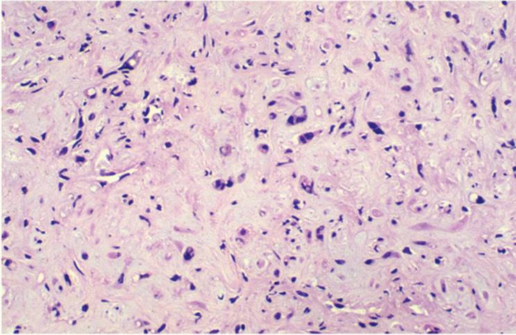
Kaposi sarcoma (KS) is a neoplastic proliferation of endothelial cells, most likely of lymphatic origin. Kaposi sarcoma appears as hemorrhagic illdefined nodules in the liver. Tumor cells are spindle shaped, often containing intracytoplasmic inclusions, most likely representing phagocytosed erythrocytes (Fig. 24.21). The nuclei are relatively bland and do not show either significant cytologic atypia or many mitoses (176). In addition, slitlike spaces, not lined by endothelial cells, contain extravasated red blood cells. Hemosiderin-laden macrophages and lymphocytic cells are present. Immunostains for endothelial markers, including factor VIII, CD31, and CD34, are rarely positive, but human herpesvirus 8 is often demonstrable.
Differentiating between angiosarcoma and Kaposi sarcoma is important. Patients with hepatic KS are almost always seropositive for human immunodeficiency virus (HIV) and are usually in the advanced stage of acquired immune deficiency syndrome (AIDS). They often already have multiple lesions elsewhere in the body.
Bacillary angiomatosis, like KS, occurs in AIDS patients with HIV and is caused by Bartonella quintana/henselae organisms, which are demonstrated with Warthin-Starry stain and also by polymerase chain reaction or electron microscopy (112,174,176).
|
|
|
FIGURE 24.21 Kaposi sarcoma in the liver in a patient with acquired immune deficiency syndrome (hematoxylin-eosin, original magnification ×200). |
Lymphoproliferative Disorders
The liver is often secondarily involved in leukemias and Hodgkin (HL) and non-Hodgkin (NHL) lymphomas. NHL is more prevalent and can be associated with AIDS, autoimmune hepatitis, and HBV and HCV infection. EBV-associated posttransplantation lymphoproliferative disorder (PTLD) occurs after liver transplantation (Fig. 24.22) (91,139,172,200).
Hepatic HL is usually seen when there is involvement of other sites, particularly the spleen. Primary HL of the liver is virtually nonexistent. Liver biopsy findings include various nonspecific and nondiagnostic lymphoid infiltrates and changes indicative of the vicinity of a mass lesion (21,91,117,124,177,185). Unexplained liver granulomas in patients with persistent febrile illness should raise a suspicion for HL (170,171). Cholestasis in the absence of obstruction may be prominent. Lymphoproliferative processes can cause acute liver failure and may clinically mimic fulminant hepatic failure resulting from acute hepatitis, which can be an important consideration in the evaluation of liver transplantation patients (198).
|
|
|
FIGURE 24.22 B-cell lymphoma, large cell type in an immunocompromised patient (hematoxylin-eosin, original magnification ×200). |
|
|
|
FIGURE 24.23 Histiocytosis X. Note numerous eosinophils (hematoxylin-eosin, original magnification ×200). |
Histiocytosis X
Histiocytosis X is an encompassing term for the spectrum of three diseases: Hand-Schüller-Christian, Letterer-Siwe, and eosinophilic granuloma of bone. Rarely, patients with histiocytosis X may present with a clinical picture mimicking sclerosing cholangitis, with jaundice and portal hypertension (187).
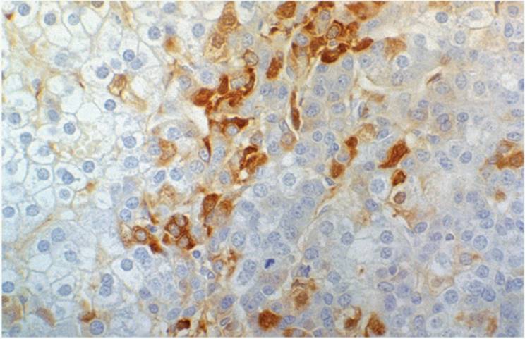
Abundant strongly S-100-positive Langerhans-type cells are seen, usually with many eosinophilic leukocytes (51) (Figs. 24.23, 24.24). Ultrastructurally, Langerhans cells contain Birbeck granules. Langerhans cells are present in the portal tracts but also extend into and replace variable portions of liver parenchyma. In addition, interlobular bile ducts show bile duct epithelial changes similar to those of primary sclerosing cholangitis, with focal ductular reaction (88,111,155,187). Liver transplantation has been performed (30).
Other Rare Tumors
Rare primary malignant neuroendocrine tumors, including hepatic gastrinoma and apudoma, have been described in the liver and in the biliary tree (10,138,168). Liver cell carcinoma may be ectopic in the abdominal cavity (8,37,108). Primary hepatic malignant melanoma has also been reported (45). Some rare primary malignant tumors of the liver include undifferentiated (embryonal, mesenchymal) sarcoma (Fig. 24.25) (60,107), choriocarcinoma (7), endodermal (yolk sac) tumor (60,72,142), adrenal or pancreatic rest tumor (32), rhabdomyosarcoma (129), leiomyosarcoma (Fig. 24.26) (55,123), fibrosarcoma (8), osteosarcoma (191), liposarcoma (Fig. 24.27) (101), malignant schwannoma (114), hemangiopericytoma (195), malignant histiocytoma (6,94), and squamous cell carcinoma (70).
|
|
|
FIGURE 24.24 Histiocytosis X. Langerhans-type cells are strongly positive for S-100 (immunoperoxidase, specific antibody for S-100, original magnification ×200). |
|
|
|
FIGURE 24.25 Mesenchymal undifferentiated sarcoma (hematoxylin-eosin, original magnification ×200). |
|
|
|
FIGURE 24.26 Primary leiomyosarcoma of the liver (hematoxylin-eosin, original magnification ×200). |
METASTATIC TUMORS
The liver is a particularly common site for metastases from almost any primary site. Liver biopsies are often performed to determine if a liver mass is primary or secondary. Fine-needle aspiration of the liver is generally not very useful in the diagnosis of acute or chronic liver diseases but is valuable in tumor diagnosis (93), particularly with appropriate immunohistochemical studies (110).
|
|
|
FIGURE 24.27 Primary myxoid liposarcoma of the liver (hematoxylin-eosin, original magnification ×200). |
|
|
|
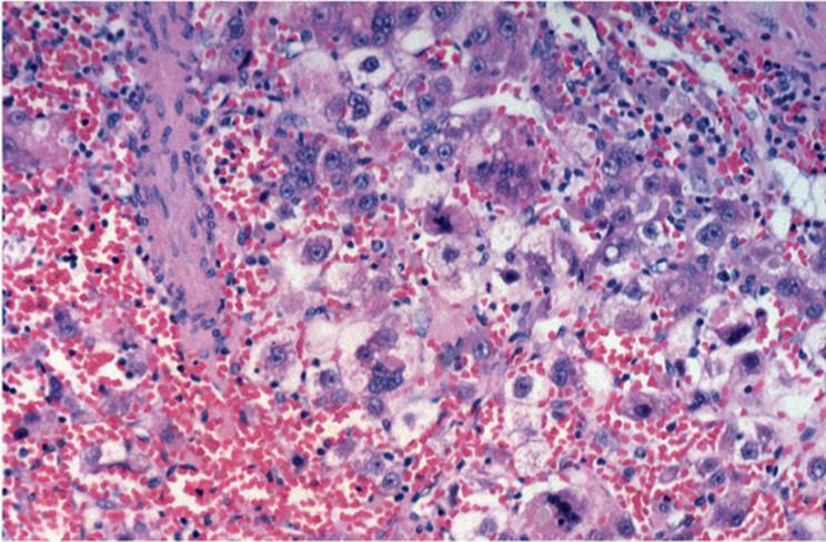
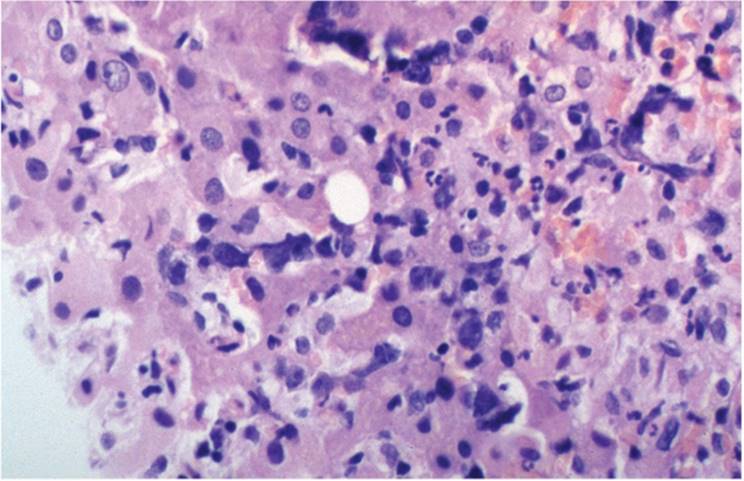
FIGURE 24.28 Liver adjacent to metastatic lesion, showing sinusoidal dilatation, cholestasis, and polymorphonuclear leukocytes (hematoxylin- eosin, original magnification ×200). |
Changes Associated with Mass Lesions
Liver parenchyma adjacent to a mass, neoplastic or nonneoplastic, shows typical changes, including bile ductular reaction with associated periductular acute inflammatory cell response, zone 1 sinusoidal dilatation, and cholestasis (Fig. 24.28) (63).
REFERENCES
1. Abbondanzo SL, Manz HJ, Klappenbach RS, et al. Hepatocellular carcinoma in an 11-year-old girl with Fanconi's anemia. Report of a case and review of the literature. Am J Pediatr Hematol Oncol 1986;8:334-337.
2. Adams PC. Hepatocellular carcinoma in hereditary hemochromatosis. Can J Gastroenterol 1993;7:37-41.
3. Aihara T, Noguchi S, Sasaki Y, et al. Clonal analysis of regenerative nodules in hepatitis C virus-induced liver cirrhosis. Gastroenterology 1994;107:1805-1811.
4. Aihara T, Noguchi S, Sasaki Y, et al. Clonal analysis of precancerous lesion of hepatocellular carcinoma. Gastroenterology 1996;111:455-461.
5. Akwari OE, Tucker A, Seigler HF, et al. Hepatobiliary cystadenoma with mesenchymal stroma. Ann Surg 1990;211:18-27.
6. Albert-Flor JJ, O'Hara MF, Weaver F, et al. Malignant fibrous histiocytoma of the liver. Gastroenterology 1985;39:890-893.
7. Alonso JF, Saez C, Perez P, et al. Primary pure choriocarcinoma of the liver. Pathol Res Pract 1992;188:375-377.
8. Alrenga DP. Primary fibrosarcoma of the liver. Case report and review of the literature. Cancer 1975;36:446-449.
9. An C, Petrovic LM, Reyter I, et al. The application of neural network technology to the study of large cell dysplasia and hepatocellular carcinoma. Hepatology 1997;26:1224-1231.
10. Angeles A, Quintanilla-Martinez L, Larriva-Sahd J. Primary carcinoid of the common bile duct. Am J Clin Pathol 1991;96:341-344.
11. Anthony PP, Vogel CL, Barker LF. Liver cell dysplasia: a premalignant condition. J Clin Pathol 1973;26:217-233.
12. Anthony PP. Liver cell dysplasia: what is its significance? Hepatology 1987;7:394-396.
13. Azizah N, Paradinas FJ. Cholangiocarcinoma coexisting with developmental liver cysts: a distinct entity different from liver cystadenocarcinoma. Histopathology 1980;4:391-400.
14. Barsky SH, Linnoila I, Triche TJ, et al. Hepatocellular carcinoma with carcinoid features. Hum Pathol 1984;15:892-894.
15. Bassendine MF. Alcohol: a major risk factor for hepatocellular carcinoma? J Hepatol 1986;2:513-519.
16. Beasly RP. Hepatitis B virus. The major etiology of hepatocellular carcinoma. Cancer 1988;61:1942-1956.
17. Berman MA, Burnham JA, Sheahan DG. Fibrolamellar carcinoma of the liver: an immunohistochemical study of nineteen cases and a review of the literature. Hum Pathol 1988;19:784-794.
18. Berman MM, Libbey NP, Foster JH. Hepatocellular carcinoma. Polygonal cell type with fibrous stroma—an atypical variant with favorable prognosis. Cancer 1980;46:1448-1455.
19. Bloustein PA. Association of carcinoma of congenital cystic conditions of the liver and bile ducts. Am J Gastroenterol 1977;67:40-46.
20. Bonetti F, Chilosi M, Pisa R, et al. Epithelial membrane antigen expression in cholangiocarcinoma. An useful immunohistochemical tool for differential diagnosis with hepatocarcinoma. Virchows Archiv A 1983;401:307-313.
21. Bruguera M, Caballero T, Carreas E, et al. Hepatic sinusoidal dilatation in Hodgkin's disease. Liver 1987;7:76-80.
22. Buchanan TF Jr, Huvos AG. Clear-cell carcinoma of the liver. A clinicopathologic study of 13 patients. Am J Clin Pathol 1974;61:529-539.
23. Burns CD, Kuhns JG, Wieman J. Cholangiocarcinoma in association with multiple biliary microhamartomas. Arch Pathol Lab Med 1990;114:1287-1289.
24. Caballero T, Aneiros J, Lopez-Caballero J, et al. Fibrolamellar hepatocellular carcinoma. An immunohistochemical and ultrastructural study. Histopathology 1985;9:445-456.
25. Chen C-J, Liang K-Y, Vajng A-S, et al. Effects of hepatitis B virus, alcohol drinking, cigarette smoking and familial tendency on hepatocellular carcinoma. Hepatology 1991;13:398-406.
26. Chen KTK. Adenocarcinoma of the liver. Association with congenital hepatic fibrosis and Caroli's disease. Arch Pathol Lab Med 1981;105:294-295.
27. Cheng WS, Govindarajan S, Redeker AG. Hepatocellular carcinoma in a case of Wilson's disease. Liver 1992;12:42-45.
28. Chisari FV, Klopchin K, Moriyama T, et al. Molecular pathogenesis of hepatocellular carcinoma in hepatitis B virus transgenic mice. Cell 1989;59:1145-1156.
29. Christensen WN, Boitnott JK, Kuhajda FP. Immunoperoxidase staining as a diagnostic aid for hepatocellular carcinoma. Mod Pathol 1989;2:8-12.
30. Concepcion W, Esquivel CO, Terry A, et al. Liver transplantation in Langerhans' cell histiocytosis (histiocytosis X). Semin Oncol 1991;18:24-28.
31. Conti JA, Kemeny N. Type Ia glycogenosis associated with hepatocellular carcinoma. Cancer 1992;69:1320-1322.
32. Contreras P. Altieri E, Liberman C, et al. Adrenal rest tumor of the liver causing Cushing's syndrome. J Clin Endocrinol Metab 1985;60:21-28.
33. Craig JR, Peters RL, Edmondson HA. Tumors of the liver and intrahepatic bile ducts. In: Atlas of Tumor Pathology, 2nd series, fascicle 26. Washington DC: Armed Forces Institute of Pathology, 1989.
34. Crawford JM. Pathologic assessment of liver cell dysplasia and benign liver tumors: differentiation from malignant tumors. Semin Diagn Pathol 1990;7:115-128.
35. Cross SS, Variend S. Combined hepatoblastoma and yolk sac tumor of the liver. Cancer 1992;69:1323-1326.
36. Darioca PJ, Tuthill R, Reed RJ. Cholangiocarcinoma arising in congenital hepatic fibrosis. Arch Pathol 1975;99:592-595.
37. Davison FD, Fagan EA, Portmann B, et al. HBV-DNA sequences in tumor and nontumor tissue in a patient with the fibrolamellar variant of hepatocellular carcinoma. Hepatology 1990;12:676-679.
38. Dehner LP., Manivel JC. Hepatoblastoma: an analysis of the relationship between morphologic subtypes and prognosis. Am J Pediatr Hematol Oncol 1988;10:301-307.
39. Dehner LP, Snover DC, Sharp HL, et al. Hereditary tyrosinemia type 1 (chronic form). Hum Pathol 1989;20:149-158.
40. Deprez C, Vangansbeke D, Fastrez R, et al. Nuclear DNA content, proliferative index, and nuclear size determination in normal and cirrhotic liver, and in benign and malignant primary and metastatic hepatic tumors. Am J Clin Pathol 1993;99:558-565.
41. Desmet V, De Vos R. Ultrastructural characteristics of novel epithelial cell types identified in human pathologic liver specimens with chronic ductular reaction. Am J Pathol 1992;16:1327-1333.
42. De Souza A, Yamada T, Mills JJ, et al. Imprinted genes in liver carcinogenesis. FASEB J 1997;11:60-67.
43. Devaney K, Goodman ZD, Ishak KG. Hepatobiliary cystadenoma and cystadenocarcinoma. A light microscopic and immunohistochemical study of 70 patients. Am J Surg Pathol 1994;18:1078-1091.
44. Deugnier Y, Guyader D, Crantock L, et al. Primary liver cancer in genetic hemochromatosis: a clinical, pathological and pathogenetic study of 54 cases. Gastroenterology 1993;104:228-234.
45. Deugnier Y, Turlin B, Lehry D, et al. Malignant melanoma of the hepatic and common bile ducts. Arch Pathol Lab Med 1991;115:915-917.
46. Eckstein RP, Bambach CP, Stiel D, et al. Fibrolamellar carcinoma as a cause of bile duct obstruction. Pathology 1988;20:326-331.
47. Edmondson HA, Steiner PE. Primary carcinoma of the liver. A study of 100 cases among 48,900 necropsies. Cancer 1954;7:462-403.
48. Eriksson S, Carlson J, Velez R. Risk of cirrhosis and primary liver cancer in alpha 1-antitrypsin deficiency. N Engl J Med 1986;314:736-739.
49. Falk H, Thomas LB, Popper H, et al. Hepatic angiosarcoma associated with androgenic-anabolic steroids. Lancet 1979;2:1120-1123.
50. Farinati F, Faginoli S, De Maria N, et al. Anti-HCV positive hepatocellular carcinoma in cirrhosis. J Hepatol 1992;14:183-187.
51. Favara BE, Jaffe R. Pathology of Langerhans cell histiocytosis. Hematol Oncol Clin North Am 1987;1:75-97.
52. Fellows IW, Stewart M, Jefcoate WJ, et al. Hepatocellular carcinoma in primary hemochromatosis in the absence of cirrhosis. Gut 1988;29:1603-1606.
53. Ferrell LD, Crawford JM, Dhillon AP, et al. Proposal for standardized criteria for the diagnosis of benign, borderline and malignant hepatocellular lesions arising in chronic advanced liver disease. Am J Surg Pathol 1993;17:1113-1123.
54. Ferrell L, Wright T, Lake J, et al. Incidence and diagnostic features of macroregenerative nodules vs. small hepatocellular carcinoma in cirrhotic liver. Hepatology 1992;16:1372-1381.
55. Fong JA, Ruebner BH. Primary leiomyosarcoma of the liver. Hum Pahol 1974;5:115-119
56. Fortwengler HP Jr, Jones D, Espinosa E, et al. Evidence for endothelial cell origin of vinyl chloride-induced hepatic angiosarcoma. Gastroenterology 1981;80:1415-1419.
57. Fujimoto J, Okamoto E, Yamanak N, et al. Flow cytometric DNA analysis of hepatocellular carcinoma. Cancer 1991;67:939-944.
58. Furuya K, Nakamura M, Yamamoto Y, et al. Macroregenerative nodule of the liver: a clinicopathologic study of 345 autopsy cases of chronic liver disease. Cancer 1988;61:99-105.
59. Gaffey MJ, Joyce JP, Carlson GS, et al. Spontaneous regression of hepatocellular carcinoma. Cancer 1990;65:2779-2783.
60. Gallivan MVE, Lack EE, Chun B, et al. Undifferentiated (embryonal) sarcoma of the liver. Pediatr Pathol 1983;1:291-300.
61. Gardiner GW, Lajoie G, Keith R. Hepatoid adenocarcinoma of the papilla of Vater. Histopathology 1992;20:541-544.
62. Geller SA, Nichols WS, Kim SS, et al. Hepatocarcinogenesis is the sequel to hepatitis in the Z#2 alpha 1-antitrypsin transgenic mouse. Hepatology 1994;19:389-397.
63. Geller SA, Dhall D, Alsabeh R. Application of immunochemistry to liver and gastrointestinal neoplasms. Arch Pathol Lab Med 2008;132:490-499.
64. Gerber MA, Thung SN, Bodenheimer HC, et al. Characteristic histologic triad in liver adjacent to metastatic neoplasm. Liver 1986;6:85-88.
65. Goldblum JR, Bartos RE, Carr KA, et al. Hepatitis B and alterations of the p53 tumor suppressor gene in hepatocellular carcinoma. Am J Surg Pathol 1993;17:1244-1251.
66. Goldstein RM, Stone M, Tillery GW, et al. Is liver transplantation indicated for cholangiocarcinoma? Am J Surg 1993;166:768-772.
67. Gonzales-Crussi F, Upton MP, Maurer HS. Hepatoblastoma. Attempt at characterization of histologic subtypes. Am J Surg Pathol 1982;6:599-612.
68. Goodman ZD, Ishak KG, Langloss JM, et al. Combined hepatocellular-cholangiocarcinoma. A histologic and immunohistochemical study. Cancer 1985;55:124-135.
69. Gourley WK, Kumar D, Bouton MS, et al. Cystadenoma and cystadenocarcinoma with mesenchymal stroma of the liver. Immunohistochemical analysis. Arch Pathol Lab Med 1992;116:1047-1050.
70. Grasl-Kraupp B, Ruttkay-Nedecky B, Mullauer L, et al. Inherent increase in apoptosis in liver tumors: implications for carcinogenesis and tumor regression. Hepatology 1997;25:906-913.
71. Gresham GA, Rue LW. Squamous cell carcinoma of the liver. Hum Pathol 1985;16:413-416.
72. Haagsma EB, Smit GPA, Niezen-Koning KE, et al. Type IIIb glycogen storage disease associated with end-stage cirrhosis and hepatocellular carcinoma. Hepatology 1997;25:537-541.
73. Hart WR. Primary endodermal sinus (yolk sac) tumor of the liver. First reported case. Cancer 1975;35:1453-1458.
74. Hasebe C, Sekiya C, Satoh H, et al. A case of alpha-fetoprotein-producing gallbladder carcinoma. Acta Hepatol Jpn 1984;25:1180-1186.
75. Hoch-Ligeti C. Angiosarcoma of the liver associated with diethylstilbestrol. JAMA 1978;240:1510-1511.
76. Honda N, Cobb C, Lechago J. Bile duct carcinoma associated with multiple von Meyenburg complexes in the liver. Hum Pathol 1986;17:1287-1290.
77. Hood DL, Bauer TW, Leibel SA, et al. Hepatic giant cell carcinoma. An ultrastructural and immunohistochemical study. Am J Clin Pathol 1990;93:111-116.
78. Hou P-C. The relationship between primary carcinoma of the liver and infestation with Clonorchis sinensis. J Pathol Bacteriol 1956;72:239-246.
79. Hurlmann J, Gardiol D. Immunohistochemistry in the differential diagnosis of liver carcinomas. Am J Surg Pathol 1991;15:280-288.
80. International Working Party. Terminology of nodular hepatocellular lesions. Hepatology 1995;22:983-993.
81. Ishak KG, Anthony PP, Sobin LH. Histological typing of tumors of the liver. In: World Health Organization International Histological Classification of Tumors, 2nd Ed. Berlin: Springer-Verlag, 1994.
82. Ishak KG, Glunz PR. Hepatoblastoma and hepatocarcinoma in infancy and childhood. Report of 4 cases. Cancer 1967;20:396-422.
83. Ishak KG, Sesterhenn IA, Goodman ZD, et al. Epithelioid hemangioendothelioma of the liver: a clinicopathologic and follow-up study of 32 cases. Hum Pathol 1984;15:839-852.
84. Ishak KG. Malignant mesenchymal tumors of the liver. In: Okuda K, Ishak KG, eds. Neoplasms of the Liver. Tokyo: Springer-Verlag, 1987:159-176.
85. Ishikura H, Ishiguro T, Enatsu C, et al. Hepatoid adenocarcinoma of the renal pelvis producing alpha-fetoprotein of hepatic type and bile pigment. Cancer 1991;67:3051-3056.
86. Ishikura H, Kanda M, Nosaka K, et al. Hepatoid adenocarcinoma: a distinctive histological subtype of alpha-fetoprotein-producing lung carcinoma. Virchows Archiv A 1990;417:73-80.
87. Ishikura H, Scully RE. Hepatoid carcinoma of the ovary. Cancer 1987;60:2775-2784.
88. Ito Y, Kojiro M, Nakashima T, Mori T. Pathomorphologic characteristics of 102 cases of Thorotrast-related hepatocellular carcinoma, cholangiocarcinoma, and hepatic angiosarcoma. Cancer 1988;62:1153-1162.
89. Iwai M, Kashiwadani M, Okuno T, et al. Cholestatic liver disease in a 20-year old woman with histiocytosis X. Am J Gastroenterol 1988;83:164-168.
90. Jaffe ES. Malignant lymphomas: pathology of hepatic involvement. Semin Liver Dis 1987;7:257-268.
91. Jameson CF. Primary hepatocellular carcinoma in hereditary hemorrhagic telangiectasia. Histopathology 1989;15:550-552.
92. Jovanovic R, Jagirdar J, Thung SN, et al. Blood-group-related antigen Lewis(x) and Lewis(y) in the differential diagnosis of cholangiocarcinoma and hepatocellular carcinoma. Arch Pathol Lab Med 1989;113:139-142.
93. Kakizoe S, Kojiro M, Nakashima T. Hepatocellular carcinoma with sarcomatous change. Cancer 1987;59:310-316.
94. Kaklamani E, Trichopoulos D, Tzonou A, et al. Hepatitis B and C and their interaction in the origin of hepatocellular carcinoma. JAMA 1991;265:1974-1976.
95. Katsuda S, Kawahara E, Matsui Y, et al. Malignant fibrous histiocytoma of the liver: a case report and review of the literature. Am J Gastroenterol 1988;83:1278-1282.
96. Kaufman SS, Wood P, Shaw B, et al. Hepatocarcinoma in a child with the Alagille syndrome. Am J Dis Child 1987;141:698-700.
97. Kawahara E, Kitamura T, Ueda H, et al. Hepatocellular carcinoma arising in the abdominal cavity. Acta Pathol Jpn 1988;38:1575-1581.
98. Kawakita N, Seki S, Sakaguchi H, et al. Analysis of proliferating hepatocytes using a monoclonal antibody against proliferating nuclear antigen/cyclin in embedded tissue from various liver diseases fixed in formalin. Am J Pathol 1992;140:513-520.
99. Kawano Y. Localization of hepatitis B surface antigen in hepatocellular carcinoma. Acta Pathol Jpn 1983;33:1087-1093.
100. Kew MC, McKnight A, Hodkinson H, et al. The role of membranous obstruction of the inferior vena cava and hepatocellular carcinoma in Southern African blacks. Hepatology 1989;9:121-125.
101. Kim CM, Koike K, Saito I, et al. HBx gene of hepatitis B virus induces liver cancer in transgenic mice. Nature 1991;351:317-320.
102. Kim YI, Yu ES, Lee KW, et al. Dedifferentiated liposarcoma of the liver. Cancer 1985; 60:2785-2790.
103. Kingston JE, Herbert A, Draper GJ, et al. Association between hepatoblastoma and polyposis coli. Arch Dis Child 1983;58:959-962.
104. Kolars JC. Aflatoxin and hepatocellular carcinoma: a useful paradigm for environmentally induced carcinogenesis. Hepatology 1992;16:848-851.
105. Koneru B, Flye MW, Busuttil RW, et al. Liver transplantation for hepatoblastoma: the American experience. Ann Surg 1991;213:118-121.
106. Koo J, Ho J, Wong J, et al. Mucoepidermoid carcinoma of the bile duct. Ann Surg 1982;196:140-148.
107. Kung ITM, Chan S-W, Gung K-H. Fine-needle aspiration in hepatocellular carcinoma. Combined cytologic and histologic approach. Cancer 1991;67:673-680.
108. Lack EE, Scloo BL, Azumi N, et al. Undifferentiated (embryonal) sarcoma of the liver. Clinical and pathologic study of 16 cases with emphasis on immunohistochemical features. Am J Surg Pathol 1991;15:1-16.
109. Laferla G, Kaye SB, Crean GP. Hepatocellular and gastric carcinoma associated with familial polyposis coli. J Surg Oncol 1988;38:19-21.
110. Lai Y-S, Thung SN, Gerber MA, et al. Expression of cytokeratins in normal and diseased liver and in primary liver carcinomas. Arch Pathol Lab Med 1989;113:134-138.
111. Lau S, Prakash S, Geller SA, et al. Comparative immunohistochemical profile of hepatocellular carcinoma, cholangiocarcinoma, and metastatic adenocarcinoma. Hum Pathol 2002;33:1175-1181.
112. Leblanc A, Hadchouel M, Jehan P, et al. Obstructive jaundice in children in histiocytosis X. Gastroenterology 1981;80:134-139.
113. Leboit PE, Berger TG, Egbert BM, et al. Bacillary angiomatosis: the histopathology and differential diagnosis of a pseudoneoplastic infection in patients with human immunodeficiency virus disease. Am J Surg Pathol 1989;13:909-920.
114. LeBrun DP, Silver MM, Freedman MH, et al. Fibrolamellar carcinoma of the liver in a patient with Fanconi anemia. Hum Pathol 1991;22:396-398.
115. Lederman SM, Martin EC, Laffey KT, et al. Hepatic neurofibromatosis, malignant schwannoma and angiosarcoma in von Recklinghausen's disease. Gastroenterology 1987;92:234-239.
116. Lee GR, Tsamndas AC, Demetris AJ. Large cell change (liver cell dysplasia) and hepatocellular carcinoma in cirrhosis: matched case-control study, pathological analysis and pathogenic hypothesis. Hepatology 1997;26:1415-1423.
117. Lefkowitch JH, Apfelbaum TF. Liver cell dysplasia and hepatocellular carcinoma in non-A, non-B hepatitis. Arch Pathol Lab Med 1987;111:170-173.
118. Leslie KO, Colby TV. Hepatic parenchymal lymphoid aggregates in Hodgkin's disease. Hum Pathol 1984;15:808-809.
119. Lithner F, Wetterberg L. Hepatocellular carcinoma in patients with acute intermittent porphyria. Acta Med Scand 1984;215:271-274.
120. Ljubimova JY, Petrovic LM, Wilson SE, et al. Gene expression of HGF, its receptor (cmet), c-myc, and albumin in cirrhotic and neoplastic human liver tissue. J Histochem Cytochem 1997;45:79-87.
121. Lok ASF, Lai C-L. Factors determining the development of hepatocellular carcinoma in hepatitis B surface antigen carriers. Cancer 1988;61:1287-1291.
122. Lones MA, Shintaku IP, Weiss LM, et al. Post-transplant lymphoproliferative disorder in liver allograft biopsies: a comparison of three methods for the demonstration of Epstein-Barr virus. Hum Pathol 1997;28:533-539.
123. Ma CK, Zarbo RJ, Frierson HF, et al. Comparative immunohistochemical study of primary and metastatic carcinomas of the liver. Am J Clin Pathol 1993;99:551-557.
124. Maki HS, Hubert BC, Sajjad SM, et al. Primary hepatic leiomyosarcoma. Arch Surg 1987;122:1193-1196.
125. Man KM, Drejet A, Keefe EB, et al. Primary sclerosing cholangitis and Hodgkin's disease. Hepatology 1993;18:1127-1131.
126. Manivel C, Wick MR, Abenoza P, et al. Teratoid hepatoblastoma. Cancer 1986;57:2168-2174.
127. Manning JT Jr, Ordonez NG, Barton JH. Endothelial cell origin of thorium oxide-induced angiosarcoma of liver. Arch Pathol Lab Med 1983;107:456-458.
128. Manowski Z, Silver MM, Roberts EA, et al. Liver cell dysplasia and early liver transplantation in hereditary tyrosinemia. Mod Pathol 1990;3:694-701.
129. Matsukuma K, Tsukamoto N. Alpha-fetoprotein-producing endometrial adenocarcinoma. Gynecol Oncol 1988;29:370-377.
130. McArdle JP, Hawley I, Shevland J, et al. Primary rhabdomyosarcoma of the adult liver. Am J Surg Pathol 1989;13:961-965.
131. Melia WH, Johnson PJ, Neuberger J, et al. Hepatocellular carcinoma in primary biliary cirrhosis. Gastroenterology 1984;87:660-663.
132. Michalopoulos G. Liver regeneration and growth factors: old puzzle and new perspectives. Lab Invest 1992;67:413-415.
133. Miura K, Shirasawa H. Primary carcinoid tumor of the liver. Am J Clin Pathol 1988;89:561-564.
134. Monroe PS, Riddell RH, Siegler M, et al. Hepatic angiosarcoma. Possible relationship to long-term oral contraceptive ingestion. JAMA 1981;246:64-65.
135. Morinaga S, Yamagouchi M, Watanabe I, et al. An immunohistochemical study of hepatoblastoma producing human chorionic gonadotropin. Cancer 1983;51:1647-1652.
136. Mzeako UC, Goodman ZD, Ishak KG. Comparison of tumor pathology with duration of survival of North American patients with hepatocellular carcinoma. Cancer 1995;76:579-588.
137. Naccarato R, Farinati F. Hepatocellular carcinoma, alcohol and cirrhosis: facts and hypothesis. Dig Dis Sci 1991;36:1137-1142.
138. Nagasue N, Kohno H, Chang Y, et al. Androgen and estrogen receptors in hepatocellular carcinoma and the surrounding liver in women. Cancer 1989;63:112-116.
139. Nagasue N, Yamanoi A, Takemoto Y, et al. Comparison between diploid and aneuploid hepatocellular carcinomas: a flow cytometric study. Br J Surg 1992;79:667-670.
140. Nagasue N, Kohno H, Hayashi T, et al. Lack of intratumoral heterogeneity in DNA ploidy pattern of hepatocellular carcinoma. Gastroenterology 1993;105:1449-1454.
141. Nagorney DM, Adson MA, Weiland LH, et al. Fibrolamellar hepatoma. Am Surg 1985;149:113-119.
142. Nakajima T, Knodo Y, Miyazaki M, et al. A histopathologic study of 102 cases of intrahepatic cholangiocarcinoma: histologic classification and modes of spreading. Hum Pathol 1988;19:1228-1234.
143. Nakanuma Y, Ohta G. Expression of Mallory bodies in hepatocellular carcinoma in man and its significance. Cancer 1986;62:558-563.
144. Nakanuma Y, Terada T, Doishita K, et al. Hepatocellular carcinoma in primary biliary cirrhosis: an autopsy study. Hepatology 1990;11:1010-1016.
145. Nakanuma Y, Terada T, Ueda K, et al. Adenomatous hyperplasia of the liver as a precancerous lesion. Liver 1993;13:1-9.
146. Nalpas B, Driss F, Pol S, et al. Association between HCV and HBV infection in hepatocellular carcinoma and alcoholic liver disease. J Hepatol 1991;12:70-74.
147. Narita T, Moriyama Y, Ito Y. Endodermal sinus (yolk sac) tumor of the liver. A case report and review of the literature. J Pathol 1988;155:41-47.
148. Nojima T, Kojima T, Kato H, et al. Alpha-fetoprotein-producing acinar cell carcinoma of the pancreas. Hum Pathol 1992;23:828-830.
149. Ojanguren I, Ariza A, Llatos M, et al. Proliferating cell nuclear antigen in normal, regenerative and neoplastic liver. Hum Pathol 1993;24:905-908.
150. Okuda K. Hepatocellular carcinoma: recent progress. Hepatology 1992;15:948-963.
151. Omata M, Petres RL, Tatter D. Sclerosing hepatic carcinoma: relationship to hypercalcemia. Liver 1981;1:33-49.
152. Ordonez NG, Manning JT Jr. Comparison of alpha-1-antitrypsin and alpha-1-antichymotrypsin in hepatocellular carcinoma: an immunoperoxidase study. Am J Gastroenterol 1984;79:959-963.
153. Orsatti G, Thiese ND, Thung SN, et al. DNA image cytometric analysis of macroregenerative nodules (adenomatous hyperplasia) of the liver: evidence in support of their preneoplastic nature. Hepatology 1993;17:621-627.
154. Otsuru A, Nagataki S, Koji T, et al. Analysis of alpha-fetoprotein gene expression in hepatocellular carcinoma and liver cirrhosis by in situ hybridization. Cancer 1988;62:1105-1112.
155. Parker-Ponder K. Analysis of liver development, regeneration, and carcinogenesis by genetic marking studies. FASEB J 1996;10:673-684.
156. Pirovino M, Jeaneret C, Lang RH, et al. Liver cirrhosis in histiocytosis X. Liver 1988;8:293-298.
157. Pistoi S, Morello D. Prometheus myth revisited: transgenic mice as a powerful tool to study liver regeneration. FASEB J 1996;10:819-828.
158. Pliskin A, Cauling H, Stenger RJ. Primary squamous cell carcinoma originating in congenital cysts of the liver. Arch Pathol Lab Med 1985;116:105-107.
159. Polio J, Enriquez RE, Chow A, et al. Hepatocellular carcinoma in Wilson's disease. J Clin Gastroenterol 1989;11:220-224.
160. Popper H, Thomas LB, Telles NC, et al. Development of hepatic angiosarcoma in man induced by vinyl chloride, thorotrast, and arsenic. Comparison with cases of unknown etiology. Am J Pathol 1978;92:349-369.
161. Rabinowitz M, Imperial JC, Schade RR, et al. Hepatocellular carcinoma in Alagille's syndrome: a family study. J Pediatr Gastroenterol Nutr 1989;8:26-30.
162. Riikonen P, Tuominen L, Seppa A, et al. Simultaneous hepatoblastoma in identical male twins. Cancer 1990;66:2429-2431.
163. Ringe B, Wittekind C, Weimann A, et al. Results of hepatic resection and transplantation for fibrolamellar carcinoma. Surg Gynecol Obstet 1992;175:299-305.
164. Rojter SE, Villamil FG, Petrovic LM, et al. Malignant vascular tumors of the liver presenting as liver failure and portal hypertension. Liver Transplant Surg 1995;1:156-161.
165. Roth JA, Berman E, Befler D, et al. A black hepatocellular carcinoma with Dubin-Johnson-like pigment and Mallory bodies: a histochemical and ultrastructural study. Am J Surg Pathol 1982;6:375-382.
166. Rubel LR, Ishak KG. Thorotrast-associated cholangiocarcinoma. An epidemiologic and clinicopathologic study. Cancer 1982;50:1408-1415.
167. Ruck P, Harms D, Kaiserling E. Neuroendocrine differentiation in hepatoblastoma: an immunohistochemical investigation. Am J Surg Pathol 1990;14:847-855.
168. Ruck P, Keiserling E. Melanin-containing hepatoblastoma with endocrine differentiation. Cancer 1993;72:361-368.
169. Rugge M, Sonego F, Militello C, et al. Primary carcinoid tumor of the cystic and common bile ducts. Am J Surg Pathol 1992;16:802-807.
170. Ruiz J, Sangro B, Cuende JI, et al. Hepatitis B and C viral infections in patients with hepatocellular carcinoma. Hepatology 1992;16:637-641.
171. Sacks E, Donaldson SS, Gordon J, et al. Epithelioid granulomas associated with Hodgkin's disease. Cancer 1979;41:562-567.
172. Saito K, Nakanuma Y, Ogawa S, et al. Extensive hepatic granulomas associated with peripheral T-cell lymphoma. Am J Gastroenterol 1991;86:1243-1246.
173. Sakamoto M, Hisrohashi S, Shimosato Y. Early stages of multistep hepatocarcinogenesis: adenomatous hyperplasia and early hepatocellular carcinoma. Hum Pathol 1991;22:172-178.
174. Sato Y, Fujiwara K, Kakagawa S, et al. A case of spontaneous regression of hepatocellular carcinoma with bone metastases. Cancer 1985;56:667-671.
175. Schneiderman DJ, Arenson DM, Cello JP, et al. Hepatic disease in patients with acquired immune deficiency syndrome. Hepatology 1987;7:925-930.
176. Schrimacher P, Rogler CE, Dienes HP. Current pathogenetic and molecular concepts in viral liver carcinogenesis. Virchows Archiv B 1993;63:71-89.
177. Scoazec J-Y, Degott C, Reynes M, et al. Epithelioid hemangioendothelioma of the liver: an ultrastructural study. Hum Pathol 1989;20:673-681.
178. Scoazec J-Y, Degott C, Brousse N, et al. Non-Hodgkin's lymphoma presenting a primary tumor of the liver: presentation, diagnosis and outcome in eight patients. Hepatology 1991;13:870-875.
179. Scully PA, Steinman HK, Kennedy C, et al. AIDS-related Kaposi's sarcoma displays differential expression of endothelial surface antigens. Am J Pathol 1988;130:244-251.
180. Sell S. Is there a liver stem cell? Cancer Res 1990;50:3811-3815.
181. Sheu J-C, Huang G-T, Chou H-C, et al. Multiple hepatocellular carcinomas at the early stage have different clonality. Gastroenterology 1993;105:1471-1476.
182. Simonetti RG, Camma C, Fiorello F, et al. Hepatitis C virus infection as a risk factor for hepatocellular carcinoma in patients with cirrhosis. Ann Intern Med 1992;116:97-102.
183. Slersema PD, Ten Kate FJW, Mulder PGH, et al. Hepatocellular carcinoma in porphyria cutanea tarda: frequency and factors related to its occurrence. Liver 1992;12:56-61.
184. Strom SC, Faust JB. Oncogene activation and hepatocarcinogenesis. Pathobiology 1990;58:153-167.
185. Theise ND, Schwartz M, Miller C, et al. Macroregenerative nodules and hepatocellular carcinoma in forty four sequential adult liver explants with cirrhosis. Hepatology 1992;16:949-955.
186. Thomas FB, Clausen KP, Greenberger NJ. Liver disease in multiple myeloma. Arch Intern Med 1973;132:195-202.
187. Thomas RM, Berman JJ, Yetter RA, et al. Liver cell dysplasia: a DNA aneuploid lesion with distinct morphologic features. Hum Pathol 1992;23:496-503.
188. Thompson HH, Pitt HA, Lewin KJ, et al. Sclerosing cholangitis and histiocytosis X. Gut 1984;25:526-530.
189. Trao K, Ohkawa S, Shimizu A, et al. The male preponderance in incidence of hepatocellular carcinoma in cirrhotic patients may depend on the higher DNA synthetic activity of cirrhotic tissue in men. Cancer 1993;72:369-374.
190. Ueki T, Fujimoto J, Suzuki T, et al. Expression of hepatocyte growth factor and its receptor, the c-met proto-oncogene in hepatocellular carcinoma. Hepatology 1997;25:619-631.
191. Van Eyken P, Sciot R, Patereson A, et al. Cytokeratin expression in hepatocellular carcinoma: an immunohistochemical study. Hum Pathol 1988;19:562-568.
192. Von Hochstetter AR, Hattenschwiler J, Vogt M. Primary osteosarcoma of the liver. Cancer 1987;60:2312-2317.
193. Wakely PE Jr, Krummel TM, Johnson DE. Yolk sac tumor of the liver. Mod Pathol 1991;4:121-125.
194. Wakely PE Jr, Silverman JF, Geisinger KR, et al. Fine needle aspiration biopsy cytology of hepatoblastoma. Mod Pathol 1990;3:688-693.
195. Weihing RR, Shintaku IP, Geller SA, et al. Hepatobiliary and pancreatic mucinous cystadenocarcinomas with mesenchymal stroma: analysis of estrogen and progesterone receptors and expression of tumor associated antigens. Mod Pathol 1997;10:372-379.
196. Weitzner S. Primary hemangiopericytoma of the liver associated with hypoglycemia. Am J Dig Dis 1970;15:673-678.
197. Wenming C, Mengchao W. The biopathologic characteristics of DNA content of hepatocellular carcinomas. Cancer 1990;66:498-501.
198. Wennerberg AE, Nalesnik MA, Coleman WB. Hepatocyte paraffin 1: a monoclonal antibody that reacts with hepatocytes and can be used for differential diagnosis of hepatic tumors. Am J Pathol 1993;143:1050-1054.
199. Woolf GM, Petrovic LM, Rojter SE, et al. Acute liver failure due to lymphoma: a diagnostic concern when considering liver transplantation. Dig Dis Sci 1994;39:1351-1358.
200. Yu M-W, You S-L, Chang AS, et al. Association between hepatitis C virus antibodies and hepatocellular carcinoma in Taiwan. Cancer Res 1991;51:5621-5625.
201. Zafrani ES, Gaulard P. Primary lymphoma of the liver. Liver 1993;13:57-61.