Liver transplantation (LT) is the treatment of choice for fulminant and subfulminant liver failure, cirrhosis, congenital biliary diseases, several metabolic diseases, and some primary neoplasms of the liver (30,37,45,57). Changes in liver biopsy must be evaluated in the context of the posttransplant setting, including effects of immunosuppressive therapy, as well as other therapies. Three distinct posttransplantation periods are recognized (Table 25.1).
WEEK ONE FOLLOWING LIVER TRANSPLANTATION
Primary Graft Failure
Primary graft nonfunction/failure (PGF) occurs in as many as 9% of transplants. Significant donor liver macrovesicular steatosis (30% to 50%) is an important contributing factor (5,8,33,53). PGF is unlikely when donor livers with more than 30% steatosis are excluded (14,49,54,63). If PGF does not occur after transplantation with more than 30% macrovesicular steatosis, 5-year survival is not affected (4). Other factors contributing to primary graft failure include a relatively small allograft, use of older donor livers, and prolonged cold ischemic preservation time greater than 12 hours. Biopsy shows extensive ischemic coagulative.
Histopathologic Changes Associated with Postperfusion State
Biopsies taken at the end of the transplantation operation (postperfusion) can show transient, nonimmunologically mediated cellular injury, thought to have no significant prognostic value, including so-called surgical hepatitis (see Chapter 7), with focal liver cell necrosis or liver cell dropout and focal, usually mild, macrovesicular steatosis.
Preservation (Harvesting) Injury
Various factors contribute, including donor hypotension immediately prior to death. Ischemia also occurs during donor liver harvesting or transport. Posttransplantation hypotension or hepatic artery or portal vein thrombosis can also lead to the changes of harvesting injury. These changes can persist for as long as 3 months.
|
TABLE 25.1 Posttransplantation Histopathologic Complications |
||||||||||||||||||||||||||||||||||
|
||||||||||||||||||||||||||||||||||
PATHOLOGY. The earliest changes are in perivenular zone 3 (centrilobular), with hepatocyte balloon degeneration and variable architectural disarray with canalicular and hepatocellular cholestasis (Fig. 25.1). Acidophilic bodies are common, and mitoses may be frequent (32). True zone 3 necrosis can be seen. In severe cases, there may be hemorrhagic and confluent necrosis. When portal tracts are affected, bile ductular reaction (proliferation) is accompanied by a variable number of polymorphonuclear leukocytes around both the bile ducts and proliferated bile ductules (ischemic cholangitis) (e-Figs. 25.1-25.3).
Features suggesting bile duct stenosis or obstruction may be the first sign of arterial thrombosis. Biopsy is not specifically diagnostic, and arterial visualization with Doppler ultrasound and angiography may be needed. Children are especially at high risk (31,69).
Centrilobular necrosis is seen in as many as 30% of liver allografts. Etiologic and pathogenic factors are not completely understood. Acute allograft rejection (AAR) can manifest as isolated perivenular and subendothelial terminal hepatic venule (central vein) inflammation without portal tract inflammation or bile duct damage, not affected by calcineurin inhibitors (32).
|
|
|
FIGURE 25.1 Preservation (harvesting) injury several days following orthotopic liver transplantation. Note ballooning of the hepatocytes in zone 3 and the associated cholestasis (hematoxylin-eosin, original magnification ×200). |
Hyperacute Rejection (Antibody-Mediated Rejection)
Hepatic hyperacute rejection is exceedingly rare and usually occurs with ABO blood group incompatibility (17). Preformed antibodies preferentially affect arterial endothelial cells. Hyperacute rejection occurs in the first few days after transplantation with the development of liver failure (1,17,35). Irregular areas of coagulative necrosis, resembling those of eclampsia and preeclampsia, are seen (see Chapter 21).
PATHOLOGY. Zone 1 coagulative necrosis with fibrinoid necrosis of hepatic arterioles is typical (13), with scattered areas of hemorrhage and infarction and focal or widespread deposition fibrin thrombi (Fig. 25.2). Immunofluorescent studies demonstrate immunoglobulins IgG, IgM, C3, and C1q in sinusoids. Early changes of acute cellular rejection can also be seen.
ONE WEEK TO THREE MONTHS FOLLOWING TRNSPLANTATION
Acute Allograft Rejection (AAR)
The classic triad of AAR is (a) a mixed portal tract inflammatory infiltrate, (b) variable degrees of bile duct injury, and (c) endothelialitis (venulitis). AAR is less common with current immunosuppressive therapy than in past years and is usually mild. Multiple episodes of AAR, associated with patient noncompliance or intercurrent viral infections, are also less frequently seen (1,35,36).
|
|
|
FIGURE 25.2 Hyperacute rejection affecting a porcine allograft transplanted into a young woman with fulminant hepatic failure, showing extensive hemorrhage and infarction (hematoxylin-eosin, original magnification ×200). |
PATHOLOGY. The portal tract inflammatory infiltrate includes mostly mature lymphocytes, proliferating lymphoblasts, histiocytes, and a variable number of eosinophils (Fig. 25.3, e-Figs. 25.4-25.11). Eosinophils are virtually always present, and their absence should make the diagnosis suspect. Polymorphonuclear leukocytes (PMNs) are also occasionally seen, especially in children. With significant numbers of portal PMNs, early posttransplantation ischemic injury (ischemic cholangitis) should be considered (47).
|
|
|
FIGURE 25.3 Acute allograft rejection 2 weeks after transplantation. Typical features are present, including mixed immunoinflammatory infiltrate rich in eosinophils with a moderate degree of bile duct injury (hematoxylin-eosin, original magnification ×200). |
|
|
|
FIGURE 25.4 Acute allograft rejection, showing bile duct injury (hematoxylin-eosin, original magnification ×400). |
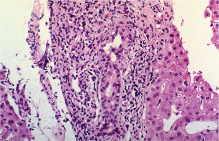
The exuberant inflammatory infiltrate of severe AAR spills into adjacent liver parenchyma, sometimes with true portal-portal bridging and extensive lobular necrosis. Despite this, graft failure rarely occurs. Increased number of CD8+ T cells are seen, particularly in the portal tracts, as well as an increase of CD45RO+ (memory) T cells.
Interlobular bile ducts show variable degrees of injury (e-Figs. 25.7, 25.9-25.11), ranging from mild to severe, including vacuolation of the epithelial cytoplasm, overlapping nuclei, nuclear pyknosis, infiltration of the epithelium with lymphocytes and occasional eosinophils, and true epithelial necrosis with subsequent disruption of the basement membrane (Fig. 25.4).
Endothelialitis involves mostly portal veins, but terminal hepatic venules (central veins) can also show changes (e-Figs. 25.4-25.7). Endothelialitis can be subtle, with only endothelial cell swelling and focal subendothelial lymphocytic infiltration (Fig. 25.5). When fully developed, there is lifting and detachment of endothelial cells undermined by lymphocytic infiltrate followed by sloughing into the lumen.
In addition to the typical AAR features, zone 3 hepatocyte ballooning (preservation injury) can be seen. The most important and clinically relevant indicator of AAR severity is the response to supplemental immunosuppression. Endothelialitis resolves first; then the immunoinflammatory infiltrate diminishes and the bile duct injury becomes less severe. Rebiopsy after treatment is useful to ensure adequate immunosuppression and to avoid excessive immunosuppression.
|
|
|
FIGURE 25.5 Acute allograft rejection, showing endothelialitis (hematoxylin-eosin, original magnification ×200). |
Earlier criteria for histologic grading of AAR were based on the degree of bile duct injury, severity and extent of portal infiltrate, and endothelialitis. One of the earliest grading systems (75) has four categories of rejection: (a) relatively mild mixed portal inflammatory infiltrate and bile duct injury involving less than 50% of all interlobular bile ducts and without endothelialitis; (b) grade 1 rejection, similar to the first category, but also showing endothelialitis; (c) grade 2 rejection, with more than 50% of interlobular bile ducts showing injury; and (d) grade 3 rejection, including all features from the previous category, but also demonstrating arteritis and paucity of bile ducts with associated ballooning of hepatocytes in zone 3 of the acinus.
A multicenter study attempted to standardize the grading and nomenclature of AAR, with moderate to excellent intraobserver and interobserver agreement (14,18). Recently, an international panel of pathologists reached consensus regarding a standardized nomenclature and grading system of allograft rejection in the liver (Table 25.2) (37,38).
MORE THAN THREE MONTHS FOLLOWING LIVER TRANSPLANTATION
Chronic Allograft Rejection
Chronic (ductopenic) rejection, affecting both bile ducts and arteries, may become obvious as early as a few months following transplantation (48,52,60).
PATHOLOGY. Vascular rejection implies changes in the intima of medium-sized arteries, which lead to obliterative endarteritis or “foamy” arteriopathy (48) (Fig. 25.6, e-Figs. 25.12-25.14). These changes, usually not seen in biopsy material, include intimal accumulation of histiocytes and subsequent fibrosis and lumen obliteration. Zone 3 (centrolobular) ballooning and, eventually, perivenular fibrosis are seen. These changes are similar to those of schemia of other causes, which must be excluded. Chronic vascular rejection may be reversible after therapy (33,75).
Potential etiologic roles for cytomegalovirus (CMV) and human leukocyte antigen incompatibility have been suggested but are controversial (5,50).
|
TABLE 25.2 Banff Schema for Grading Liver Allograft Rejection |
||||||||||||
|
||||||||||||
|
|
||||||||||||
|
FIGURE 25.6 Chronic ductopenic rejection, showing transplant (foamy) subintimal arteriopathy (hematoxylin-eosin, original magnification ×200). |
||||||||||||
|
|
|
FIGURE 25.7 Chronic ductopenic rejection (vanishing bile duct syndrome). Note complete absence of the original interlobular bile ducts as well as the absence of bile ductular proliferation (hematoxylin-eosin, original magnification ×200). |
Chronic ductopenic rejection (vanishing bile duct syndrome, VBDS) manifests as progressive bile duct injury and subsequent reduction in the number of bile ducts (paucity) with eventual loss of virtually all interlobular bile ducts (Fig. 25.7, e-Fig. 25.15) (48,52). This develops with or without features of chronic vascular rejection. The biopsy shows virtually complete absence of interlobular bile ducts, without significant bile ductular proliferation, but with extensive zone 3 canalicular cholestasis (e-Fig. 25.16). Inflammatory infiltrate is usually not prominent.
Early paucity may not be easily appreciated unless markers for biliary epithelium, including CK19 and keratin AE 1,3, are used (Fig. 25.8). An acute form of chronic ductopenic rejection can develop within a month of liver transplantation (Fig. 25.9). Criteria for grading and reporting of chronic rejection have been proposed (Table 25.3) (39).
RECURRENT DISEASES
Hepatitis B
The recurrence rate of hepatitis B virus (HBV) infection was once high, with severe progressive disease, ultimately resulting in graft failure (40,51). In the last 20 years, however, hepatitis B immunoglobulin (HBIG) posttransplantation clears hepatitis B surface antigen (HBsAg) from serum, with HBV DNA still detectable in serum or circulating mononuclear cells, potentially sustaining a low level of viral replication (58).
|
|
|
FIGURE 25.8 Ductopenic rejection, showing complete absence of interlobular bile ducts (immunoperoxidase, using antibody against keratin AE1/3, original magnification ×200). |
In recurrence, various histologic changes are seen. As many as 80% show chronic hepatitis changes including cirrhosis, which can develop within a few years. In approximately 10% there are only nonspecific changes. Fibrosing cholestatic hepatitis (FCH) is a unique and usually rapidly progressive and fatal form of recurrent HBV infection.
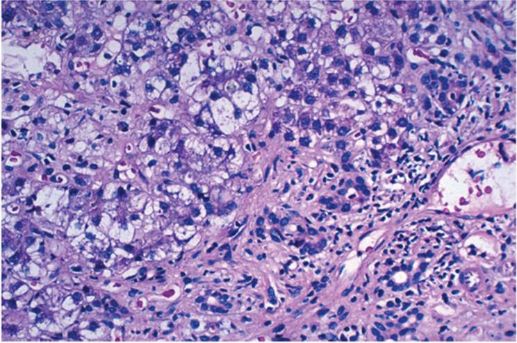
PATHOLOGY. Recurrent HBV in the allograft may be indistinguishable from HBV in the nontransplant patient. Typical chronic hepatitis, with many “ground glass” hepatocytes filled with HBsAg can be seen (see Chapter 8) (Fig. 25.10). FCH variant has portal, periportal, and perisinusoidal/pericellular fibrosis, canalicular and intracellular cholestasis, severe ballooning of hepatocytes, relatively mild portal inflammatory infiltrate, and usually, significant expression of HBsAg and HbcAg in hepatocytes (Fig. 25.11). Excessive intracellular accumulation of HBsAg may be directly cytopathic (16).
|
|
|||||||||||||||||||||||||||||||||
|
FIGURE 25.9 Ductopenic rejection developing within a month after transplantation, showing loss of interlobular bile ducts as well as portal tract edema (hematoxylin-eosin, original magnification ×200). |
|||||||||||||||||||||||||||||||||
|
TABLE 25.3 Rejection Activity Indexa |
|||||||||||||||||||||||||||||||||
|
|||||||||||||||||||||||||||||||||
|
|
|
FIGURE 25.10 Recurrent hepatitis B virus infection, showing features of chronic hepatitis with numerous “ground glass” (hepatitis B surface antigen-containing) cells (hematoxylin-eosin, original magnification ×200). |
Patients who undergo transplantation for fulminant HBV or with simultaneous HBV and hepatitis delta virus (HDV) infection have a recurrence rate of 50% to 70%. Paradoxically, the clinical course is usually relatively mild. In contrast with the nontransplantation population, patients develop recurrent HDV without associated HBV markers of infection, suggesting that HDV replicates in the absence of HBV and is not necessarily cytopathic, instead requiring HBV to cause liver cell injury (46). Coagulative necrosis of zone 1 is seen, with associated fibrinoid necrosis of hepatic arterioles (13,15).
|
|
|
FIGURE 25.11 Recurrent hepatitis B virus infection with histopathologic features of fibrosing cholestatic cholangitis (hematoxylin-eosin, original magnification ×100). |
Hepatitis C
Hepatitis C virus (HCV) recurrence following liver transplantation is virtually universal and is confirmed by serologic tests or polymerase chain reaction (PCR). Reinfection is usually with the same HCV strain as the original disease. Although the natural history of the posttransplant recurrent HCV hepatitis is variable, recurrent HCV hepatitis is generally more rapidly progressive when compared to the patients who underwent LT years ago (68). Newer therapies, such as pegylated interferon, may be altering this. Risk factors associated with more aggressive HCV recurrence include treated prior acute allograft rejection, cytomegalovirus (CMV) infection, and use of OKT3.
Five risk factors possibly contributing to severe recurrent disease within 2 years are the following: donor age older than 50 years, HCV genotype 1b, use of OKT3, mycophenolate mofetil (MMF) immunosuppression induction, and short-term prednisone and azathioprine (AZT) use (68).
Some patients develop HCV as an acquired infection following transplantation, either from blood products or from the organ donor (23,62). Recurrent HCV is often relatively mild (22). In spite of this, 5% to 10% of patients develop cirrhosis and, ultimately, graft failure. In some patients, HCV recurs within weeks of transplantation, and in others recurrence takes months to years. Levels of circulating HCV are much higher in individuals experiencing early recurrence (10,20,21). Furthermore, immunosuppression may contribute to high viral levels. HCV can be directly cytopathic for bile duct epithelium, but there is no correlation between levels of circulating HCV and degree of liver cell or bile duct injury. A donor-recipient match at one or two human leukocyte antigen DQ loci has been associated with more severe recurrent HCV. Other factors include cytokine gene polymorphism and chemokine effects (68). Patients infected with HCV type 1 genotype seem to have higher recurrence risk. As many as 30% of treated patients have features of ductopenic rejection.
PATHOLOGY. Typical chronic HCV changes are seen in as many as 80% of patients with recurrent HCV. Various findings not typically associated with HCV in the nontransplantation setting can also be seen, including severe bile duct injury and bile ductular proliferation, mimicking large duct obstruction (23). Cholestasis and ischemic changes may be prominent. When HCV recurs early in the posttransplantation period, before 6 weeks, the features are quite subtle (Fig. 25.12) and differentiation from AAR can be difficult (Table 25.4) (61,67,68). Multiple serial posttransplantation biopsies can be helpful as portal tract inflammation, lymphoid aggregate formation, and lobular activity become more evident (25,27). Early HCV recurrence can be predominantly lobular hepatitis with little or no portal inflammation.
|
|
||||||||||||||||||||||||||||||||||||||||||||||||
|
FIGURE 25.12 Recurrent hepatitis C virus infection developing within 3 months of transplantation. Note sinusoidal dilatation and only focal lobular inflammation (hematoxylin-eosin, original magnification ×200). |
||||||||||||||||||||||||||||||||||||||||||||||||
|
TABLE 25.4 Histopathologic Features Useful to Differentiate Acute Cellular Rejection from Early Recurrent Hepatitis C (2-3 Months following LT) |
||||||||||||||||||||||||||||||||||||||||||||||||
|
||||||||||||||||||||||||||||||||||||||||||||||||
|
|
|
FIGURE 25.13 Recurrent hepatitis C virus infection occurring more than a year following transplantation and histologically indistinguishable from chronic hepatitis C in the nontransplantation setting (hematoxylin-eosin, original magnification ×200). |
One of the most important features of HCV is bile duct injury (Fig. 25.13) (Table 25.5). The precise mechanism for this is unclear.
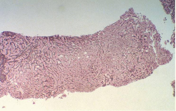
Nodular regenerative hyperplasia (NRH) has been described in the setting of recurrent HCV infection (Fig. 25.14).
|
TABLE 25.5 Differentiating Early Recurrent HCV (<3 Months following LT) from AAR Liver Biopsy Findings |
||||||||||||||||||||||||||||||
|
||||||||||||||||||||||||||||||
|
|
|
FIGURE 25.14 Nodular hyperplasia in a patient with recurrent hepatitis C virus infection (reticulin silver preparation, original magnification ×100). |
Primary Biliary Cirrhosis
Primary biliary cirrhosis (PBC) can recur (6,34,59,64), generally after many years. It can be exceedingly difficult to distinguish early recurrent PBC from AAR because of the similarity of bile duct injury. Typical early granulomatous bile duct injury and features of florid bile duct injury are usually not seen. Mild bile duct injury, with predominantly mononuclear inflammatory infiltrate, is more usual. Recurrent PBC progresses slowly, and advanced stages are uncommon.
Primary Sclerosing Cholangitis
Primary sclerosing cholangitis (PSC) also recurs (31). Infectious and noninfectious causes, including ischemia, CMV, and technical complications, may cause secondary sclerosing cholangitis, which can be difficult to distinguish from PSC. If these and other potential etiologic factors can be excluded, the finding of imaging and pathology features of PSC (Fig. 25.15) can be considered diagnostic. Features compatible with PSC that become apparent at least a year after LT are considered more significant, since the other potential factors (including ischemia) are less likely at that time (28,40,74). Recurrent PSC is generally histologically indistinguishable from usual PSC.
Other Conditions
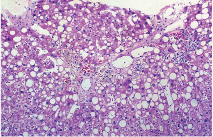
Recurrence of alcoholic liver disease, Budd-Chiari syndrome, and autoimmune hepatitis are well recognized (30,37). Some patients with recurrent autoimmune hepatitis can have concurrent HCV. Nonalcoholic steatohepatitis (NASH)/nonalcoholic fatty liver disease (NAFLD) also recurs, especially in patients who have undergone prior jejunoileal bypass (Fig. 25.16). The course may be more rapidly progressive.
|
|
|
FIGURE 25.15 Recurrent primary sclerosing cholangitis. Note the characteristic periductal concentric fibrosis (Masson trichrome, original magnification ×200). |
Extrahepatic biliary atresia has not been shown to recur, but metabolic diseases, including glycogenosis IV, may.
Hepatocellular Malignancies
Malignant tumors, including hepatocellular carcinoma and hepatoblastoma, can recur (39,57). Cholangiocarcinomas can recur relatively quickly with a 2-year posttransplant survival less than 50%. Epithelioid hemangioendothelioma, even when extensive, is not a contraindication for LT. Long-term survival occurs when the tumor is limited to the liver, even with lymph node metastasis. Metastatic neoplasms, even if solitary, can also recur relatively quickly, and transplantation is generally considered inappropriate (26).
|
|
|
FIGURE 25.16 Recurrent nonalcoholic steatohepatitis in a patient who had previously undergone jejunoileal bypass (hematoxylin-eosin, original magnification ×200). |
Hepatitis G and Hepatitis E in Liver Transplantation Patients
Hepatitis G virus (HGV) can be present in as many as 20% of HCV patients, but apparently has little or no clinical impact (3,12,24). It has recently been suggested that hepatitis E is associated with clinically chronic disease in the posttransplant setting, but HEV is quite rare (41).
OPPORTUNISTIC INFECTIONS
Infection with opportunistic organisms is a common problem because all liver transplantation patients undergo immunosuppressive therapy. The most common viral infections are CMV, herpes simplex virus and varicella zoster virus, adenovirus (especially in children), and Epstein-Barr virus (EBV).
Viruses
CMV is the most commonly encountered posttransplantation infectious agent (34,38). Scattered collections of PMNs (microabscesses) can be seen (Fig. 25.17), but they are not necessarily CMV and confirmation with immunostains is needed. Their presence, however, should be taken as presumptive evidence of CMV pending confirmation. Characteristic intranuclear viral inclusions can also be seen (Fig. 25.18, e-Figs. 25.17-25.19).
|
|
|
FIGURE 25.17 Microabscesses in the posttransplantation allograft. A specific etiologic factor was not identified (hematoxylin-eosin, original magnification ×200). |
|
|
|
FIGURE 25.18 Cytomegalovirus hepatitis occurring during the first week after liver transplantation, showing characteristic cell enlargement and typical intranuclear inclusion (hematoxylin-eosin, original magnification ×200). |
Other viral infections, such as herpes simplex (e-Figs. 25.20, 25.21), can be seen. Herpes has varying degrees of liver cell necrosis, and viral inclusions may not always be appreciated until immunostains are used. In adenovirus, necrosis may be extensive (9) (e-Fig. 25.22).
Fungal Infection and Bacterial Infections
Fungal infection is only rarely seen in the posttransplantation liver biopsy.
Bacterial infection, with associated septicemia and sepsis, develops in patients after LT. Similar to the nontransplantation setting, a mild mononuclear and polymorphonuclear portal infiltrate is seen, with focal bile ductular proliferation.
Other Infectious Agents
Toxoplasma gondii, Pneumocystis carinii, and Bartonella quintana can occur in the posttransplantation setting.
Posttransplantation Lymphoproliferative Disorders
Most posttransplantation lymphoproliferative disorders (PTLDs) occurring after liver transplantation are associated with EBV infection and can occur as early as 4 to 6 weeks following transplantation. The incidence in the pediatric population is approximately 8% and in the adult population is 1% to 2%. EBV may develop de novo or may represent viral reactivation. The presence of the viral genome can be confirmed by in situ hybridization or polymerase chain reaction of serum or tissue. In situ hybridization for EBER-1 gene in tissue is sufficiently sensitive and specific for the detection of the viral genetic material. Immunostain for latent membrane protein seems to be somewhat less sensitive (44).
PATHOLOGY. The findings are similar to those in the nontransplantation setting. Sinusoidal and portal tract infiltration with B lymphocytes, including immunoblastic forms, is characteristic. Initially, B-cell proliferation is polyclonal and, at this stage, will resolve with reduction of immunosuppression and antiviral therapy. Monoclonal proliferation is likely to be malignant and unresponsive to treatment. Most cases of PTLD are usually of recipient origin. Rarely, proliferating lymphoid cells of donor origin have been demonstrated. T-cell PTLD can also occur.
Histopathologic features of EBV-associated PTLD may be difficult to recognize and differentiate from AAR, especially in the early, polyclonal phase (2,42). Portal tracts are mildly to moderately expanded with a predominantly mononuclear lymphoid infiltrate composed of small, mature lymphocytes and transformed cells, including lymphoblasts and immunoblasts (Fig. 25.19, e-Figs. 25.23-25.26). Plasma cells may be prominent. The absence of eosinophils is striking and may be helpful in excluding AAR. Bile duct injury, if present, is minimal, with only mild irregularity of epithelial cells, without evidence of lymphocytic infiltration or bile duct disruption, despite being surrounded by a dense lymphoid infiltrate. The lymphoid infiltrate often has angiocentric (perivenular) distribution, mimicking endothelialitis, but no true endothelialitis. The histopathologic features may be exceedingly subtle, may mimic bile duct obstruction with focal bile ductular proliferation, or may be quite patchy and not seen in any given biopsy (2,65,66). Immunohistochemical and molecular methods for demonstrating EBV should be performed (Fig. 25.20, e-Fig. 25.26) (44).
|
|
|
FIGURE 25.19 Posttransplantation Epstein-Barr virus-related lymphoproliferative disorder (hematoxylin-eosin, original magnification ×200). |
|
|
|
FIGURE 25.20 Posttransplantation Epstein-Barr virus-related lymphoproliferative disorder with numerous cells demonstrating positive reaction for EBER-1 gene (in situ hybridization with a probe for EBER-1 gene, original magnification ×200). |
FUTURE DEVELOPMENTS
Although LT is only about 30 years old, newer modalities are being applied, with their own sets of problems. There is growing use of split donor livers, use of living-related donor livers, heterotopic grafts, and xenografts using porcine and baboon livers as bridge grafts until a donor is obtained (8,19). Various bioartificial liver devices are being evaluated, also serving as therapeutic bridges in critically ill patients with fulminant or subfulminant liver failure.
LIVER BIOPSY FOLLOWING BONE MARROW TRANSPLANTATION
Acute Graft Versus Host Disease
Graft versus host disease (GVHD) is commonly associated with bone marrow transplantation but can develop following orthotopic liver transplantation (11). Changes primarily involve skin and colon, and the liver is affected in only the most severe cases. Rarely, liver is the single affected organ (55,70,71). GVHD is an immunologically mediated condition and develops from the effects of the immunocompetent donor cells on liver cells.
|
|
|
FIGURE 25.21 Graft versus host disease in a patient who had undergone liver transplantation, showing bile duct epithelial necrosis (hematoxylin-eosin, original magnification ×200). |
PATHOLOGY. Acute GVHD manifests as interlobular bile duct injury and cholestasis (55,71) (Fig. 25.21, e-Figs. 25.27-25.29). Bile duct epithelium appears irregular and shows cytoplasmic vacuolation, overlapping nuclei, and nuclear pleomorphism sometimes with increased nuclear/cytoplasmic ratio. Numerous apoptotic nuclei with scattered nuclear debris may be seen. Endothelialitis of both portal and hepatic veins is seen in as many as approximately 10% of patients, as well as true venous occlusion. Endothelialitis is not invariable, as it is in AAR, and hence does not seem to have the same diagnostic and prognostic importance. Focal liver cell necrosis with acidophilic body formation occurs. Kupffer cells and sinusoidal endothelial cells may contain abundant hemosiderin.
There may be associated nodular hyperplasia (NH) (see Chapter 23) with portal hypertension (72). Furthermore, patients with GVHD may have concomitant hepatitis, either viral or drug induced, and establishing the correct diagnosis may be quite difficult.
Chronic Graft Versus Host Disease
Chronic GVHD usually develops after more than 100 days following bone marrow transplantation, and manifestations may be seen in multiple organs.
PATHOLOGY. Chronic GVHD is characterized by progressive bile duct injury and ultimate bile duct loss with marked cholestasis (Fig. 25.22). Eventually, there is portal and septal fibrosis. A few patients become cirrhotic (38,43). Arteriopathic changes, frequently seen in ductopenic allograft rejection, are not common.
|
|
|
FIGURE 25.22 Chronic graft versus host disease involving the liver in a patient following bone marrow transplantation (hematoxylin-eosin, original magnification ×200). |
Differential diagnosis includes various types of hepatitis. However, in most hepatitis severed bile duct and injury and bile duct loss are not seen, even with marked hepatocellular injury and necrosis. GVHD can, of course, have concurrent hepatitis.
OTHER FINDINGS. Venoocclusive disease (VOD) and NH occur in patients who have undergone cytoreductive therapy. VOD is most common during the first 30 days after bone marrow transplantation. VOD and HN can have similar manifestations, and differentiation generally requires liver biopsy. Furthermore, although NH does not necessarily have a significant immediate effect, patients develop portal hypertension (72).
LIVER BIOPSY FOLLOWING RENAL TRANSPLANTATION
After renal transplantation, various conditions affecting the liver have been described, including steatosis, NH, peliosis hepatis, infection with hepatitis B and C viruses, and hepatocellular carcinoma (53). Secondary hemosiderosis is common in patients who underwent hemodialysis or received multiple transfusions. After long-standing hemodialysis, silicone from the dialysis tubing may elicit a foreign-body reaction, with subsequent hepatic fibrosis. Silicone may be seen as a birefringent crystalloid foreign material in the tissue.
Drug-induced changes, including cholestasis, sinusoidal dilatation, and VOD associated with azathioprine, are also well recognized after renal transplantation (53). Superimposed bacterial infection with associated septicemia and sepsis and various opportunistic infections may also occur.
REFERENCES
1. Adams DH, Neuberger JM. Patterns of graft rejection following liver transplantation. J Hepatol 1990;10:113-119.
2. Alshak NS, Jimenez AM, Gedebou M, et al. Epstein-Barr virus infection in liver transplantation patients: correlation of histopathology and semi-quantitative Epstein-Barr virus-DNA recovery using polymerase chain reaction. Hum Pathol 1993;24:1306-1312.
3. Alter HJ. The cloning and clinical implications of HGV and HGBV-C. N Engl J Med 1996;334:1536-1537.
4. Angele MK, Rentsch M, Wittmann B, et al. Effect of steatosis on liver function and organ survival after liver transplantation. Am J Surg 2008;195(2):214-220.
5. Arnold JC, Portman BC, O'Grady JG, et al. Cytomegalovirus infection persists in the liver graft in the vanishing bile duct syndrome. Hepatology 1992;16:285-292.
6. Balan V, Batts KP, Porayko MK, et al. Histologic evidence of recurrence of primary biliary cirrhosis after liver transplantation. Hepatology 1993;18:1392-1398.
7. Bizzolon T, Palazzo U, Ducerf C, et al. Pilot study of the combination of interferon alpha and ribavirin as therapy of recurrent hepatitis C after liver transplantation. Hepatology 1997;26:500-505.
8. Broelsch CE, Whitington PF, Emond JC, et al. Liver transplantation in children from living related donors: surgical techniques and results. Ann Surg 1994;214:428-439.
9. Cames B, Rahier J, Burtomboy G, et al. Acute adenovirus hepatitis in liver transplant patients. J Pediatr 1992;120:33-37.
10. Chazouilleres O, Kim M, Combs C, et al. Quantitation of hepatitis C virus RNA in liver transplant recipients. Gastroenterology 1994;106:994-999.
11. Collins RH Jr, Cooper B, Nikaein A, et al. Graft-versus-host disease in a liver transplant recipient. Ann Intern Med 1992;116:391-392.
12. Cotler SJ, Gretch DR, Bronner MP, et al. Hepatitis G virus co-infection does not alter the course of recurrent hepatitis C virus infection in liver transplantation recipients. Hepatology 1997;26:432-437.
13. Craig FE, Gulley ML, Banks PM. Posttransplant lymphoproliferative disorders. Am J Clin Pathol 1993;99:265-276.
14. D'Alessandro AM, Kalayoglu M, Sollinger HW, et al. The predictive value of donor liver biopsies for the development of primary nonfunction after orthotopic liver transplantation. Transplantation 1991;51:157-163.
15. David E, Rahier J, Pucci A, et al. Recurrence of hepatitis D (delta) in liver transplants: histopathologic aspects. Gastroenterology 1991;104:1122-1128.
16. Davies SE, Portmann BC, O'Grady JG, et al. Hepatic histologic findings after transplantation for chronic hepatitis B virus infection, including a unique pattern of fibrosing cholestatic hepatitis. Hepatology 1992;13:150-157.
17. Demetris AJ, Jaffe R, Tzakis A, et al. Antibody-mediated rejection of human orthotopic liver allografts: a study of liver transplantation across ABO blood group barriers. Am J Pathol 1988;132:489-502.
18. Demetris AJ, Belle SH, Hart J, et al. Intraobserver and interobserver variation in the histopathologic assessment of liver allograft rejection. Hepatology 1991;14:949-951.
19. Demetriou AA, Whiting J, Levenson SM, et al. New method of hepatocyte transplantation and extracorporeal liver support. Ann Surg 1986;204:259-271.
20. Faust TW. Recurrent primary biliary cirrhosis, primary sclerosing cholangitis, and autoimmune hepatitis after transplantation. Liver Transplant 2001;7:99-108.
21. Ferey C, Samuel D, Thiers V, et al. Reinfection of liver graft by hepatitis C virus after liver transplantation. J Clin Invest 1992;89:1361-1365.
22. Ferey C, Gigou M, Samuel D, et al. The course of hepatitis C virus infection after liver transplantation. Hepatology 1994;20:1137-1143.
23. Ferey C, Gigou M, Samuel D, et al. Influence of the genotypes of hepatitis C virus on the severity of recurrent liver disease after liver transplantation. Gastroenterology 1995;108: 1088-1096.
24. Freese DK, Snover DC, Sharp HL, et al. Chronic rejection after liver transplantation: a study of clinical, histopathological and immunological features. Hepatology 1991;13: 882-891.
25. Fried MW, Khudyakov YE, Smallwood GA, et al. Hepatitis G co-infection in liver transplantation recipients with chronic hepatitis C and nonviral chronic liver disease. Hepatology 1997;25:1271-1276.
26. Gane EJ, Portmann BC, Naoumov NV, et al. Long-term outcome of hepatitis C infection after liver transplantation. N Engl Med 1996;334:815-820.
27. Geubel AP, Cnudde A, Ferrant A, et al. Diffuse biliary tract involvement mimicking primary sclerosing cholangitis after bone marrow transplantation. J Hepatol 1990;10:23-28.
28. Graziadei IW, Wiesner RH, Batts KP, et al. Recurrence of primary sclerosing cholangitis following liver transplantation. Hepatology 1999;29:1050-1056.
29. Greenson JK, Svoboda-Newman SM, Merion RM, et al. Histologic progression of recurrent hepatitis C in liver transplant allografts. Am J Surg Pathol 1996;20:731-738.
30. Gretch DR, Bacchi CE, Corey L, et al. Persistent hepatitis C virus infection after liver transplantation: clinical and virological features. Hepatology 1995;22:1-10.
31. Halff G, Todo S, Tzakis AG, et al. Liver transplantation for the Budd-Chiari syndrome. Ann Surg 1990;202:43-49.
32. Harrison RF, Davies MH, Goldin RD, et al. Recurrent hepatitis B in liver allografts: a distinctive form of rapidly developing cirrhosis. Histopathology 1993;23:21-28.
33. Hartman GG, Gordon R, Lerut J, et al. Intrahepatic bile duct strictures in a liver allograft recipient mimicking recurrent primary sclerosing cholangitis. Transplant Int 1991;4: 191-192.
34. Hertzler G, Milikan WJ. The surgical pathologist's role in liver transplantation. Arch Pathol Lab Med 1991;115:273-382.
35. Hubscher SG, Buckels JAC, Elias E, et al. Reversible vanishing bile duct syndrome after liver transplantation. Report of 6 cases. Transplant Proc 1991;23:1415-1416.
36. Hubscher SG, Elias E, Buckels JAC, et al. Primary biliary cirrhosis: histologic evidence of disease recurrence after liver transplantation. J Hepatol 1993;18:173-184.
37. International Working Party. Terminology of hepatic allograft rejection. Hepatology 1995; 22:648-655.
38. International Panel. Banff schema for grading liver allograft rejection; an international consensus document. Hepatology 1997;25:658-664.
39. International Panel. Update of the international Banff schema for liver allograft rejection: working recommendations for the histopathologic staging and reporting of chronic rejection. Hepatology 2000;31:792-799.
40. Jeyarajah DR, Netto GJ, Lee SP, et al. Recurrent primary sclerosing cholangitis after orthotopic liver transplantation: is chronic rejection part of the process? Transplantation 1998;66:1300-1306.
41. Kamar N, Selves J, Mansuy JM, et al. Hepatitis E virus and chronic hepatitis in organtransplant recipients. N Engl J Med 2008;358(8):811-817.
42. Keeffe EB. Milestones in liver transplantation for alcoholic liver disease. Liver Transplant Surg 1997;3:197-199.
43. Knapp AB, Crawford JM, Rappaport JM, et al. Cirrhosis as a consequence of graft-versus-host disease. Gastroenterology 1997;92:513-519.
44. Koneru B, Flye MM, Busittil RW, et al. Liver transplantation for hepatoblastoma: the American experience. Ann Surg 1991;213:118-121.
45. Lake JR, Wright T, Ferrell L, et al. Hepatitis C and B in liver transplantation. Transplant Proc 1993;25:2006-2009.
46. Langnas AN, Castaldo P, Markin RS, et al. The spectrum of Epstein-Barr infection with hepatitis following liver transplantation. Transplant Proc 1991;23:1513-1514.
47. Lee RG. Recurrence of alcoholic liver disease after liver transplantation. Liver Transplant Surg 1997;3:292-296.
48. Lones MA, Shintaku IP, Weiss LM, et al. Posttransplant lymphoproliferative disorder in liver allograft biopsies: a comparison of three methods for the demonstration of Epstein-Barr virus. Hum Pathol 1997;28:533-539.
49. Lucey MR, Merion RM, Henley KS, et al. Selection for and outcome of liver transplantation in alcoholic liver disease. Gastroenterology 1992;102:1736-1741.
50. Lucey MR, Graham DM, Martin P, et al. Recurrence of hepatitis B and delta hepatitis after orthotopic liver transplantation. Gut 1992;33:1390-1396.
51. Ludwig J, Batts KP, MacCarty RL. Ischemic cholangitis in hepatic allografts. Mayo Clin Proc 1992;67:519-526.
52. Ludwig J, Wiesner RH, Batts KP, et al. The acute vanishing bile duct syndrome (acute irreversible rejection) after orthotopic liver transplantation. Hepatology 1987;7:476-483.
53. Manez R, White LT, Linden P, et al. The influence of HLA matching on cytomegalovirus hepatitis and chronic rejection after liver transplantation. Transplantation 1993;55: 1067-1071.
54. Markin RS, Wisecarver JL, Radio SJ, et al. Frozen section evaluation of donor livers before transplantation. Transplantation 1990;56:1403-1409.
55. Marubbio AT, Danielson B. Hepatic veno-occlusive disease in renal transplant patient receiving azathioprine. Gastroenterology 1975;69:739-743.
56. Mason AL, Wick M, White HM, et al. Increased hepatocyte expression of hepatitis B virus transcription in patients with features of cholestatic fibrosing hepatitis. Gastroenterology 1993;105:237-244.
57. Matsumoto Y, McCaughan GW, Painter DM, et al. Evidence that portal tract microvascular destruction precedes bile duct loss in human liver allograft rejection. Transplantation 1993;56:69-75.
58. McDonald GB, Sharma P, Matthews DE, et al. Venoocclusive disease of the liver after bone marrow transplantation: diagnosis, incidence and predisposing factors. Hepatology 1984;4:116-122.
59. McPeake JR, O'Grady JG, Zaman S, et al. Liver transplantation for primary hepatocellular carcinoma: tumor size and number determine outcome. J Hepatol 1993;18:226-234.
60. Milkiewicz P, Hubscher SG, Skiba G, et al. Recurrence of autoimmune hepatitis after liver transplantation. Transplantation 1999;68:253-256.
61. Nalesnik MA. Lymphoproliferative disease in organ transplant recipients. Springer Semin Immunopathol 1991;13:199-216.
62. Neuberger J. Recurrent primary biliary cirrhosis. Liver Transplant 2001;7:596-599.
63. Noack KB, Wiesner RH, Batts K, et al. Severe ductopenic rejection with features of vanishing bile duct syndrome: clinical, biochemical and histologic evidence for spontaneous resolution. Transplant Proc 1991;23:1448-1451.
64. Palazzo J, Lundquist K, Mitchell D, et al. Rapid development of lymphoma following liver transplantation in a recipient with hepatitis B and primary hemochromatosis. Am J Gastroenterol 1993;88:102-104.
65. Paya C, Wiesner R, Hermans P, et al. Lack of association between cytomegalovirus infection, HLA matching and the vanishing bile duct syndrome after liver transplantation. Hepatology 1992;16:66-70.
66. Pereira BJG, Milford EL, Kirkman RL, et al. Prevalence of hepatitis C virus RNA in organ donors positive for hepatitis C antibody and in the recipients of their organs. N Engl J Med 1992;327:910-915.
67. Petrovic LM, Villamil FG, Vierling JM, et al. Comparison of histopathology in acute allograft rejection and recurrent hepatitis C infection after liver transplantation. Liver Transpl Surg 1997;3:398-406.
68. Petrovic LM. Early recurrent hepatitis C virus infection after liver transplantation Liver Transplantation. 2006;12(11 Suppl 2):S32-37.
69. Ploeg RJ, D'Alessandro AM, Knechtle SJ, et al. Risk factors for primary dysfunction after liver transplantation: a multivariate analysis. Transplantation 1993;55:807-813.
70. Randhawa PS, Jaffe R, Demetris AJ, et al. The systemic distribution of Epstein-Barr virus genomes in fatal posttransplantation lymphoproliferative disorders. An in situ hybridization study. Am J Pathol 1991;138:1027-1033.
71. Randhawa PS, Jaffe R, Demetris AJ, et al. Expression of Epstein-Barr virus-encoded small RNA (by the EBER-1 gene) in liver specimens from transplant recipients with posttransplantation lymphoproliferative disease. N Engl J Med 1992;327:1710-1714.
72. Roberts JP, Ascher NL, Lake J, et al. Graft vs. host disease after liver transplantation: a report of four cases. Hepatology 1991;14:274-281.
73. Sanchez-Urdazpal L, Gores GJ, Ward EM, et al. Diagnostic features and clinical outcome of ischemic-type biliary complications after liver transplantation. Hepatology 1993;17:605-609.
74. Sheng R, Campbell WI, Zajki AB, et al. Cholangiographic features of biliary stricture liver transplantation for primary sclerosing cholangitis: evidence of recurrent disease. AJR Am J Roentgenol 1996;166:1109-1113.
75. Snover DC, Freese DK, Sharp HL, et al. Liver allograft rejection. An analysis of the use of the biopsy in determining outcome of rejection. Am J Surg Pathol 1987;11:1-10.