
Benjamin W. Friedman
Head pain accounts for 2 million emergency department (ED) visits annually in the United States (1). The vast majority of headaches presenting to the ED do not threaten life or limb. Most often, they are acute manifestations of an episodic headache disorder or a self-limited, infectious or inflammatory condition readily amenable to treatment. Management of the patient with headache in the ED requires distinguishing the small fraction of patients with serious illness who require immediate diagnosis and hospitalization from the great majority in whom the clinical focus is directed toward headache-specific, short-term pain management, prophylaxis against recurrence, explanation, reassurance, and careful follow-up, while minimizing throughput time and use of healthcare resources.
CLASSIFICATION OF HEAD PAIN
The simplest means of categorizing headaches is to dichotomize into primary headaches (e.g., migraine) versus “organic” or secondary headaches. The latter are so named because they are secondary to some underlying cause (e.g., subarachnoid hemorrhage) (2).
The pain-sensitive structures of the head include the blood vessels and all extracranial structures of the head and neck. The only intracranial structures sensitive to pain are the vasculature of the brain, the dura mater, the venous sinuses, and cranial and cervical nerves. The parenchyma of the brain itself and much of its meningeal coverings are not pain sensitive.
For many years, traditional teaching held that there were three major mechanisms of head pain: Vasodilatation within the cranial vasculature, causing a “vascular” headache; spasm of the neck and scalp muscles, causing a “muscle contraction” headache; and traction on blood vessels or inflammation around or within these and other pain-sensitive structures, causing a “traction–inflammatory” headache. More recently, traditional concepts of the etiology of headache, particularly of migraine, have given way to explanations based on animal models and neuroimaging in which abnormal release of vasoactive neuropeptides actuates nociceptive pathways. It is now clear that vasodilation is not the primary mechanism of headache. Headache is best thought of as a cranial and cervical neuropathy causing activation of secondary and tertiary nociceptive pathways within the brainstem (3).
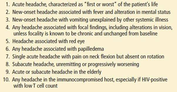
For emergency clinicians, thorough headache management addresses the twin challenges of accurate diagnosis and effective treatment (Table 153.1). Secondary headaches, particularly malignant secondary headaches, must be considered and excluded. This often can be accomplished based on the description of the acute headache and the patient’s past headache history. For select patients, a diagnostic workup is indicated (Table 153.2). Patients with an acute exacerbation of a recurrent primary headache disorder should be treated with disease-specific therapies rather than nonspecific analgesics.
TABLE 153.1
Differential Diagnosis of Headache

TABLE 153.2
Ten Headaches to Worry About
CLINICAL PRESENTATION AND DIFFERENTIAL DIAGNOSIS
Secondary Headaches
Meningitis/Encephalitis
The headache of meningitis tends to be generalized, though it is the constellation of signs and symptoms rather than the headache itself that remains key to diagnosis (4). Alterations in mental status are typical for bacterial meningitis, as is fever. The absence of fever, neck stiffness, and altered mental status practically eliminates bacterial meningitis from the differential diagnosis, though the individual components of this triad have poor predictive ability (5). Classically, the headache of meningitis worsens with sharp, sudden head movements—so called jolt accentuation. Patients with encephalitis may present with isolated headache, but evidence of brain parenchymal involvement, such as altered mental status, seizures, or focal neurologic findings should be apparent. In the United States, patients with Lyme meningitis frequently recall the classic erythema migrans rash and often present with a concomitant seventh cranial nerve palsy, while in Europe, painful radiculopathy is more common (6). Opportunistic infections are discussed below.
Subarachnoid Hemorrhage
Headache of SAH is the prototypic “first or worst” headache of one’s life, of sudden onset, often associated with nausea, vomiting, neck stiffness, and a transient alteration of consciousness. In patients with focal findings or sustained abnormalities of consciousness, the need for a neuroimaging study is apparent. However, some patients present without focal neurologic findings. In these patients, SAH may be difficult to distinguish from a first attack of migraine.
An especially difficult challenge for the emergency physician is identification of the “sentinel leak” that precedes major hemorrhage in one-third of patients. Detection of this premonitory bleed presents an opportunity to prevent a neurologic catastrophe. Clinical decision rules may allow the clinician to risk stratify patients (7).
Hypertension
Elevated blood pressure and headache have long been linked in the public mind. The causative pathway remains uncertain. It is clear that both hypertension and headache may both be present in malignant processes such as preeclampsia, posterior reversible encephalopathy syndrome, and reversible cerebral vasoconstriction syndrome. However, most headaches in hypertensive patients are either migraine or tension type and are only rarely due to elevated blood pressure. Mild-to-moderate elevations in blood pressure are not generally believed to be a cause of headache. By in large, it is felt that only acute elevation of diastolic pressure of at least 25% above baseline or sustained diastolic elevations in excess of 130 mm Hg have the potential to cause headache (2).
Narrow-Angle Glaucoma
Ocular or periorbital pain characterizes headache caused by narrow-angle glaucoma. The diagnosis of acute glaucoma also requires diminished visual acuity, a pupil that is typically fixed in midposition, an edematous (“steamy”) cornea, perilimbal injection, and increased intraocular pressure. Vomiting may be the dominant feature, especially in the elderly, who may also present with altered mentation.
Sinusitis
Acute sinusitis presents with frontal headache or facial pain, tenderness, and fullness. The diagnosis of acute sinus headache requires symptoms of a recent or intercurrent upper respiratory infection. Specifically, patients should report rhinorrhea or nasal congestion, stuffiness or blockage, and fever. Chronic sinusitis does not cause acute headache. Migraine, too, may present with nasal congestion or rhinorrhea. Physicians should consider this latter diagnosis in all patients who presents with recurrent headaches (8).
Cervical Artery Dissection
Spontaneous dissection of the cervical arteries is an uncommon syndrome, often associated with surprisingly minor trauma. It is marked by sudden onset of headache associated with facial or neck pain and focal ischemic symptoms. The pain and its location are determined by the location of the dissection in the carotid or vertebrobasilar system. In the carotid system, the pain is anterior and may be accompanied by monocular symptoms, Horner syndrome, and other focal findings. With dissection in the posterior circulation, the pain is occipital, and brainstem findings predominate.
Pituitary Apoplexy
Acute hemorrhage or infarction of the pituitary gland is a rare cause of sudden-onset headache that typically occurs in patients with underlying (although often not diagnosed) pituitary adenoma. This headache may be accompanied by decreased visual acuity, temporal visual field cuts, ocular palsies, and hormonal dysfunction such as acute adrenal crisis.
Cerebral Venous Thrombosis
Thrombosis of the venous system of the brain is an uncommon cause of headache that is often difficult to diagnose. The onset may be sudden or gradual. Both noncontrast head CT and spinal fluid analysis are often normal, so diagnosis is often delayed until after hospital admission. Magnetic resonance venography is useful in making the diagnosis. Risk factors include traditional thromboembolic risk factors, pregnancy and the postpartum period, trauma, and contiguous sources of infection.
Brain Tumor
Headache is the initial symptom of primary brain tumor in half of all cases, though by the time patients present to medical attention, they have often developed other signs suggestive of a malignant CNS process. Headache is an uncommon presenting symptom of a metastatic undiagnosed non-CNS malignancy. Brain tumor headache is nonspecific and cannot be used to differentiate brain tumor from benign headache etiologies. Initially brain tumor headache is often mild to moderate in intensity, relieved by over-the-counter analgesics, and diffuse. Brain tumor headache may be intermittent rather than progressive and while it may awaken the patient at night, worsen with Valsalva maneuver, or present with nausea and vomiting, it is often not associated with these signs of increased intracranial pressure (9).
Temporomandibular Joint Syndrome
Headache precipitated by chewing or jaw movements may be due to temporomandibular joint syndrome. Definitive diagnosis requires evidence of joint pathology on imaging studies, though may be suspected if physical examination reveals a tender joint (2).
Headache Associated with Low Cerebrospinal Fluid Pressure
Low cerebrospinal fluid pressure headaches occur most commonly in the setting of recent lumbar puncture or spinal anesthesia. Noniatrogenic causes of low CSF pressure headaches are increasingly recognized. Low CSF pressure headaches are characteristically orthostatic. There is worsening of pain, dizziness, and nausea when the patient is upright and improvement on lying down. Diplopia with cranial nerve palsies may be present. The history is often diagnostic. If an LP is performed, an opening CSF pressure of <30 mm H2O strongly suggests this diagnosis.
Idiopathic Intracranial Hypertension (Pseudotumor Cerebri)
Headache in idiopathic intracranial hypertension is associated with high CSF pressures in the absence of intracranial structural disease. This is characteristically a disease of young obese women. Because of overlapping demographics, idiopathic intracranial hypertension may be misdiagnosed as migraine. Pain is often postural and associated with pulsatile tinnitus, nausea, vomiting, and eventually progressive visual loss, which may occur rapidly. Patients may report transient visual obscurations. Papilledema is present in about 90% of patients. The diagnosis is confirmed by CSF pressures >250 mm H2O and unremarkable neuroimaging (10).
Temporal (Giant Cell) Arteritis
Headache is the dominant complaint in temporal (giant cell) arteritis, a progressive, systemic, inflammatory disorder. The syndrome is exceedingly rare in patients younger than 50 years of age. It is accompanied by constitutional symptoms such as anorexia and weight loss. The onset is gradual, with polymyalgia, proximal weakness, masseter claudication, and periocular discomfort developing over months to years. The diagnosis is suggested by firm, tender, temporal and occipital arteries and an elevated erythrocyte sedimentation rate (>50 mm/h), although about 25% of cases have only mild elevations of the sedimentation rate. Major complications include monocular blindness and stroke (11).
Headache in Immunocompromised HIV-Positive Patients
Headache in immunocompromised patients with HIV is often caused by a benign recurrent headache disorder. However, the clinician should consider infectious etiologies, particularly cryptococcal meningitis, which may present with no more than an indolent subacute headache that seems like a persistent tension-type headache. HIV itself may cause headache, particularly as T cell counts dwindle. The list of other opportunistic CNS infections includes mycobacteria (Mycobacterium tuberculosis and Mycobacterium avium-intracellulare), spirochetes (Treponema pallidum), viruses (herpes simplex, herpes zoster, cytomegalovirus, and JC virus), and protozoa (Toxoplasma). Noninfectious causes of headache include CNS lymphoma (primary and secondary) and adverse reaction to antiretroviral or prophylactic therapy (4).
Posttraumatic Headache
Posttraumatic headache begins within 1 week of traumatic brain injury. While if often resolves within several months, it will persist in a minority of patients as either a chronic or episodic headache. Headache is often part of a postconcussive syndrome that may include dizziness, neurocognitive impairment, depression, irritability, and emotional lability. Phenotypically, it may take the form of migraine, tension type, or cervicogenic headache. Prognosis is varied and not related to initial mechanism or severity of injury. History of pre-existing headache disorder, and psychiatric and social stressors will impact outcome (12).
Headache in Pregnancy
In general, pregnancy decreases the frequency of migraine attacks in patients with a history of migraine. This becomes more apparent in the latter two trimesters. Benign causes of headache predominate by far among women who complain of headache in pregnancy. Headache may be the presenting symptom of preeclampsia or eclampsia. These headaches have a vascular throbbing quality. An assessment of blood pressure, proteinuria, and reflexes should occur in all patients with headache in the final trimester of pregnancy. In addition, the latter half of pregnancy and the first several weeks postpartum are considered a thrombophilic period. Clinicians should consider the diagnosis of venous sinus thrombosis for de novo headaches that present in this time period.
Acute Headache Associated with Metabolic Disorders
Of all patients presenting to the ED with headache, a little more than one-third have a systemic infection, usually associated with fever, causing their head pain. Other metabolic causes of headache include hypoglycemia, hypoxia, carbon monoxide poisoning, and hypercarbia.
Primary Headache Disorders
The two most common causes of headache visit in emergency practice are the highly prevalent primary headache disorders: Migraine and tension-type headache. The acute migraine or tension-type headache that causes the patient to visit the ED is a symptom of an underlying episodic headache disorder, which ultimately requires attentive long-term management. For these headaches a diagnostic workup is generally not needed. The goal of the emergency clinician is to reassure the patient of the benign nature of the headache, provide durable short-term relief, and assist the patient to access continuing headache care.
Migraine
Migraine affects 12% of the US population with a 3:1 female predominance. If left untreated, an acute migraine attack generally lasts between 4 and 72 hours (2). Classically described features include unilateral location, pulsating quality, moderate or severe intensity, aggravation by routine physical activity, and association with nausea, photophobia, and phonophobia, although not all of these features are required for diagnosis. Constitutional symptoms occur primarily during the prodrome and the “postdrome.” These include mood changes, fatigue, myalgias, food cravings, and irritability. Neurologic symptoms occur mainly during the migraine aura (which occurs in a minority of patients) and include visual phenomena such as bright sparkling lights (scintillating scotomata), jagged lines (fortification spectra), and geometric figures. A unique characteristic of migraine visual auras is their propensity to move across the visual field and to vary in color or intensity, sometimes shifting from positive to negative phenomena (scotomata). Less common migraine auras include motor abnormalities (hemiparesis, ophthalmoplegia, aphasia), sensory dysesthesias, and brainstem disturbances (vertigo, ataxia).
Cluster Headache
In contrast to migraine, cluster headache is uncommon, affecting 0.1% of the population. Prevalence is three to four times higher in men than woman and associated with tobacco use. The first episode of cluster headache may be indistinguishable from SAH, carotid artery dissection, or acute orbital, periorbital, or ocular disease. Unilateral pain is localized to the orbital, supraorbital, and/or temporal areas. Attacks are associated with one or more of the following ipsilateral signs: Conjunctival injection, lacrimation, nasal congestion, rhinorrhea, forehead and facial sweating, miosis, ptosis, and eyelid edema. Restlessness and agitation are common features. Attacks characteristically occur in clusters lasting for weeks or months, with multiple extremely painful attacks occurring several times each day (13).
Tension-Type Headache
Tension-type headache is the most common type of primary headache, with a prevalence in the general population of nearly 40% (14). The pain is typically bilateral, pressing or tightening in quality, of mild-to-moderate intensity, and does not worsen with routine physical activity. There is no nausea, but photophobia or phonophobia may be present. Tenderness of the pericranial musculature may accompany this headache (2).
Trigeminal Neuralgia
Trigeminal neuralgia is the prototypic form of facial neuralgia, characterized by repetitive, unilateral, lancinating pain in the distribution of at least one branch of the trigeminal nerve. Attacks may be triggered by some minor stimulus, such as a light touch on the face. Sensory deficits and bilateral symptoms are suggestive of a specific underlying anatomic lesion (2).
Coital Headache
Headache that occurs immediately before, during, or shortly after orgasm is termed coital or orgasmic headache. The pain is often severe and may persist for several hours. If there is no prior history of coital headache, SAH must be excluded. Reversible cerebral vasoconstriction syndrome can cause recurrent coital headaches.
Chronic Headache
Patients presenting to the ED with chronic headache (i.e., headache that has been present for months to years) are part of what has been called the “last straw” syndrome (15). Chronic headaches can be divided clinically into several different groups, each requiring a different diagnostic and therapeutic strategy. These include patients with features of chronic tension-type headache, chronic migraine (most often transformed from an episodic pattern to a chronic daily pattern), and, rarely, tumor, subdural hematoma, idiopathic intracranial hypertension, and posttraumatic headache. A comorbid association exists between headache and psychiatric disorders including depression, and anxiety disorder, but current opinion does not favor a causal relationship. Some patients with severe tension-type headache or frequent migraines overuse nonnarcotic analgesics resulting in worsening of the frequency of the underlying headache attacks.
ED EVALUATION
A meticulous history is the key to headache diagnosis. Only in unusual circumstances does the neurologic examination, laboratory testing, or neuroimaging reveal an abnormality that was completely unsuspected after a careful history.
Headache History
Temporal Profile
Headaches should be classified into one of the following four categories.
1. Single acute: Is this a “first/worst” headache of sudden onset?
2. Recurrent acute: What are the frequency, pattern, and duration of individual headache episodes?
3. Subacute: Is the headache progressively worsening?
4. Chronic: Has there been a pattern break or is this a “last straw” visit to the ED?
Associated Symptoms
In particular, look for transient loss of consciousness at onset, nausea, vomiting, neck stiffness, unilateral lacrimation or rhinorrhea, photophobia, phonophobia, or visual changes.
Circumstances of Onset
For single acute headaches, determine exactly what the patient was doing when the headache began. For recurrent headache, identify typical precipitants.
Prodromes and Aura
Prodromes such as a shift in appetite, mood, sleep patterns, or energy level, may precede the headache by several days and suggest migraine. Visual or sensory aura, which precede the headache by no more than 1 hour and generally resolve with headache onset, defines the subtype of migraine with aura.
Location of Pain
Hemicranial, hemifacial, and unilateral ocular and periorbital pain have greater discriminating value than holocranial or bilateral pain.
Quality and Severity of Pain
Quality and severity of pain have limited value in identifying the type of headache and correlate poorly with the seriousness of underlying disease.
Age at Onset
For many patients, migraine begins in the second decade of life, with a peak prevalence in the fourth decade of life. New-onset headache in the elderly is worrisome and deserves a thorough evaluation.
Recent Trauma
Recent history of trauma is relevant to chronic subdural hematomas and posttraumatic headaches.
Relief Measures
Headaches that are substantially relieved by lying down are usually posttraumatic or low CSF pressure headaches. Patients with migraine seek dark, quiet spaces, while those with cluster prefer to pace.
Family History
Migraine, and to a lesser extent, a predisposition to SAH are inherited.
Medical History
Ask about concurrent illnesses, medications (including oral contraceptives), hospitalizations, allergies, smoking, alcohol, drug use, and risk factors for HIV.
Occupational History
Note exposure to carbon monoxide, fumes, solvents, and other toxins.
Psychosocial History
Search especially for signs and symptoms of depression, anxiety, and stress.
Multiplicity
Many patients suffer from different kinds of headaches. To minimize confusion, it is important to determine this early and to obtain parallel but distinct histories of each headache type.
Physical Examination
General Examination
After obtaining vital signs, the remainder of the nonneurologic examination should be goal directed, including the eyes, neck, face, and scalp. Visual acuity should be checked, visual fields assessed, and the eye examined for ptosis, conjunctival injection (perilimbal vs. diffuse), corneal clouding, and pupillary size, reactivity, and equality. Any suggestion of acute glaucoma mandates measurement of intraocular pressure.
A funduscopic examination may identify signs of increased ICP. Spontaneous venous pulsations (SVPs) are lost when the ICP exceeds about 250 mm H2O, though absence of SVPs in patients with normal ICP occurs in about 20% of the normal population. Bilateral papilledema is a highly specific indicator of increased ICP but may lag behind acute elevations of ICP by as much as 1 week (10).
The neck should be examined for pain with flexion, which indicates meningeal irritation secondary to chemical or infectious meningitis. Tension-type headache may produce tightness in the paracervical muscles with discomfort on flexion. Unlike the nuchal rigidity of meningitis, tension-type headache causes pain with chin-to-shoulder rotation of the head. Because it may take several hours for subarachnoid blood to reach the cervical area of the meninges and several more hours for it to produce a chemical meningitis, absence of nuchal rigidity early in the course of an SAH does not reliably exclude this entity. Tender areas along the spine may indicate a cervical etiology of headache.
The sinuses, temporomandibular joints, and scalp should be palpated for tenderness, paying special attention to the temporal and occipital arteries in older patients. In posttraumatic headache, hemotympanum, CSF otorrhea, infraorbital ecchymoses, posterior auricular ecchymoses, and CSF rhinorrhea indicate basilar skull fracture.
Neurologic Examination
Mental status assessment is a critically important feature of the neurologic examination that is commonly overlooked. Focality is detected most readily by pronator drift, asymmetry of cranial nerves II to VIII, and abnormalities of gait and station. In general, sensory examination (other than cranial nerve V), assessment of reflexes, and motor testing of individual muscle groups are not efficient use of the emergency physician’s time in the evaluation of the patient with headache.
Laboratory Testing
As with the physical examination, laboratory tests are usually normal. In patients with a history strongly suggestive of a particular kind of headache (e.g., migraine), no laboratory tests are necessary. An exception to this rule is the erythrocyte sedimentation rate and C-reactive protein in patients suspected of having temporal arteritis (16). In certain situations, an assessment of carboxyhemoglobin level will indicate the diagnosis.
Imaging
When a diagnosis can be made on the basis of the history and a normal clinical examination, no imaging is needed. Patients with new-onset headache often require CT or magnetic resonance imaging at the time of their ED visit. Those with subacute and chronic headache may have an imaging procedure scheduled electively.
Optimal imaging modality depends on availability. For most patients at most EDs, a noncontrast head CT can be obtained in an expedited manner, and provides valuable information about space-occupying lesions and acute bleeds. MRI is less readily available on an emergent basis, but is generally more sensitive than CT, and does not expose patients to ionizing radiation. A noncontrast head CT is the preferred test for evaluation of acute SAH, as small amounts of acute blood are more difficult to detect on the MRI. Detection of subarachnoid hemorrhage on CT is dependent on the amount of subarachnoid blood, resolution of the scanner, and skill of the interpreter and is inversely related to time interval from onset of bleeding to scan. Sensitivity of noncontrast head CT for subarachnoid blood approaches 100% if performed within 6 hours of headache onset and greater than 90% if performed with 24 hours (17). Noncontrast head CT is inadequately sensitive for subarachnoid blood beyond 24 hours.
The sensitivity of MRI is inferior to that of CT during the first 24 hours after an SAH, and improves thereafter. In addition to late identification of SAH, MRI is preferred for imaging the posterior fossa (cerebellum and brainstem), and detecting nonhemorrhagic stroke.
Digital subtraction angiography is the traditional gold standard for evaluation of aneurysms and arteriovenous malformations causing subarachnoid hemorrhage. CT angiography (CTA) can be performed rapidly, has a sensitivity of approximately 95% for the detection of aneurysms, and is playing an increasing role in the diagnostic evaluation of patients with subarachnoid hemorrhage (18). MRA has performance characteristics similar to CTA but may be less available and less feasible depending on patient condition.
Lumbar Puncture
There are two primary indications for an emergency LP in a patient with headache: Suspicion of CNS infection or substantial suspicion of subarachnoid hemorrhage. The other circumstance in which an LP is the diagnostic standard is in idiopathic intracranial hypertension.
The risk of herniation after LP in patients with increased ICP or a mass lesion is low but has not been precisely quantified. A subset of young, healthy, neurologically intact ED patients can safely undergo LP without screening CT (19). Postural headache is a common occurrence after diagnostic LP. Despite common teaching, no evidence exists from randomized trials to suggest bed rest is more effective than immediate mobilization after LP. The role of fluid supplementation in the prevention of postural puncture headache remains uncertain. Several meta-analyses have found the use of smaller diameter and atraumatic-tip needles to be associated with a lower incidence of postural puncture headache than larger and bevel-tip needles, respectively. If a beveled needle is used, the bevel should be aligned parallel to the dural fibers (i.e., pointed toward the patient’s hip) (20).
KEY TESTING
• Only a minority of ED patients with headache require diagnostic evaluation. For the emergency clinician, deciding whether or not to test is equally important as deciding which test to order.
• Laboratory values, while suggestive, are rarely helpful. The exception to this is an erythrocyte sedimentation rate and C-reactive protein. When these values are normal, the diagnosis of temporal arteritis is practically excluded.
• Neuroimaging is useful to exclude disease in a minority of patients. Benefits of CT scan such as availability, rapidity of test, and sensitivity for acute blood must be weighed against risk of radiation sequelae. Benefits of MRI such as increased sensitivity for space-occupying lesions, demyelinating processes, and ischemia must be weighed against considerations such as throughput time and costs to the healthcare system.
ED MANAGEMENT AND DISPOSITION
Treatment of many of the secondary headaches mentioned above is discussed in corresponding chapters elsewhere in this text. Below, we review management and disposition of the primary headaches and select secondary headaches not discussed elsewhere.
Migraine
Table 153.3 lists parenteral therapies for acute migraine (21). Historically, emergency clinicians used opioids to treat acute migraine. A growing consensus favors the use of the disease-specific agents discussed below, rather than nonspecific analgesics, with the goals of decreasing ED recidivism, improving functionality after treatment, and preventing progression of the underlying episodic migraine disorder to a chronic persistent illness (22).
TABLE 153.3
Parenteral Therapies for Acute Migraine
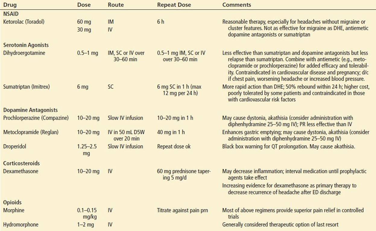
The established efficacy and selective pharmacology of the triptans have earned this group of drugs a central role in the abortive management of migraine over the past decade. Subcutaneous sumatriptan, the putative gold standard, is highly effective if administered early after headache onset. This medication is best avoided in patients with cardiovascular risk factors and those who are pregnant. Unpleasant adverse events such as palpitations and flushing frequently accompany this medication, and headache recurrence after ED discharge occurs in nearly two-third of patients who initially experience relief. This medication should be reserved for those who have had a previous favorable experience with it.
Newer triptans offer additional dosage forms and longer half-lives, though to date, sumatriptan is the only parenteral option. ED use of oral triptans may be limited by nausea, vomiting, and delayed gastrointestinal absorption, a frequent feature of migraine attacks.
Dihydroergotamine, a more tolerable form of the original ergot alkaloid, is best administered as a slow intravenous drip following pretreatment with an antiemetic, preferably one of the antiemetics with antimigraine properties discussed below. Dihydroergotamine should not be used in those with cardiovascular risk factors, those who are pregnant, or taking macrolide antibiotics or antiretrovirals.
The intravenous dopamine antagonists metoclopramide, droperidol, and prochlorperazine can be used as monotherapy or as adjunctive therapy in combination with other abortive agents (21). The efficacy of these agents has been established in multiple systematic reviews (21) though the therapeutic mechanism of action is not well understood. The dopamine antagonists are well tolerated and highly effective with few practical contraindications. Akathisia, which may at times be severe enough to interfere with the therapeutic plan, can be mitigated with the coadministration of 25 to 50 mg of intravenous diphenhydramine.
Parenteral nonsteroidal anti-inflammatory drugs or salicylates may be used as primary or adjunctive therapy for acute migraine. Caution should be exercised in patients who have taken substantial doses of over-the-counter medication prior to ED presentation, which is not uncommon.
Corticosteroids such as dexamethasone decrease the frequency of headache recurrence after ED discharge, though with a number-needed-to-treat of 10, this class of medication may not be appropriate for every patient (21). Unfortunately, there is no evidence to guide optimal dosage and duration of therapy with corticosteroids. There is a belief that patients who present with status migrainosus (unremitting migraine for greater than 72 hours) are best served by a course of corticosteroids lasting at least several days.
Cluster
High-flow 100% oxygen is highly effective for cluster headache and should be administered as soon as the patient with cluster headache enters the ED (13). Dihydroergotamine (DHE) and sumatriptan are also effective in the ED management of cluster. However, concerns about coronary vasospasm limit the use of these drugs in patients with any cardiovascular risk factors. Limited evidence suggests the dopamine antagonists may be effective as well. On discharge, patients with an exacerbation of cluster should receive verapamil, starting at 80 mg tid, if there are no contraindications. A tapering course of steroids should be prescribed, and close follow-up arranged (13).
Tension-Type Headache
Many patients with tension-type headache can be managed with oral analgesics, such as nonsteroidal medication, aspirin, or acetaminophen. Those who require parenteral medication will often respond to migraine medications. The parenteral dopamine antagonists are highly effective for tension-type headache. Subcutaneous sumatriptan should be reserved for those patients with tension-type headache who have previously experienced migraine headache.
Trigeminal Neuralgia
Trigeminal neuralgia syndrome responds best to carbamazepine, which has a number-needed-to-treat of less than two (23). Oxcarbazepine or baclofen may be used as second-line agents. Nonsteroidals or opioids may be necessary until other medications take effect. Facial nerve blocks with long-acting local anesthetics will provide immediate but short-lived relief in selected patients. All patients should be referred to specialty care for a diagnostic workup, which may reveal an anatomic cause such as multiple sclerosis, tumor, or neurovascular compression.
Coital Headache
Once subarachnoid hemorrhage has been excluded, coital headache patients require explanation, reassurance, and referral. It is reasonable to offer the patient a therapeutic trial of indomethacin 50 mg PO, taken 1 hour before intercourse.
Posttraumatic Headache
Once the diagnosis has been confirmed by a normal CT or MRI, posttraumatic headache patients should be educated and reassured. Confirmation that this is indeed “real” and that it will improve with time is an important therapeutic intervention. Patients should be referred to a neurologist.
Headache Associated with Low Cerebrospinal Fluid Pressure
Traditional management of post-LP headache includes fluid replacement and bed rest. Other treatments include administration of intravenous caffeine and performance of an epidural blood patch, although a solid evidence base for these practices is lacking (24). Unless individuals are incapacitated by the headache, they can generally be managed as outpatients.
Idiopathic Intracranial Hypertension
Acute symptoms can be relieved with lumbar puncture, with the goal of removing enough CSF to normalize pressure. Acute headache may be relieved with migraine medication. Control of pain does not treat the underlying threat to the patient’s vision. Patient should be prescribed acetazolamide and referred for specialty care. Modest reduction in weight may improve prognosis. If acuity worsens in spite of maximal medical management, an optic sheath decompression or ventricular shunt will be needed (10).
Temporal Arteritis
An erythrocyte sedimentation rate and C-reactive protein should be used to exclude temporal arteritis in patients older than 50 who present with new-onset headache. Because untreated patients are at risk of blindness, all patients with the working diagnosis of temporal arteritis should immediately receive corticosteroids and be referred for temporal artery biopsy.
Headache in Immunocompromised HIV Patients
Immunocompromised HIV-positive patients presenting with headache are at high risk for opportunistic infections. Emergency clinicians should have a low threshold for initiating a diagnostic workup, including neuroimaging to look for mass lesions, lumbar puncture to evaluate for meningitis, and a serum cryptococcal antigen, which is highly sensitive for cryptococcal meningitis (4).
Chronic Headache
Individuals with chronic headache are among the most challenging to manage in a busy ED because their underlying pain disorder cannot be solved from the acute setting. Though tempting to treat the acute pain with parenteral opioids, this course of action will not improve the patient’s long-term course. Within the heterogeneous group of patients with chronic headache, one must try to identify treatable subsets, such as those with medication overuse headache and untreated psychiatric comorbidities, and make the appropriate referrals. Comprehensive headache management programs with a multidisciplinary approach and individualized treatment plan have demonstrated some benefit in a select group of frequent ED users with chronic headache who are motivated to attend specialized headache classes (25). In general, patients with chronic headache, including chronic tension-type headache, chronic migraine, and medication overuse headache can benefit most from the expertise of a headache or pain specialist.
CRITICAL INTERVENTIONS
• Consider full headache differential diagnosis in all patients who present to ED with headache.
• Perform head CT and LP for patients who present with acute onset of “worst headache” to exclude subarachnoid hemorrhage.
• Administer antibiotics promptly in suspected bacterial meningitis.
• Treat pain appropriately and promptly.
• Help patients access suitable outpatient care.
Common Pitfalls
• Failure to diagnose the sentinel leak of SAH until a major bleed occurs subsequently.
• Misdiagnosis of migraine as hypertension or chronic sinusitis.
• Failure to diagnose and initiate treatment in patients with ongoing cluster headache because the acute headache has resolved prior to evaluation by the emergency clinician.
• Failure to diagnose and treat migraine appropriately either because a large tension-type component is present or because the headache is not hemicranial.
• Presuming that response to specific medications necessarily defines the etiology of a headache. The headache of brain tumors, aneurysms, and SAH may respond to a migraine regimen.
REFERENCES
1. Goldstein JN, Camargo CA Jr, Pelletier AJ, et al. Headache in United States emergency departments: Demographics, work-up and frequency of pathological diagnoses. Cephalalgia. 2006;26(6):684–690.
2. Headache Classification Subcommittee of the International Headache Society. Classification of Headache Disorders. 2nd ed. Cephalalgia: 2004;24(Suppl 1):1–151.
3. Goadsby PJ, Charbit AR, Andreou AP, et al. Neurobiology of migraine. Neuroscience. 2009;161(2):327–341.
4. Gladstone J, Bigal ME. Headaches attributable to infectious diseases. Curr Pain Headache Rep. 2010;14(4):299–308.
5. Attia J, Hatala R, Cook DJ, et al. The rational clinical examination. Does this adult patient have acute meningitis? JAMA. 1999;282(2):175–181.
6. Pachner AR, Steiner I. Lyme neuroborreliosis: Infection, immunity, and inflammation. Lancet Neurol. 2007;6(6):544–552.
7. Perry JJ, Stiell IG, Sivilotti ML, et al. Clinical decision rules to rule out subarachnoid hemorrhage for acute headache. JAMA. 2013;310(12):1248–1255.
8. Cady RK, Dodick DW, Levine HL, et al. Sinus headache: A neurology, otolaryngology, allergy, and primary care consensus on diagnosis and treatment. Mayo Clin Proc. 2005;80(7):908–916.
9. Valentinis L, Tuniz F, Valent F, et al. Headache attributed to intracranial tumours: A prospective cohort study. Cephalalgia. 2010;30(4):389–398.
10. Friedman DI. Pseudotumor cerebri presenting as headache. Expert Rev Neurother. 2008;8(3):397–407.
11. Waldman CW, Waldman SD, Waldman RA. Giant cell arteritis. Med Clin North Am. 2013;97(2):329–335.
12. Seifert TD, Evans RW. Posttraumatic headache: A review. Curr Pain Headache Rep. 2010;14(4):292–298.
13. Nesbitt AD, Goadsby PJ. Cluster headache. BMJ. 2012;344:e2407.
14. Schwartz BS, Stewart WF, Simon D, et al. Epidemiology of tension-type headache. JAMA. 1998;279(5):381–383.
15. Edmeads J. Emergency management of headache. Headache. 1988;28(10):675–679.
16. Kermani TA, Schmidt J, Crowson CS, et al. Utility of erythrocyte sedimentation rate and C-reactive protein for the diagnosis of giant cell arteritis. Semin Arthritis Rheum. 2012;41(6):866–871.
17. Perry JJ, Stiell IG, Sivilotti ML, et al. Sensitivity of computed tomography performed within six hours of onset of headache for diagnosis of subarachnoid haemorrhage: Prospective cohort study. BMJ.2011;343:d4277.
18. Brown SC, Brew S, Madigan J. Investigating suspected subarachnoid haemorrhage in adults. BMJ. 2011;342:d2644.
19. Edlow JA, Panagos PD, Godwin SA, et al. Clinical policy: Critical issues in the evaluation and management of adult patients presenting to the emergency department with acute headache. Ann Emerg Med.2008;52(4):407–436.
20. Turnbull DK. Shepherd DB. Post-dural puncture headache: Pathogenesis, prevention and treatment. Br J Anaesth. 2003;91(5):718–729.
21. Sumamo Schellenberg E, Dryden DM, Pasichnyk D, et al. Acute migraine treatment in emergency settings. Comparative Effectiveness Review No. 84. Rockville (MD): Agency for Healthcare Research and Quality (US); 2012. Available online at http://www.ncbi.nlm.nih.gov/books/NBK115368/.
22. Evans RW, Friedman BW. Headache in the emergency department. Headache. 2011;51(8):1276–1278.
23. Gronseth G, Cruccu G, Alksne J, et al. Practice parameter: The diagnostic evaluation and treatment of trigeminal neuralgia (an evidence-based review): Report of the Quality Standards Subcommittee of the American Academy of Neurology and the European Federation of Neurological Societies. Neurology. 2008;71(15):1183–1190.
24. Basurto Ona X, Martínez García L, Solà I, et al. Drug therapy for treating post-dural puncture headache. Cochrane Database Syst Rev. 2011;(8):CD007887.
25. Harpole LH, Samsa GP, Jurgelski AE, et al. Headache management program improves outcome for chronic headache. Headache. 2003;43(7):715–724.