
Jonathan A. Edlow
Headache is a common presenting complaint in patients presenting to the emergency department (ED). Subarachnoid hemorrhage (SAH), which comprises less than 1% of ED headaches, is a true medical emergency in that effective time-dependent treatment exists (1,2). Deciding which patients to evaluate for SAH and the components of that evaluation is therefore a critical issue for emergency physicians. The classic presentation of a thunderclap headache in a patient with a stiff neck and an abnormal mental status presents little difficulty; however approximately 40% of patients present awake, alert, and neurologically intact. Misdiagnosis in this group results in worse outcomes (1,2).
This chapter is limited to atraumatic SAH, the most common cause of which is a ruptured cerebral aneurysm or arteriovenous malformation (AVM). Approximately 2% of the general population harbors cerebral aneurysms and yet the incidence of SAH is ∼8 to 10 per 100,000 (2,3). Therefore, the vast majority of aneurysms never rupture. The fact that the vast majority of aneurysms are incidental has important diagnostic implications for emergency physicians (4).
Although most unruptured aneurysms are asymptomatic, they can present with mass lesion (classically the pupil-involving third nerve palsy), headache, seizure, and cerebral ischemia. Presentation of an unruptured aneurysm as isolated thunderclap headache without SAH is rare. Aneurysmal rupture is associated with a surge in intracranial pressure (ICP) that can lead to decreased cerebral perfusion resulting in syncope, any neurologic deficit or in some cases, sudden death. Mortality rates on the first day and first month are 12% and 40% respectively (1–3).
Blood from a ruptured aneurysm is nearly always found at the base of the brain in the basal cisterns. Emergency physicians should be aware of two other patterns (Fig. 154.1A–C). The first is a perimesencephalic SAH, in which the blood is localized to the prepontine cistern. These bleeds are nonaneurysmal and are thought to be due to venous bleeding. Angiography is negative and patient outcomes are excellent (5). The second type is the convexal SAH in which the blood is found in the subarachnoid space high up on the hemispheres. These bleeds are also nonaneurysmal and are usually due to amyloid angiopathy, reversible cerebral vasoconstriction syndrome (RCVS), or hypertension (6). The outcomes are a function of the underlying condition more than the SAH by itself (6).

FIGURE 154.1 A: White arrows show subarachnoid blood in the basal cisterns, the typical location for a patient with an aneurysmal SAH. The red arrows show mild early hydrocephalus. B: White arrowsshow blood in the interpeduncular cistern (blood was also present in the prepontine cistern) in a patient with a perimesencephalic hemorrhage. He had a negative angiogram. C: Arrows show blood in the subarachnoid space high on the convexities in a patient with a bleed due to amyloid angiopathy. This location is also typical for reversible cerebral vasoconstriction bleeds. D: This is a sagittal cut of source image from a CT angiogram showing an aneurysm of the anterior communicating artery (yellow arrows), a very common location for an aneurysm.
Both the American Heart Association (AHA, 2012) and the European Stroke Organization (ESO, 2013) have published recent evidence-based guidelines on SAH including detailed recommendations about the management beyond the ED phase of care (3,7).
CLINICAL PRESENTATION
There is a wide spectrum of presentations ranging from a nonacute onset headache in a neurologically intact patient to sudden death. This “bell-shaped curve” range of presentations is important for emergency physicians to know. On the right side of the curve are ill patients with an abnormal mental status and other clear-cut focal neurologic deficits. Although the specific diagnosis may not be immediately obvious, the fact that they require diagnostic testing is clear. Computed tomography (CT) of the brain is almost always an initial part of that workup and will usually reveal the problem in severely affected SAH patients. In the middle of the bell-shaped curve are patients presenting with the classic abrupt onset of a severe, unusual headache. Often, they will have had syncope at onset, be vomiting, or have prominent neck pain or stiffness. In this group too, the need for a diagnostic evaluation for SAH is clear.
Importantly however, physicians must understand the left-hand side of the curve. These patients complain of various less typical (but well described in the literature) symptoms. The headache of SAH usually is abrupt in onset, severe in intensity, and unlike other prior headaches in quality; however any of these aspects may be absent in an individual patient. Of this latter group, physicians ultimately must use their clinical judgment to decide which patients need a workup for SAH. This clinical judgment must be informed by knowledge of the range of presentations.
These presentations include abrupt onset isolated neck pain (suggesting neck strain or cervical arthritis), neck pain and low-grade fever or meningismus (viral syndrome or viral meningitis), retro-orbital headache (sinusitis), headache with an elevated blood pressure, chest pain or an abnormal electrocardiogram (hypertensive emergency, Takosubo’s cardiomyopathy, or other cardiac cause), and prominent vomiting (gastrointestinal conditions) and headache with confusion or mildly altered level of consciousness (intoxication or psychiatric diagnosis) (1,2,8–10). The severity of the SAH is often conveyed by using a clinical severity score (Table 154.1).
TABLE 154.1
Common Grading Scales Used in SAH Patients

One misconception is that the onset of symptoms is always with strenuous activity. In fact, onset during sleep or quiet activity is common (1,2). The headache may not be “worst of life.” As a general rule, one must consider the likelihood of other possibilities in the differential diagnosis, understanding that one cannot confidently make a diagnosis of migraine or tension-type headache after a first ever headache. Although patients often use the term “sinusitis,” this is an unusual cause of an acute severe headache (9).
Historically, misdiagnosis of SAH is reported to occur ∼20% of cases (1,2). Even in the modern era of near-universal CT access in EDs and educational focus on misdiagnosis of SAH, the rate of misdiagnosis of SAH in the ED remains 5% to 7% (2,10,11).
Based on a prospective cohort study of nearly thunderclap headache patients presenting to six Canadian EDs, a sensitive clinical decision rule has been proposed (12). Of the 2000 patients, 130 (6.5%) had SAH, making the study population representative of the spectrum seen in practice. Using recursive partitioning, they developed three clinical decision rules all of which were 100% sensitive for SAH that would have reduced the rates of investigations from 83% (baseline) to between 64% and 74% (12). The one rule that did not include the variable “arrival by ambulance” identified all SAH patients (all had at least one of the four).
• Age >40 years
• Complaint of neck pain or stiffness
• Witnessed loss of consciousness
• Onset with exertion
This clinical rule validated well in an independent population of patients although it was used somewhat differently in the second study (13).
The concept of warning bleeds is also important to consider. Some patients with SAH will have had a severe unusual headache in the days to weeks prior to being diagnosed with their SAH. These headaches have been called warning (or sentinel) bleed (or headache). Evidence suggests that these are actually small SAHs that were either not recognized by the patient or by the physician and so no evaluation (or an incomplete evaluation) was done. In a pooled analysis of 813 patients with thunderclap headache who had a negative CT and LP, none were subsequently diagnosed with SAH or died suddenly during follow-up (14). This suggests the standard workup will correctly diagnose warning bleeds.
One final point should be made regarding which patients to evaluate for SAH is the patients’ response to analgesics administered for their headache. Because there is a final common pathway for pain sensation in the head, a patient’s favorable response to any analgesic, narcotic or otherwise, including triptans, should not be used to make diagnostic inferences. The headache of patients with serious causes including SAH can improve or resolve with various analgesics including triptans.
DIFFERENTIAL DIAGNOSIS
Even among patients presenting with a thunderclap headache, only ∼10% will have an SAH, most commonly from a ruptured aneurysm or AVM (2). The majority of patients with thunderclap headache have migraine and tension-type headache. A very small but important minority will have far less common but serious and treatable causes such as arterial dissection, cerebral venous sinus thrombosis (CVST), RCVS, pituitary apoplexy, and other vasculopathies.
The presence of multiple thunderclap headaches in a neurologically intact patient is nearly pathognomonic for RCVS (15). Table 154.2 shows the differential diagnosis of important treatable and serious causes of headache, of thunderclap headache, and of the vascular lesions responsible for nontraumatic SAH. Table 154.3 summarizes reasons for misdiagnosis.
TABLE 154.2
Differential Diagnoses Relevant to Patients with SAH

TABLE 154.3
Reasons for Misdiagnosis of SAH

ED EVALUATION
For decades, the standard ED evaluation has been CT followed by lumbar puncture (LP) if the CT is nondiagnostic. As of mid-2013, in guidelines published by ACEP, the AHA and the ESO, this recommendation stands (3,7,16). Large studies confirm the safety and the sensitivity of this protocol (14,17).
A key point is to realize that the sensitivity of CT and LP changes with the time elapsed from the onset of the bleed and the performance of the test (1,2,8). CT is an excellent test early but the sensitivity degrades rapidly with time. LP and cerebrospinal fluid (CSF) analysis shows blood early in the course of a SAH. Increasing numbers of RBCs in the fourth tube and low percent change in RBCs from tube 1 to tube 4 are the most useful to predict SAH (18,19). Xanthochromia (the yellow color of CSF due to hemoglobin breakdown) takes time to develop but is usually present for up to 2 weeks after the bleed. Nearly all North American hospital laboratories use visual inspection for xanthochromia (20). Studies support the notion that measuring xanthochromia by visual inspection is a valuable method and that spectrophotometry, while more sensitive, lacks specificity (21,22).
Newer data about older techniques (early CT) and greater availability and data about newer techniques—CT angiography (CTA), magnetic resonance imaging (MRI), and MR angiography (MRA)—are beginning to impact the diagnostic paradigm for SAH (Fig. 154.1D). Each of these testing strategies has both advantages and limitations compared to the time-honored CT followed by LP (Table 154.4). Although official guidelines do not reflect it, the preponderance of data suggest that a normal CT performed within 6 hours of headache onset and interpreted by an experienced attending level radiologist excludes SAH with close to 100% sensitivity and that LP is no longer required in this setting (23–25).
TABLE 154.4
Diagnostic Strategies

A recent study reported instances of CT-negative LP-positive patients (CT done within 6 hours) but the data as reported make it unclear how many of their “SAH” patients truly had SAH nor how many patients in the early presenting group had positive CT scans. Another study of early CT-negative patients found no patients with aneurysmal SAH among those presenting early (26).
For the strategies that include an angiogram, it is important to recognize that many of these aneurysms will be incidental. Approximately 2% of the general population has cerebral aneurysms. Since most patients with SAH have a positive CT, a “positive” CTA in a CT-negative patient makes it even more likely that the aneurysm is incidental. A CTA or MRA that diagnoses (what is actually) an incidental lesion can lead to an unnecessary procedure on the one hand, or a lot of anxiety and subsequent imaging on the other (4). For CTA, there are also considerations of radiation and dye complications (4).
Whatever diagnostic algorithm is used, one must factor in time from headache onset and understand the limitations of the tests.
KEY TESTING
• Noncontrast brain CT
• LP if CT is nondiagnostic
• Consider other testing algorithms; however each has limitations that must be explicitly addressed (Table 154.4)
ED MANAGEMENT
As always, initial attention to airway, breathing and circulation takes priority. Most patients with SAH do not require intubation. If an SAH patient needs airway control, standard rapid sequence intubation is used. For patients being transferred to another facility, one must consider the possible need for airway control over the duration of time of the transfer.
With regard to blood pressure control, the data are poor quality in terms of a specific target blood pressure. The 2012 AHA recommends that the systolic blood pressure be kept at less than 160 mm Hg and the 2013 European ESO recommends that the systolic pressure be kept below 180 mm Hg. However many practitioners aim for a lower number. Both the AHA and ESO caution that the data are poor and that more important than a specific one-size-fits-all pressure, the clinicians must balance issues such as cerebral perfusion, risk of rebleeding, premorbid blood pressure, and other factors. Whatever target is used, use an antihypertensive intravenous easy-to-titrate drug such as nicardipine, esmolol, or labetalol.
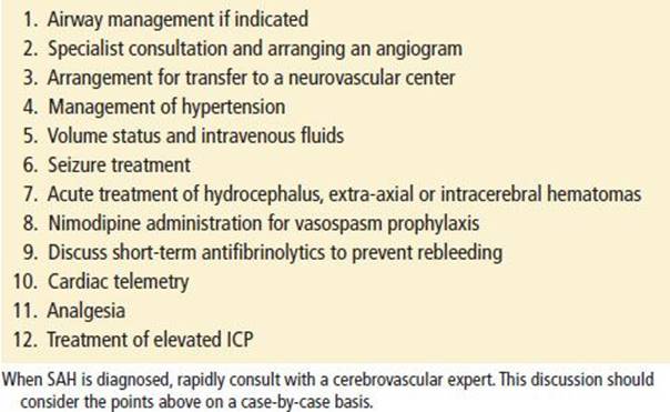
With the ABCs covered, and the causative vascular lesion defined as above, there are a number of other issues that the emergency physician must consider. Pain and anxiety should be treated. Patients should be placed on telemetry and have a 12-lead electrocardiogram. Intravenous access should be established and blood should be sent for testing to the laboratory for routine hematologic and chemistry tests as well as tests of clotting and troponins. Other management issues should all be discussed and resolved with the accepting neurovascular specialist. These include treatment of seizures and hydrocephalus, vasospasm prophylaxis, the timing and choice of cerebrovascular imaging, and administration of short-term antifibrinolytics (Table 154.5).
TABLE 154.5
Treatment Approach
If a patient with SAH deteriorates while still in the ED, repeat the CT. There may be little to do for a massive rebleed or large territorial cerebral infarction from vasospasm; however, in some cases, acute hydrocephalus or a space-occupying bleed may be amenable to surgical intervention. In cases in which the CT is unchanged, consider nonconvulsive status epilepticus, cerebral hypoperfusion from overly aggressive lowering of the blood pressure or side effects of analgesics or anxiolytics.
Controversies
• Avoiding LP in the evaluation of SAH in patients who present typically with thunderclap headache and have a noncontrast CT scan performed within 6 hours of headache onset that is read as negative by an attending radiologist
• Ideal level of blood pressure control is unclear
• Seizures should be treated; however, prophylactic anticonvulsants are not indicated
• Administration of short-term antifibrinolytics (after diagnosis but before definitive treatment) to prevent early rebleeding may be considered
CRITICAL INTERVENTIONS
• Control airway using standard rapid sequence technique if clinically indicated
• Whatever diagnostic algorithm is used, follow it to its completion
• When controlling blood pressure, factor in premorbid blood pressure and current cerebral perfusion pressure
• Repeat CT scan for SAH patients who deteriorate while in the ED to identify a reversible cause
• Patients with SAH should be admitted to a facility with experience in SAH
DISPOSITION
Patients with SAH should be admitted to an ICU. Based on data over the past decade, the AHA recommends that patients with aneurysmal SAH be treated in a high-volume center (defined as one that manages >35 cases per year) (3). The improved outcomes likely result not only from the technical expertise of the interventionalist and/or surgeon, but also the multidisciplinary neurointensive care that is available. Increasing numbers of SAH patients are being treated by endovascular coiling.
Common Pitfalls
• Failure to consider SAH in a patient with headache because they “do not appear ill” or “have a normal level of consciousness and neurologic examination,” that is, failure to understand the spectrum of presentations
• Failure to fully workup patients with thunderclap headache for SAH
• Assuming that a patient with a severe headache whose pain has improved with analgesics does not have a serious cause of headache
• Failure to understand the limitations of CT, LP, and MRI, especially with regard to “timing” of the test compared to the onset of symptoms
• Failure to refer a patient with an SAH to a high-volume neurovascular center where patient outcomes are demonstrably better
REFERENCES
1. Edlow JA, Caplan LR. Avoiding pitfalls in the diagnosis of subarachnoid hemorrhage. N Engl J Med. 2000;342(1):29–36.
2. Edlow JA, Malek AM, Ogilvy CS. Aneurysmal subarachnoid hemorrhage: Update for emergency physicians. J Emerg Med. 2008;34(3):237–251.
3. Connolly ES Jr, Rabinstein AA, Carhuapoma JR, et al. Guidelines for the management of aneurysmal subarachnoid hemorrhage: A guideline for healthcare professionals from the American Heart Association/American Stroke Association. Stroke. 2012;43(6):1711–1737.
4. Edlow JA. What are the unintended consequences of changing the diagnostic paradigm for subarachnoid hemorrhage after brain computed tomography to computed tomographic angiography in place of lumbar puncture? Acad Emerg Med. 2010;17(9):991–995; discussion 996–997.
5. Canovas D, Gil A, Jato M, et al. Clinical outcome of spontaneous non-aneurysmal subarachnoid hemorrhage in 108 patients. Eur J Neurol. 2012;19(3):457–461.
6. Beitzke M, Gattringer T, Enzinger C, et al. Clinical presentation, etiology, and long-term prognosis in patients with nontraumatic convexal subarachnoid hemorrhage. Stroke. 2011;42(11):3055–3060.
7. Steiner T, Juvela S, Unterberg A, et al. European stroke organization guidelines for the management of intracranial aneurysms and subarachnoid haemorrhage. Cerebrovasc Dis. 2013;35(2):93–112.
8. Edlow JA, Samuels O, Smith WS, et al. Emergency neurological life support: Subarachnoid hemorrhage. Neurocrit Care. 2012;17(Suppl 1): S47–S53.
9. Pope JV, Edlow JA. Avoiding misdiagnosis in patients with neurological emergencies. Emerg Med Int. 2012;2012:949275.
10. Yamada T, Natori Y. Evaluation of misdiagnosed cases of subarachnoid hemorrhage and causal factors for misdiagnosis. J Stroke Cerebrovasc Dis. 2013;22(4):430–436.
11. Vermeulen MJ, Schull MJ. Missed diagnosis of subarachnoid hemorrhage in the emergency department. Stroke. 2007;38(4):1216–1221.
12. Perry JJ, Stiell IG, Sivilotti ML, et al. High risk clinical characteristics for subarachnoid haemorrhage in patients with acute headache: Prospective cohort study. BMJ. 2010;341:c5204.
13. Mark DG, Hung YY, Offerman SR, et al. Nontraumatic subarachnoid hemorrhage in the setting of negative cranial computed tomography results: External validation of a clinical and imaging prediction rule. Ann Emerg Med.2013;62(1):1–10 e1.
14. Savitz SI, Levitan EB, Wears R, et al. Pooled analysis of patients with thunderclap headache evaluated by CT and LP: Is angiography necessary in patients with negative evaluations? J Neurol Sci.2009;276(1–2):123–125.
15. Singhal AB, Hajj-Ali RA, Topcuoglu MA, et al. Reversible cerebral vasoconstriction syndromes: Analysis of 139 cases. Arch Neurol. 2011;68(8):1005–1012.
16. Edlow JA, Panagos PD, Godwin SA, et al. Clinical policy: Critical issues in the evaluation and management of adult patients presenting to the emergency department with acute headache. Ann Emerg Med.2008;52(4):407–436.
17. Perry JJ, Spacek A, Forbes M, et al. Is the combination of negative computed tomography result and negative lumbar puncture result sufficient to rule out subarachnoid hemorrhage? Ann Emerg Med.2008;51(6):707–713.
18. Czuczman AD, Thomas LE, Boulanger AB, et al. Interpreting red blood cells in lumbar puncture: Distinguishing true subarachnoid hemorrhage from traumatic tap. Acad Emerg Med. 2013;20(3):247–256.
19. Gorchynski J, Oman J, Newton T. Interpretation of traumatic lumbar punctures in the setting of possible subarachnoid hemorrhage: Who can be safely discharged? Cal J Emerg Med. 2007;8(1):3–7.
20. Edlow JA, Bruner KS, Horowitz GL. Xanthochromia. Arch Pathol Lab Med. 2002;126(4):413–415.
21. Dupont SA, Wijdicks EF, Manno EM, et al. Thunderclap headache and normal computed tomographic results: Value of cerebrospinal fluid analysis. Mayo Clin Proc. 2008;83(12):1326–1331.
22. Perry JJ, Sivilotti ML, Stiell IG, et al. Should spectrophotometry be used to identify xanthochromia in the cerebrospinal fluid of alert patients suspected of having subarachnoid hemorrhage? Stroke.2006;37(10):2467–2472.
23. Backes D, Rinkel GJ, Kemperman H, et al. Time-dependent test characteristics of head computed tomography in patients suspected of nontraumatic subarachnoid hemorrhage. Stroke. 2012;43(8):2115–2119.
24. Edlow JA, Fisher J. Diagnosis of subarachnoid hemorrhage: Time to change the guidelines? Stroke. 2012;43(8):2031–2032.
25. Perry JJ, Stiell IG, Sivilotti ML, et al. Sensitivity of computed tomography performed within six hours of onset of headache for diagnosis of subarachnoid haemorrhage: Prospective cohort study. BMJ.2011;343:d4277.
26. Horstman P, Linn FH, Voorbij HA, et al. Chance of aneurysm in patients suspected of SAH who have a ‘negative’ CT scan but a ‘positive’ lumbar puncture. J Neurol. 2012;259(4):649–652.