
Landen Rentmeester and Richard F. Clark
Antipsychotic agents encompass a class of pharmaceuticals originally designed to treat the psychotic features of mental illnesses such as schizophrenia, schizoaffective disorder or acute psychosis but have also been found useful to treat nausea, agitation, hiccoughs, and tics. These medications, especially the older typical antipsychotics, have also been called “neuroleptics” or “major tranquilizers.” Antipsychotics are generally divided into the “typical” and “atypical” agents. The “typical” agents were the first antipsychotics to be developed, and were effective in treating hallucinations, agitation, delusions, and behavioral disturbances, the “positive” symptoms of schizophrenia. These older medications are also associated with the development of motor disturbances collectively known as the extrapyramidal syndrome (EPS). The newer “atypical” antipsychotic medications were developed to minimize extrapyramidal effects but were also found to be more effective in treating the “negative” symptoms of schizophrenia, such as alogia, avolition, flattened affect, and social withdrawal (1).
Butyrophenones (e.g., haloperidol), dephenylbutylpiperidines (e.g., pimozide), and phenothiazines (e.g., thioridazine) are the three major classes of the typical antipsychotics, whereas the benzamides (e.g., metoclopramide), diazepines (e.g., olanzapine), indoles (e.g., risperidone), and quinolinones (e.g., aripiprazole) are the four major classes of atypical antipsychotics. The antipsychotic action of each of these medications is thought to be due to blockade of excessive dopamine–mediated neurotransmission in the central nervous system (CNS) (2). Antipsychotic medications also have pharmacologic effects outside of the dopaminergic pathways, blocking alpha-adrenergic (α1), histaminergic (H1), muscarinic (M1), and serotonergic (5-HT2A) receptors (3).
The atypical agents have lower and more selective (mesolimbic) D2 potency and higher M1 and/or 5-HT2A antagonist activity than the typical agents (3). Although higher doses are required for antipsychotic effect, they are less likely to cause acute dystonia and tardive dyskinesia and have minimal or no effect on prolactin secretion. Only clozapine, olanzapine, and quetiapine have significant antimuscarinic activity. Atypical antipsychotics cause EPS less frequently than the older agents; whereas lurasidone may be more likely to promote extrapyramidal symptoms than other atypical agents (4). For unclear reasons, all atypical antipsychotics can cause hyperglycemia and an FDA black box warning recommends that diabetics and those at risk for diabetes (e.g., obese patients and those with a family history of diabetes) be monitored for this complication at the start of therapy. Atypical antipsychotics, especially olanzapine and clozapine, seem to put patients at risk for “metabolic syndrome,” a constellation of weight gain, insulin resistance, glucose intolerance, and hyperlipidemia (3). Among the first-generation antipsychotics agents, chlorpromazine is most frequently associated with weight gain (4).
Most antipsychotics are much more potent H1 blockers than diphenhydramine. Antimuscarinic (M1) potency correlates directly with sedation and inversely with the incidence of EPS. Chlorpromazine, chlorprothixene, and thioridazine have potent sedative effects, whereas haloperidol, zotepine, and chlorpromazine are commonly associated with EPS (4).
Hypotension and miosis are often seen with both therapeutic dosing and overdose of both typical and atypical antipsychotics, a result of peripheral α-adrenergic blockade. Hypotensive effects are primarily orthostatic and systolic in nature. Concomitant therapy with either β-receptor agonists (e.g., epinephrine) or β-receptor blockers (e.g., propranolol) may result in severe hypotension. All the antipsychotics, particularly chlorpromazine, clozapine, and loxapine lower the seizure threshold and can induce epileptiform encephalogram (EEG) discharge patterns at therapeutic dosages and in overdose, probably because of inhibition of γ-aminobutyric acid (GABA) receptors, norepinephrine reuptake, or antimuscarinic effects.
Antipsychotics also have dose-related effects on cellular membrane ion channels (3). Phenothiazines (particularly chlorpromazine, thioridazine and its metabolite mesoridazine) block sodium channels, which in overdose may cause local anesthetic or quinidine-like (class Ia) effects and result in dysrhythmias, myocardial depression, and delayed depolarization with conduction disturbances, especially QRS prolongation. QRS prolongation is most often seen with quetiapine but is rarely an isolated finding with atypical agents (3). Droperidol, haloperidol, sertindole (Serlect), thioridazine, most atypical antipsychotics, and most other antipsychotics also block potassium channels, resulting in delayed repolarization with QT interval prolongation. Higher IV doses of the atypical agents such as ziprasidone may also cause this effect. Electrocardiogram (ECG) changes such as prolonged PR and QT intervals, ST-segment depression, T-wave abnormalities (biphasic, blunting, inversion, notching, widening), and increased U waves occur with both therapeutic and excessive doses (3,6). Reports of QT prolongation leading to torsades de pointes (polymorphic ventricular tachycardia), ventricular tachycardia (VT), and death prompted a controversial box warning for IV droperidol in 2001 and IV haloperidol in 2007 that an ECG be obtained prior to its use, that it not be given to those with QT prolongation (>440 ms in males and 450 ms in females), and for cardiac monitoring for 2 to 3 hours after its use (7). Ventricular dysrhythmias, asphyxia from aspiration, or respiratory depression from seizures have been postulated as the cause of sudden death with therapeutic doses of antipsychotics, particularly phenothiazines (6).
Gastrointestinal (GI) absorption of antipsychotics is relatively slow and unpredictable, with peak levels occurring 2 to 4 hours after ingestion. Bioavailability after parenteral administration is two to ten times greater than with oral dosing because presystemic (GI and hepatic) metabolism occurs after ingestion. Antipsychotics are highly bound to plasma proteins, are highly lipophilic, and have large volumes of distribution (10 to 40 L/kg). Thus, therapeutic serum concentrations are low (one to several hundred ng/mL), and drug accumulates in the brain and fat and readily crosses the placenta.
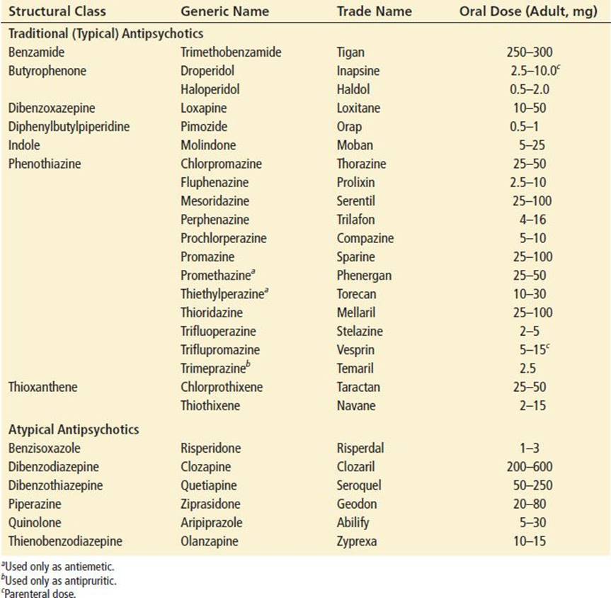
Therapeutic doses vary widely (see Table 338.1) and are adjusted on the basis of clinical response and side effects, not by monitoring drug levels. Pharmacologic effects generally last 24 hours or more, allowing once or twice daily dosing. Elimination occurs slowly by hepatic metabolism, with serum concentration half-lives averaging 20 to 40 hours. Hepatic metabolism yields multiple metabolites, some of which are pharmacologically active (aripiprazole, risperidone, quetiapine, thioridazine and clozapine are known to have active metabolites). Metabolites are eliminated by urinary and, to some extent, biliary excretion and can be detected in the urine for more than a month after the cessation of chronic therapy. Concomitant use of agents that inhibit or stimulate hepatic cytochrome oxidases (CYP isoenzymes) can increase or decrease, respectively, the blood level and effect of these agents.
TABLE 338.1
Antipsychotic Agents
Side effects of therapeutically dosed antipsychotics include antimuscarinic sequelae (see Chapter 344, “Anticholinergic Agents”), extrapyramidal symptoms (EPS), neuroleptic malignant syndrome (NMS), hypotension, and sedation (3,4). Clozapine can cause agranulocytosis, myocarditis, and cardiomyopathy (3).
Antipsychotic agents have a high therapeutic index. Of the thousands of overdoses reported to poison centers each year, only 4% to 6% of patients develop serious toxicity, and <1% die. Thioridazine (because of its tricyclic antidepressant-like cardiotoxicity) or mixed overdoses are involved in most successful suicides (3,5). Toxic doses for most of these agents are not well established and vary greatly.
CLINICAL PRESENTATION
Mild poisoning includes ataxia, confusion, lethargy, and slurred speech. Hypotension, both orthostatic and diastolic, may be caused by α-adrenergic blockade. Tachycardia may develop from antimuscarinic effects or as a reflex tachycardia. Miosis due to α blockade is common, but mydriasis due to antimuscarinic effects can also occur. Antimuscarinic signs and symptoms (Chapter 344, “Anticholinergic Agents”) are often present, and patients may be hyperreflexic. Prolonged PR and QT intervals and nonspecific ST-segment, T-wave, and U-wave abnormalities may be seen.
Moderate poisoning is characterized by sedation, with or without respiratory depression, and systolic hypotension accompanied by reflex tachycardia. Hyperthermia and hypothermia (less common) may be present. Paradoxical agitation, delirium, and tachypnea are reported but are likely antimuscarinic effects. Sialorrhea may be seen after clozapine overdose and at therapeutic doses.
In severe poisoning, deep coma with loss of brainstem and deep tendon reflexes, apnea, hypotension, tachycardia, and cardiac dysrhythmias (especially with thioridazine and mesoridazine) are seen. Tachydysrhythmias, rare bradydysrhythmias, and cardiac conduction delays (especially with thioridazine and mesoridazine) may occur. QT interval prolongation and torsades de pointes are reported after haloperidol, loxapine, phenothiazine, pimozide quetiapine, risperidone, sertindole, and ziprasidone overdose and during high-dose IV droperidol and haloperidol therapy (6). Seizures are rare, except with chlorpromazine, loxapine, and clozapine (3).
The onset of symptoms of poisoning occurs within 1 to 2 hours of acute ingestion, with maximal severity usually apparent in 2 to 6 hours. Children appear more susceptible to toxicity than adults. Recovery can be expected within several hours to several days, depending on the severity and drug ingested. The mortality rate from acute poisoning is <1%.
EPS may occur early (within days), or late (after 3 months or more) after beginning antipsychotic therapy, increasing the dose, or changing the agent. Dyskinesia or acute dystonic reactions (see Chapter 340), akathisia, NMS, and parkinsonism occur early whereas tardive dyskinesia or dystonias occur late. These idiosyncratic reactions are most common with older high-potency agents. Akathisia, a subjective sensation of motor restlessness, occurs in about 20% of patients treated with traditional antipsychotics and is sometimes associated with severe parkinsonism (see Chapter 162, “Parkinsonism and Other Movement Disorders”). Transient akathisia has been observed in up to 50% of those treated with IV droperidol or prochlorperazine for headache, nausea, and vomiting. Women are affected more often than men. Patients complain of feeling restless, jittery, and tense; they cannot sit or stand still and when standing may shift their weight from foot to foot as if walking in place. Vital signs remain normal. Semipurposeful or purposeless limb movements (especially of the lower extremities), frequent shifting of body position, and tremors and myoclonic jerking may be noted. Focal perioral tremor, also known as rabbit syndrome, is a rhythmic motion of the mouth and lips, resembling the chewing movements of a rabbit. In contrast to oral tardive dyskinesia, tongue movements do not occur in rabbit syndrome.
NMS can be precipitated by concurrent illness, surgery, dehydration, heat stress, acute agitation and, very rarely, by acute overdose of antipsychotic agents. It is estimated to occur in <1% of patients treated with antipsychotics for psychiatric conditions and appears to be more common with parental therapy. Of the cases linked to atypical antipsychotics, clozapine has been implicated in many of them, despite the relative rarity of extrapyramidal symptoms with this drug (3). NMS is characterized by elevated body temperature (>38°C or 100.4°F), altered mental status ranging from confusion and delirium to coma, autonomic dysfunction, increased motor activity, and laboratory abnormalities. Autonomic disturbances include hypertension (>150/100 mm Hg; diastolic or systolic blood pressure increased 20 or 30 mm Hg above baseline, respectively), tachycardia (heart rate >90 to 100 or increased by 30 beats/min), increased respiratory rate (alternatively dyspnea, hypoxemia, or respiratory failure), diaphoresis (often profuse), sialorrhea, and incontinence. Hypotension, rather than hypertension, sometimes occurs. Although “lead pipe” rigidity is the classic motor finding, lesser increases in muscle tone, akinesia, dystonia, dyskinesia, tremor, choreiform movements, opisthotonus, and parkinsonism (cogwheel rigidity) may be present. Hyperactivity of pharyngeal muscles may result in dysarthria, dysphagia, dysphonia, and trismus. Laboratory abnormalities include leukocytosis (white blood cells >14,000/mm3 without a left shift), rhabdomyolysis (creatine phosphokinase >500 IU/mL or three times normal), and markedly elevated white blood cell count. Hyponatremia, hypernatremia, or hypokalemia may be present. Onset is typically insidious, and resolution occurs slowly, sometimes taking up to 2 weeks. Complications include aspiration pneumonia, coagulopathy, renal failure, rhabdomyolysis, myocardial infarction, heart failure, dysrhythmias, and venous thromboembolism. The mortality rate of NMS ranges from 12% to 20% and is primarily related to complications. Sequelae include dysarthria, dysphagia, myoclonus, and weakness.
DIFFERENTIAL DIAGNOSIS
Antidysrhythmic, tricyclic antidepressant, antimuscarinic, anticonvulsant, opioid, and sedative–hypnotic toxicity may all cause CNS or CV effects similar to those seen in antipsychotic poisoning. CNS infection, infarction, and trauma should also be considered in the differential diagnosis of antipsychotic poisoning.
EPS from antipsychotics may be misdiagnosed as anxiety or agitation related to an underlying psychiatric disorder. EPS can occur when beginning or increasing therapy with neuroleptic or nonneuroleptic dopamine antagonists (e.g., hydroxyzine, metoclopramide, reserpine), discontinuing or decreasing therapy with a dopamine agonist (e.g., amantadine, bromocriptine, carbidopa, levodopa, lithium, pergolide, pramipexole, ropinirole) or use of a catecholamine degradation inhibitor (e.g., catechol-O-methyltransferase inhibitors such as entacapone and tolcapone). The differential diagnosis of NMS also includes dystonic reactions, lithium, monoamine oxidase inhibitor, salicylate, stimulant, and strychnine poisoning, heat stroke, malignant catatonia, malignant hyperthermia (MH), encephalitis, meningitis, pheochromocytoma, serotonin syndrome (SS), tetanus, thyrotoxicosis, and withdrawal states.
Distinguishing NMS from MH or SS (see Chapter 343, “Serotonin Reuptake Inhibitors”) is difficult as clinical features overlap. The correct diagnosis relies primarily on the drug exposure history. Onset after general anesthesia or ketamine administration strongly suggests MH. Use of proserotonin drugs is suggestive of SS. SS also begins more abruptly and resolves more quickly than NMS (over hours rather than days). Myoclonus, hyperreflexia, and shivering are rare in NMS but common in SS.
Although specific criteria have been proposed for the diagnosis of NMS, the required elements are not universally agreed on, and patients can have NMS without manifesting a classic or characteristic feature (e.g., fever or rigidity). Diagnosing patients with attenuated forms of NMS relies primarily on the clinical settings and history of drug exposure (3).
ED EVALUATION
A complete history should always be obtained from the patient and, when possible, corroborated with the person who found or brought the patient to the emergency department (ED). The name, quantity, and time of ingestion should be determined. In patients taking antipsychotics for therapeutic purposes, a history of a recent medication or dosage change, illness, or surgery is important.
The physical examination should focus on the vital signs, CV system, and neurologic function. An initial rhythm strip and subsequent 12-lead ECG should be evaluated for prolonged QRS or QTc intervals or other dysrhythmias. Pulse oximetry can monitor oxygenation and end tidal carbon dioxide monitoring can monitor adequacy of ventilation. Because phenothiazines can be radiopaque, abdominal radiographs may be useful to quantify or verify ingestion, however, the lack of x-ray visualization does not rule out ingestion or imply successful gastric decontamination. Laboratory evaluation should include a complete blood count, electrolytes, blood urea nitrogen, creatinine, and glucose to rule out concomitant abnormalities, urinalysis to check for myoglobin, and serum creatine kinase (CK) to evaluate for rhabdomyolysis. In patients with hyperthermia, liver function tests and coagulation profiles are useful to determine hepatic injury or disseminated intravascular coagulation. Computed tomography (CT) of the head and lumbar puncture should be considered as intracranial hemorrhage and meningitis may present in a similar manner as NMS. In seriously ill patients, toxicologic analysis of the urine and serum may confirm the identity of the agent, but results will often not be readily available. Quantitative drug levels are not helpful in predicting clinical toxicity or in guiding treatment. Urine drug immunoassays most often do not detect antipsychotics and are of little value in treating a patient who presents with an acute medication overdose. Although neither sensitive nor specific, the Forest, Mason, and Phenistix colorimetric urine tests may be positive with phenothiazine ingestions and can be used to rapidly screen for these agents.
KEY TESTING
• 12-lead ECG to evaluate for QRS or QTc interval abnormalities.
• Complete blood count, basic metabolic profile including magnesium, calcium, and phosphorus levels.
• Consider liver transaminase, creatine kinase, and coagulation panel.
• Head CT and lumbar puncture if altered mental status is of unclear etiology.
• Consider physostigmine challenge in the appropriate clinical setting.
ED MANAGEMENT
Advanced life-support measures should be instituted as necessary. All patients require cardiac and respiratory monitoring along with IV access. Core temperature should be monitored for evidence of hypo- or hyperthermia with appropriate cooling or heating measures. Endotracheal intubation may be required for respiratory failure, CNS depression, or when paralysis is indicated for significant hyperthermia. If paralysis is required, nondepolarizing paralytic agents such as rocuronium (0.6 to 1 mg/kg IV) or vecuronium (0.08 to 0.1 mg/kg IV) are recommended, because succinylcholine (and other depolarizing neuromuscular blockers) can cause MH and potentiate rhabdomyolysis and hyperkalemia in antipsychotic-poisoned patients.
Hypotension should initially be treated with IV crystalloids. Norepinephrine and/or phenylephrine are the drugs of choice for refractory hypotension due to their α1-adrenergic properties. Dopamine may be ineffective or even exacerbate the hypotension associated with antipsychotics agents. At modest doses, dopamine’s activity may be impaired because it is unable to enter presynaptic terminals through the blocked catecholamine reuptake pump caused by the antipsychotic, whereas at higher doses, dopamine may encounter α-blocking effects of these drugs, leading potentially to unopposed β-receptor stimulation of blood vessels and vasodilation.
Unstable tachydysrhythmias should be treated with electrical cardioversion or defibrillation for ventricular fibrillation and nonperfusing monomorphic or polymorphic VT. If the QRS complex is wide prior to or after termination of unstable tachydysrhythmias, sodium bicarbonate (1 mEq/kg IV) may be effective (see Chapter 321, “Antidysrhythmic Drugs and Local Anesthetics,” and Chapter 339, “Cyclic Antidepressants”). Amiodarone or lidocaine can also be used to treat refractory stable VT. Type Ia (disopyramide, quinidine, procainamide), Ic (encainide, flecainide, propafenone), II (β blockers), and IV (calcium channel blockers) antidysrhythmic drugs should be avoided in wide complex tachydysrhythmias. If the QTc interval is significantly prolonged (typically greater than 500 ms) or torsades de pointes is observed, magnesium (50 to 100 mg/kg IV over 1 hour), isoproterenol, or overdrive pacing are preferred. Increasing the heart rate shortens the QT interval and is recommended for preventing the recurrence of torsades de pointes. Bradydysrhythmias should be treated according to Advanced Cardiac Life-Support protocols. Complete heart block may require temporary cardiac pacing after underlying metabolic abnormalities are corrected.
Seizures should be treated with benzodiazepines (e.g., lorazepam, 0.05 mg/kg IV, or diazepam, 0.1 mg/kg IV). Barbiturates (e.g., phenobarbital, 18 to 20 mg/kg IV, or pentobarbital, 5 mg/kg IV) and propofol may be added if necessary. Patients with refractory seizures may require propofol infusion or neuromuscular paralysis to prevent complications such as hyperthermia and rhabdomyolysis. Continuous or serial EEG monitoring should be instituted during paralysis. Aggressive IV fluid resuscitation with or without alkalinization of the urine may prevent myoglobinuric renal failure in patients with rhabdomyolysis. Physostigmine can reverse the delirium resulting from muscarinic receptor blockade if the QRS length on ECG is normal and if there is no seizure activity (see Chapter 344, “Anticholinergic Agents”). This is typically dosed in adults as 1 to 2 mg infused over 5 minutes. The patient should respond within 10 minutes, but the effect will be transient, lasting 45 minutes to an hour and the dose can be repeated. Antimuscarinic reversal is classified as a return to normal mental state which provides a window of opportunity to obtain confirmation of the patient’s overdose and avoid the need for a more extensive diagnostic workup (8).
Early and aggressive treatment of muscular hyperactivity and hyperthermia in patients with NMS appears to improve outcome. Initial management includes benzodiazepines, but therapeutic paralysis may be required for patients with refractory seizures or severe hyperthermia. Active cooling measures (e.g., cooling blankets, ice packs, and evaporative cooling methods) are necessary for most severely hyperthermic patients. Antipyretics are not effective. The antipsychotic therapy must be discontinued. Although dantrolene (2.5 to 10 mg/kg/d IV) has been proposed to treat NMS, there are no convincing clinical data or theoretical reasons to suggest that it would be efficacious. The release of calcium from muscle sarcoplasmic reticulum is reduced by dantrolene through inhibition of ryanodine receptors. This mechanism is involved in the pathogenesis of MH, but does not seem to cause or contribute to NMS. However, if MH cannot be definitely excluded, dantrolene sodium can be attempted as this may be a lifesaving intervention.
Activated charcoal may be considered in patients with a secure airway and ingestion within 1 hour who do not have suspected caustic injury or are at risk for aspiration. Repeated oral doses of activated charcoal are not beneficial for these poisonings. Diuresis, dialysis, and hemoperfusion are not effective. Patients in cardiovascular collapse from severe antipsychotic overdose have responded to use of lipid emulsion therapy (Intralipid) and extracorporeal life-support and these may be considered if resources are available (9,10)
Patients with EPS who require continued antipsychotic therapy can be managed by reducing the dose, switching to an agent of lower potency, or giving an antimuscarinic agent (e.g., benztropine or diphenhydramine). Benzodiazepines, clonidine, propranolol, and short-acting barbiturates have also been used with moderate success. Patients who develop akathisia after a single therapeutic dose of antipsychotic (e.g., for migraine or vomiting) should be reassured that symptoms will resolve within 24 hours.
CRITICAL INTERVENTIONS
• Establish IV access, cardiac monitoring, and ECG for QRS and QT intervals, and frequently reevaluate the vital signs and clinical status.
• Obtain a core temperature and CK in patients with suspected NMS (altered mental status, autonomic dysfunction, and muscle hyperactivity).
• Treat muscle hyperactivity, and seizures aggressively with benzodiazepines, barbiturates, therapeutic paralysis for refractory cases.
• Use physical cooling measures for hyperthermia, and alkaline diuresis for rhabdomyolysis.
• Administer sodium bicarbonate to patients with monomorphic VT. Lidocaine or amiodarone may be used in refractory VT.
• Administer magnesium, isoproterenol, or overdrive pacing to patients with torsades de pointes.
DISPOSITION
Asymptomatic patients with an acute overdose should be observed for 6 hours. Symptomatic patients with significant CNS depression, hypotension, seizures, or dysrhythmias should be admitted to an intensive care unit (ICU) or a monitored observation area. Alert patients with a history of overdose and abnormal ECG should be monitored as ECG abnormalities can be seen with therapeutic doses and have been implicated as a cause of sudden death.
Except for NMS, EPSs are not life-threatening and do not require admission, although the symptoms may be quite stressful to patients. Medication adjustment may be appropriate, but this should be done in consultation with the patient’s psychiatrist. Patients with NMS will require ICU admission. Patients requiring transfer to a facility capable of providing intensive care should be accompanied by advanced life-support personnel and should have cardiac monitoring en route.
Common Pitfalls
• Failure to appreciate that antipsychotics can prolong QRS and QTc intervals, causing cardiac dysrhythmias.
• Failure to appreciate that atypical antipsychotics, although less likely to cause EPS than traditional agents, are more sedating and cause similar CV toxicity.
• Failure to appreciate that antipsychotics can lower seizure threshold.
• Failure to appreciate that attenuated forms of NMS can occur and to discontinue antipsychotic therapy in patients with any manifestation of NMS.
• Administration of Type Ia, Ic, II (β blockers), and IV (calcium channel blockers) antidysrhythmic agents in wide complex tachydysrhythmias.
• Failure to appreciate that dopamine is not as effective as norepinephrine or phenylephrine in treating antipsychotic-induced hypotension.
ACKNOWLEDGMENTS
The authors gratefully acknowledge the contributions of Christopher Linden, Michael Young, and Alexander Miller to the content of this chapter.
REFERENCES
1. Risch SC. Pathophysiology of schizophrenia and the role of newer antipsychotics. Pharmacotherapy. 1996;16(1 Pt 2):11–14.
2. Zhang A, Neumeyer JL, Baldessarini RJ. Recent progress in development of dopamine receptor subtype-selective agents: Potential therapeutics for neurological and psychiatric disorders. Chem Rev.2007;107(1):274–302.
3. Minns AB, Clark RF. Toxicology and overdose of atypical antipsychotics. J Emerg Med. 2012;43(5):906–913.
4. Leucht S, Cipriani A, Spineli L, et al. Comparative efficacy and tolerability of 15 antipsychotic drugs in schizophrenia: A multiple-treatments meta-analysis. Lancet. 2013;382(9896):951–962.
5. Buckley NA, White IM, Dawson AH. Cardiotoxicity more common in thioridazine overdose than with other neuroleptics. J Toxicol Clin Toxicol. 1995;33:199–204.
6. Haddad PM, Anderson IM. Antipsychotic-related QTc prolongation, torsades de pointes and sudden death. Drugs. 2002;62:1649–1671.
7. Richards JR, Schnier AB. Droperidol in the emergency department: Is it safe? J Emerg Med. 2003;24:441–447.
8. Cole JB, Stellpflug SJ, Ellsworth H, et al. Reversal of quetiapine-induced altered mental status with physostigmine: A case series. Am J Emerg Med. 2012;30(6):950–953.
9. Bartos M, Knudsen K. Use of intravenous lipid emulsion in the resuscitation of a patient with cardiovascular collapse after a severe overdose of quetiapine. Clin Toxicol (Phila). 2013;51(6):501–504.
10. Lannemyr L, Knudsen K. Severe overdose of quetiapine treated successfully with extracorporeal life support. Clin Toxicol (Phila). 2012;50(4):258–261.