
Jon B. Cole
Cyclic antidepressants (CAs) have been used to treat depression since the 1950s. Additional therapeutic use has included attention deficit disorder, anorexia nervosa, obsessive-compulsive disorder, phobias, chronic pain, migraine headaches, enuresis, and peptic ulcer disease. The term CAs refers to a family of drugs that have similar structure, pharmacology and toxicity (Table 339.1). In the 1980s, another group of antidepressants, the selective serotonin reuptake inhibitors (SSRIs; see Chapter 343), emerged and their use has superceded that of the CAs. SSRIs offer a wider margin of safety, with less central nervous system (CNS) and cardiovascular (CV) toxicity, compared with the CAs (1). During the 1990s and 2000s, the atypical antipsychotic agents (see Chapter 338) grew in popularity and are also being used to treat depression, especially in association with psychotic disorders. During this time the serotonin and norepinephrine reuptake inhibitors (SNRIs), venlafaxine and duloxetine, as well as the unique norepinephrine and dopamine reuptake inhibitor bupropion also came to prominence. The toxicity profiles of the atypical antipsychotics, SNRIs, and bupropion are dependent on the unique pharmacologic actions of each drug, but several of them share aspects that resemble CA poisoning, including coma, tachycardia, delirium, QTc prolongation, QRS prolongation, shock, and cardiac dysrhythmias. Even with these alternative drug classes, CAs are still commonly used. In 2011, 73 fatalities related to the ingestion of CAs were reported to American poison centers (2). Moreover, overdoses with CA continue to cause more toxicity than overdoses with other antidepressants (3). CAs also are still commonly prescribed in children, and CA poisoning can be potentially lethal in younger children as well (4).
TABLE 339.1
Antidepressant Agents

The pharmacologic properties of the CAs contribute to their toxicity in overdose. In general CAs are highly lipophilic, with large volumes of distribution (Vd = 10 to 50 L/kg), and are highly protein-bound (>95%). The onset of CA toxicity is typically rapid; CAs are rapidly absorbed and quickly distributed to their target organs. Most CAs are tertiary or secondary amines. Tertiary amines (e.g., amitriptyline and imipramine) are metabolized to their respective secondary amines (e.g., nortriptyline and desipramine), which are also active drugs. Tertiary amines block norepinephrine and serotonin reuptake, whereas secondary amines primarily block norepinephrine reuptake. Amoxapine and maprotiline are CAs that have unique structures that also primarily block norepinephrine reuptake. Tertiary amines have more anticholinergic and antihistaminic effects than the secondary amines. CAs have half-lives of >24 hours. Early reports described delayed recovery from toxicity but improved supportive care, particularly alkalinization therapy, is probably responsible for improving outcomes today. With good supportive care, most CA-poisoned patients make a full recovery within 1 to 3 days.
Therapeutic doses of CAs are typically <5 mg/kg but tolerance does develop with chronic use. Toxicity is often seen with ingestion >10 mg/kg, which is also considered to be a minimum fatal dose. Doses >15 mg/kg often result in severe and potentially fatal ingestions.
The CNS and CV toxicity of CAs result from their pharmacologic actions which include (1) delayed cardiac conduction from sodium-channel blockade, (2) initial sympathomimetic, and late sympatholytic actions resulting from the blockade of norepinephrine reuptake, and (3) anticholinergic, (4) antihistaminic, (5) GABAA receptor channel, (6) α1-adrenergic blocking, and (7) block cardiac potassium delayed-rectifier channels. Some CAs present with either predominantly CNS or cardiac toxicity. For example, amoxapine produces seizures, including status epilepticus, but rarely causes cardiac conduction disturbances. In general, newer and safer antidepressants, such as the SSRIs, have a relative absence of sodium-channel blockade, catecholamine reuptake blockade, and anticholinergic effects.
CLINICAL PRESENTATION
CA poisoning may progress rapidly from asymptomatic to cardiopulmonary arrest within minutes as an alert patient may have seizures, become comatose, or develop hemodynamic and cardiac instability. CA-induced seizures typically occur early and are unlikely to recur beyond 12 hours after ingestion but may deteriorate into status epilepticus. Status myoclonic activity is seen with severe poisoning. Vital signs can be unpredictable in CA poisoning. Patients are often initially tachycardic and mildly hypertensive (as a result of the blockade of norepinephrine reuptake and anticholinergic effects), or they may be hypotensive (from α1 blockade). Later, the metabolism of norepinephrine by enzymes such as catechol-O-methyltransferase (COMT) may lead to a catecholamine depletion state resulting in hypotension, bradycardia, and finally cardiogenic shock. Most patients recover in 24 to 72 hours with good supportive care but refractory hypotension is seen in most fatalities.
Cardiac dysrhythmias include both supraventricular tachydysrhythmias and ventricular dysrhythmias. A wide QRS seen with intraventricular conduction delay from sodium-channel blockade makes it difficult to distinguish ventricular tachycardia (VT) from a supraventricular tachycardia with aberrant conduction. Polymorphous VT (torsades de pointes) has also been reported though this is rare. Aspiration pneumonia is relatively common in comatose patients, and acute respiratory distress syndrome may occur. Rhabdomyolysis and compartment syndrome are additional complications particularly if patients experience status epilepticus or are comatose for a long period of time without medical care.
Patients with ingestions of <15 mg/kg often have a predominant anticholinergic syndrome with minimal cardiac conduction effects. Anticholinergic effects can also occur during recovery from severe poisoning and may last for days. Typical signs and symptoms include tachycardia, hypertension, absent bowel sounds, ileus, and urinary retention. Other anticholinergic effects, such as dilated pupils, flushed hot skin, and dry mucous membranes, however, are more variable. For example, the patient’s pupils may be large if anticholinergic effects predominate, however if α1 blockade is prominent, small pupils would be seen. Central anticholinergic findings may vary from agitation to psychosis to coma. Additional neurologic manifestations include choreoathetosis, ataxia, myoclonus, status epilepticus, and coma.
DIFFERENTIAL DIAGNOSIS
The differential diagnosis of coma, seizures, and cardiac dysrhythmias includes hypoxia, metabolic abnormalities, serotonin syndrome, and intrinsic neurologic and cardiac diseases. Other drugs with toxicities that can appear similar to CAs include the antidysrhythmics, antihistamines, antimalarials, atypical antipsychotics, dihydropyridine-type calcium-channel blockers, carbamazepine, phenothiazines, tramadol, and propoxyphene. Anticholinergic agents, camphor, cocaine, chloral hydrate, isoniazid, lithium, sympathomimetic poisoning, and drug withdrawal should also be considered.
ED EVALUATION
The history should be obtained from the patient, family, friends, roommates, and available prehospital personnel. The patient’s pharmacy records may be useful as well. If possible, the time, amount, and identity of all agents ingested should be determined. When known, the amount of CA ingested can help predict the potential for deterioration. The therapeutic dose of 5 mg/kg should be tolerated by all, including children. Observation without intervention would be the best approach for these patients. Conversely, patients who ingest 15 mg/kg (roughly 1 g for a 70-kg adult) have the potential for serious poisoning.
The physical examination should focus on the vital signs, CV, and neurologic function. Vigilant observation of mental status is required. The potential for seizures and rapid deterioration requires that IV access and continuous cardiac and oxygenation monitoring be initiated immediately. Vital signs and neurologic status should be evaluated continuously, and a limb lead QRS duration (discussion follows) should be checked every 30 minutes or if the monitor looks suspicious.
An electrocardiogram (ECG) should be performed early in the evaluation. A QRS duration >100 ms is associated with seizures, and a QRS duration >160 ms is associated with ventricular dysrhythmias. Patients with a QRS duration >120 ms who have a seizure are at great risk for sudden CV toxicity including CV collapse (5). More subtle, early ECG changes include a rightward axis of the terminal 40 ms of the QRS in the frontal plane. This manifestation can be quickly assessed by looking for the presence of a wide S wave in leads 1 and aVL, and an R wave in lead aVR (Fig. 339.1). These terminal 40-ms changes are good indicators for sodium-channel blockade, from CA poisoning, but they do not absolutely confirm or exclude the diagnosis.
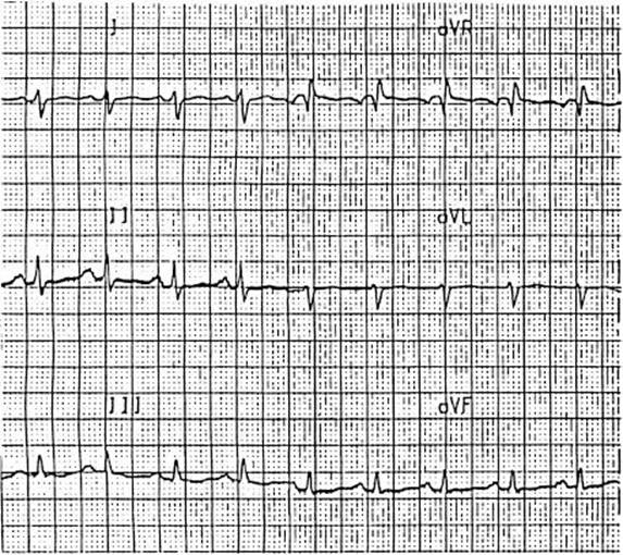
FIGURE 339.1 Terminal QRS changes in CA poisoning.
Other than a lactic acidosis secondary to seizures or shock, CAs are not known to produce biochemical disturbances. However, evaluation of patients with altered mental status or cardiotoxicity includes assessment of serum electrolytes, blood urea nitrogen, creatinine, and glucose. A blood gas, creatine phosphokinase, calcium, magnesium, phosphorus, liver chemistries, and a chest radiograph should be considered for ill patients with abnormal vital signs, significant CNS depression, seizures, or an abnormal ECG. Patients with an intentional ingestion should have a serum acetaminophen concentration to rule-out coingestion.
Drug screening for CAs has no place in the management of the acutely CA-poisoned patient. Urine qualitative immunoassays are the most common form of drug screening for CAs but many other drugs including diphenhydramine, cyclobenzaprine, quetiapine, cyproheptadine, carbamazepine, and most phenothiazines cause false positives. Although serum CA concentrations >1,000 ng/mL generally correlate with significant toxicity, levels do not assist in emergency management. Monitoring the changes on the ECG and the patient’s mental status are more clinically useful indicators of ongoing CA toxicity.
KEY TESTING
• ECG and monitoring QRS width and dysrhythmias
• Acetaminophen level to detect coingestion
• Unstable patients need other laboratories for supportive care management
• Urine drug testing and serum levels are not clinically helpful
ED MANAGEMENT
Patients with coma, seizures, hypotension, and dysrhythmias include endotracheal intubation, administration of anticonvulsants, intravenous crystalloids, and serum alkalinization with parenteral sodium bicarbonate boluses.
Parenteral benzodiazepines such as diazepam (5 to 10 mg) or lorazepam (1 to 2 mg) are the agents of choice for the treatment of seizures. They should be administered to any patient with seizures. Repeat doses may be required. When intubation is required, parenteral benzodiazepines such as lorazepam and diazepam may be useful as well for sedative and anticonvulsant effects, though propofol is a reasonable option if hypotension is not present. If repeat doses of benzodiazepines do not completely control seizures, phenobarbital or propofol should be added. Phenobarbital or propofol may be necessary for the treatment of myoclonus. In the intubated patient, a phenobarbital loading dose of 18 mg/kg is usually sufficient. The dose for propofol sedation of the intubated patient begins with a bolus infusion of 0.5 mg/kg; this can be repeated if blood pressure allows. The dose is titrated to the desired sedation level in increments of 20 to 80 μg/kg/hr as a constant infusion, titrated to the appropriate sedation level. Propofol’s rapid onset and offset make it ideal for the management of these ventilated CA-poisoned patients. Seizures should be anticipated so benzodiazepines should be kept at the bedside to minimize the rapidly produced lactic acidosis that may further compromise the cardiac and hemodynamic status. In addition, an intravenous bolus of 1 to 2 ampules (44 to 100 mEq) of hypertonic sodium bicarbonate is recommended as adjunctive treatment for seizures.
Hypotension should be treated with intravenous crystalloid boluses. This treatment should overcome hypotension from α-adrenergic blockade and mild dehydration. For more seriously poisoned patients, vasopressors will be needed. Dopamine and norepinephrine have been used; however, many cases are refractory to dopamine. Because dopamine partially acts by causing catecholamine release from sympathetic nerve terminals, it may lose some of its effectiveness when norepinephrine stores are depleted as a result of CA poisoning. Norepinephrine has been demonstrated to have greater efficacy than dopamine in successfully treating refractory hypotension (6). Dosing of norepinephrine should start at 0.04 μg/kg/min and be titrated up as needed to a mean arterial pressure of 60 mm Hg. In patients unresponsive to these measures, bedside or formal echocardiography, pulse-contour analysis to determine cardiac output and systemic vascular resistance, or the insertion of a pulmonary artery catheter may be useful to guide additional fluid and vasopressor management.
For patients in refractory shock, true extremis or cardiac arrest from CA poisoning, intravenous fat emulsion (IFE or Intralipid) has been successfully used (7). Though the mechanism of IFE is not fully elucidated, the infusion of lipid may create a “lipid sink” in the blood. Fat-soluble drugs diffuse into the lipid sink and away from the deleterious site of action. IFE was initially described and has now become standard care in the resuscitation of patients with local anesthetic overdose in the operating room. Its use subsequently expanded to other cardiotoxic drugs, including CAs (7). Results, however, have been mixed with regard to the use of IFE in CA poisoning. In animals IFE outperformed hypertonic sodium bicarbonate for treating hypotension for clomipramine (8) however was associated with worse survival and higher serum drug concentrations for amitriptyline (9). The differences in these studies may be evidence that IFE is effective for the treatment of poisoning from some CAs but not others. At this time the use of IFE in CA poisoning remains reasonable for the patient in true extremis refractory to sodium bicarbonate, norepinephrine, and usual supportive care. The dose is 1.5 mL/kg of 20% IFE over 2 to 3 minutes, followed by an infusion of 0.25 mL/kg/min over 30 minutes. The bolus dose may be repeated as needed up to two additional doses. As experience increases, the use of IFE may become better defined.
Cardiac dysrhythmias can be terminated with sodium bicarbonate. Sodium and bicarbonate have both proven to be beneficial treatments for toxins that have quinidine-like, sodium-channel blocking actions. In experimental models of amitriptyline toxicity, sodium loading and increasing pH each separately improve cardiac conduction, but both treatments together produced the greatest improvement. Although hypertonic saline, 7.5% in dextran, without bicarbonate dramatically increases blood pressure and narrows the QRS interval after nortriptyline poisoning in animals, hypertonic saline is not widely used in clinical practice. Sodium bicarbonate boluses and normal saline infusions are usually sufficient but require careful monitoring as overuse can cause severe hypernatremia, fluid overload, and alkalemia.
Dysrhythmias should be treated with at least two or three ampules (100 to 150 mEq, or a dose of 1 to 2 mEq/kg) of sodium bicarbonate. This bolus dosing should occur every 3 to 5 minutes until the QRS narrows; this dosing scheme can result in large doses of bicarbonate administration. Sodium bicarbonate has acutely terminated VT and wide-complex tachycardia. The QRS duration shortens, and the terminal 40-ms changes become less evident. Respiratory alkalosis is advocated by many clinicians, but the pH increase is not as instantaneous as with bolus bicarbonate administration, and sustained respiratory alkalosis will lead to compensatory renal bicarbonate excretion, resulting in a relative deficiency state. The target pH for alkalinization is 7.45 to 7.55, but caution is needed to avoid overshooting the desired pH or causing hypernatremia or ionized hypocalcemia. In addition, the administration of large amounts of bicarbonate for a number of hours may lead to respiratory acidosis and hypoventilation in the nonintubated patient.
Dialysis is not helpful because of high protein-binding and the large volume of distribution.
Because in overdose, CAs are, epileptogenic and benzodiazepines can be therapeutic, the use of flumazenil is in general contraindicated for patients with coma and a history or ECG suggestive for moderate or severe CA poisoning. Anticholinergic delirium is common in moderate TCA poisoning, and physostigmine has been effective in reversing anticholinergic toxicity. The use of physostigmine in CA poisoning is controversial. Because of case reports of asystole after physostigmine use in unstable CA-poisoned patients, physostigmine was avoided for many years. It was contraindicated for patients with a prolonged QRS interval or dysrhythmias suggestive of CA cardiotoxicity. More recently, one center reported uncontrolled use of physostigmine for delirium and coma with improvement in 236 of 252 retrospective and 62 of 63 prospective CA-poisoned patients. No patients in either arm suffered significant ventricular dysrhythmias or asystole (10). While physostigmine may improve delirium, coma, or agitation from the anticholinergic syndrome in those not critically ill, it is unclear if this surpasses routine intensive care. Care must be taken to administer physostigmine over 3 to 5 minutes since an IV push bolus is associated with seizures and bradycardia. Concomitant use of benzodiazepines is recommended. Benzodiazepines alone do not effectively control anticholinergic delirium unless heavily sedating doses are used, which may extend the length of stay and increase complications and intubations.
Gastrointestinal decontamination should be accomplished after the institution of any necessary life-support measures. AC in a dose of 1 g/kg is the method of choice for gastric decontamination, when safe to administer. Multiple doses of AC have not been of clinical benefit and are not recommended. Care should be taken to avoid aspiration of charcoal into the lung, which can dramatically prolong hospital stays. Though no longer routinely recommended for most poisonings, gastric lavage, may benefit some early presenting (within 1 to 2 hours) CA-poisoned patients because the toxicity of this overdose is so high.
CRITICAL INTERVENTIONS
• Establish IV access and initiate continuous cardiac and oxygen saturation monitoring.
• Administer benzodiazepines quickly and aggressively for seizures to avoid increasing acidosis.
• Use sodium bicarbonate at 1 to 2 mEq/kg IV every 3 to 5 minutes for dysrhythmias and cardiac conduction disturbances >100 ms.
• Administer intravenous fluids and norepinephrine for hypotension.
• Administer propofol or phenobarbital for refractory seizures or myoclonus.
DISPOSITION
The maximal severity of CA poisoning is usually apparent within 2 to 6 hours of an overdose. Patients who develop coma, hypotension, seizures, or dysrhythmias require intensive care unit admission. Those who remain or become asymptomatic including lack of sinus tachycardia after 6 hours of observation can be medically cleared for discharge or psychiatric evaluation. Patients with persistent but mild symptoms, such as lethargy or tachycardia, require additional monitoring and observation in emergency department (ED) observation units or intermediate care units are appropriate.
If critically ill and complicated poisoned patients are transferred, either an air ambulance or an advanced cardiac life-support–equipped crew, accompanied by a nurse or physician, is required to treat ongoing complications.
Common Pitfalls
• Failure to appreciate that awake and stable-appearing patients with recent CA overdose may rapidly deteriorate.
• Failure to recognize the subtle electrocardiographic changes consistent with CA poisoning.
• Failure to wait for conduction delay or wide QRS before starting bicarbonate therapy.
• Failure to aggressively prevent and treat seizures and correct the resultant metabolic acidosis.
ACKNOWLEDGMENTS
The author would like to thank William J. Cimikoski and Keith K. Burkhart who contributed to this chapter in previous editions.
REFERENCES
1. Hawton K, Bergen H, Simkin S, et al. Toxicity of antidepressants: rates of suicide relative to prescribing and non-fatal overdose. Br J Psychiatry. 2010;196(5):354–358.
2. Bronstein AC, Spyker DA, Cantilena LR, et al. 2011 Annual Report of the American Association of Poison Control Centers’ National Poison Data System (NPDS): 29th Annual Report. Clin Toxicol (Phila). 2012;50:911–1164.
3. White NC, Litovitz T, Clancy C. Suicidal antidepressant overdoses: A comparative analysis by antidepressant type. J Med Toxicol.2008;4:238–249.
4. Jensen PS, Bhatara VS, Vitiello B, et al. Psychoactive medication prescribing practices for US children: Gaps between research and clinical practice. J Am Acad Child Adolesc Psychiatry. 1999;38:557–565.
5. Taboulet P, Michard F, Muszynski J, et al. Cardiovascular repercussions of seizures during cyclic antidepressant poisoning. J Toxicol Clin Toxicol. 1995;33:205–211.
6. Tran TP, Panacek EA, Rhee KJ, et al. Response to dopamine vs norepinephrine in tricyclic antidepressant-induced hypotension. Acad Emerg Med. 1997;4:864–868.
7. Levine M, Brooks DE, Franken A, et al. Delayed-onset seizure and cardiac arrest after amitriptyline overdose, treated with intravenous lipid emulsion therapy. Pediatrics. 2012;130:e432–e438.
8. Harvey M, Cave G. Intralipid outperforms sodium bicarbonate in a rabbit model of clompiramine toxicity. Ann Emerg Med. 2007;49(2):178–185.
9. Perichon D, Turfus S, Gerostamolous D, et al. An assessment of the in vivo effects of intravenous lipid emulsion on blood drug concentration and haemodynamics following oro-gastric amitriptyline overdose. Clin Toxicol.2013;51(4):208–215.
10. Rasimas JJ, Sachdeva K, Salama AM, et al. A review of bedside toxicologic experience with physostigmine and flumazenil. Clin Toxicol. 2010;48:648.