
DOORWAY INFORMATION
Opening Scenario
Riva George, a 35-year-old female, comes to the hospital complaining of pain in her right calf.
Vital Signs
BP: 130/70 mm Hg Temp: 99.9°F (37.7°C)
RR: 13/minute HR: 88/minute
Examinee Tasks
1. Take a focused history.
2. Perform a focused physical exam (do not perform rectal, genitourinary, or female breast exam).
3. Explain your clinical impression and workup plan to the patient.
4. Write the patient note after leaving the room.
Checklist/SP Sheet
Patient Description
Patient is a 35 yo F, married with two children.
Notes for the SP
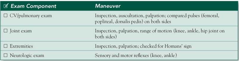
■ Exhibit pain in your calf when the doctor dorsiflexes your right ankle.
■ Place a bandage on your right leg to cover the cuts that you got after a fall.
Challenging Questions to Ask
“My father had a clot in his leg. What do you think I should do to make sure I don’t get one too?”
Sample Examinee Response
“There are several measures you can take that may prevent you from having a clot. Above all, you should avoid immobilization for long periods of time—for example, while sitting at your computer desk or on long-distance plane trips. Try to move in place and perhaps take a short walk. If you are on oral contraceptive pills, I strongly recommend that you stop taking them, as they are known to precipitate clotting. Studies have also shown that obesity increases your risk of having a clot, so I suggest that you exercise regularly and manage your diet.”
Examinee Checklist
Building the Doctor-Patient Relationship Entrance
□ Examinee knocked on the door before entering.
□ Examinee introduced self by name.
□ Examinee identified his/her role or position.
□ Examinee correctly used patient’s name.
□ Examinee made eye contact with the SP.
Reflective Listening
□ Examinee asked an open-ended question and actively listened to the response.
□ Examinee asked the SP to list his/her concerns and listened to the response without interrupting.
□ Examinee summarized the SP’s concerns, often using the SP’s own words.
Information Gathering
□ Examinee elicited data efficiently and accurately.


Connecting with the Patient
□ Examinee recognized the SP’s emotions and responded with PEARLS.
Physical Examination
□ Examinee washed his/her hands.
□ Examinee asked permission to start the exam.
□ Examinee used respectful draping.
□ Examinee did not repeat painful maneuvers.
Closure
□ Examinee discussed initial diagnostic impressions.
□ Examinee discussed initial management plans:
□ Follow-up tests.
□ Examinee asked if the SP had any other questions or concerns.
Sample Closure
Mrs. George, on the basis of your history and my physical examination, I believe it is possible that you had a blood clot. However, we will also look for other possible causes of your symptoms, such as an infection or a ruptured cyst. We will be running a few blood tests as well as some imaging studies that should help us make a final diagnosis. If your test results show a clot, we will start you on blood thinners to prevent further complications, such as the possibility of a clot traveling to your lungs. Do you have any questions for me?
History
HPI: 35 yo F c/o right calf pain of a few days' duration. The pain is constant, 8/10 in intensity, not radiating, aggravated on walking and extending the knee, and associated with swelling, redness, and warmth. It is alleviated on elevation of the foot and with ibuprofen. The patient took a 15-hour flight 1 week ago. She has a history of weight gain postpartum and cuts to the right leg secondary to a fall. She has 2 children, both normal deliveries. LMP was 2 weeks ago. The patient says she has gained 50 lbs in the past 3 years. She has been on OCPs for 2 years. No history of chest pain or shortness of breath.
ROS: Negative except as above.
Allergies: NKDA.
Medications: OCPs, ibuprofen.
PMH: None.
PSH: None.
SH: No smoking, no EtOH, no illicit drugs.
FH: Father had DVT. No history of sudden deaths in the family.
Physical Examination
Patient is in severe pain.
VS: WNL except for low-grade fever.
Chest: Clear breath sounds bilaterally; no rales or rhonchi.
Heart: RRR; normal S1/S2; no murmurs, rubs, or gallops.
Abdomen: Soft, nontender, nondistended, © BS.
Extremities: Inspection: Right calf appears red and swollen compared to left; contours of the muscles appear normal; no ulcers or pigmentation. Palpation: Right leg is warmer compared to left; pitting pedal edema © on right side; multiple healing cuts covered with bandage on right leg; dorsalis pedis pulse felt and equal on both sides; mobility normal at ankle joint, knee, and hip joint; © Homans' sign on right side. Neuro: Mental status: Alert and oriented. DTRs: Symmetric 2+. Motor/sensation: Normal. Cranial nerves: 2-12 intact. Gait: Normal.
Differential Diagnosis


CASE DISCUSSION
Patient Note Differential Diagnoses
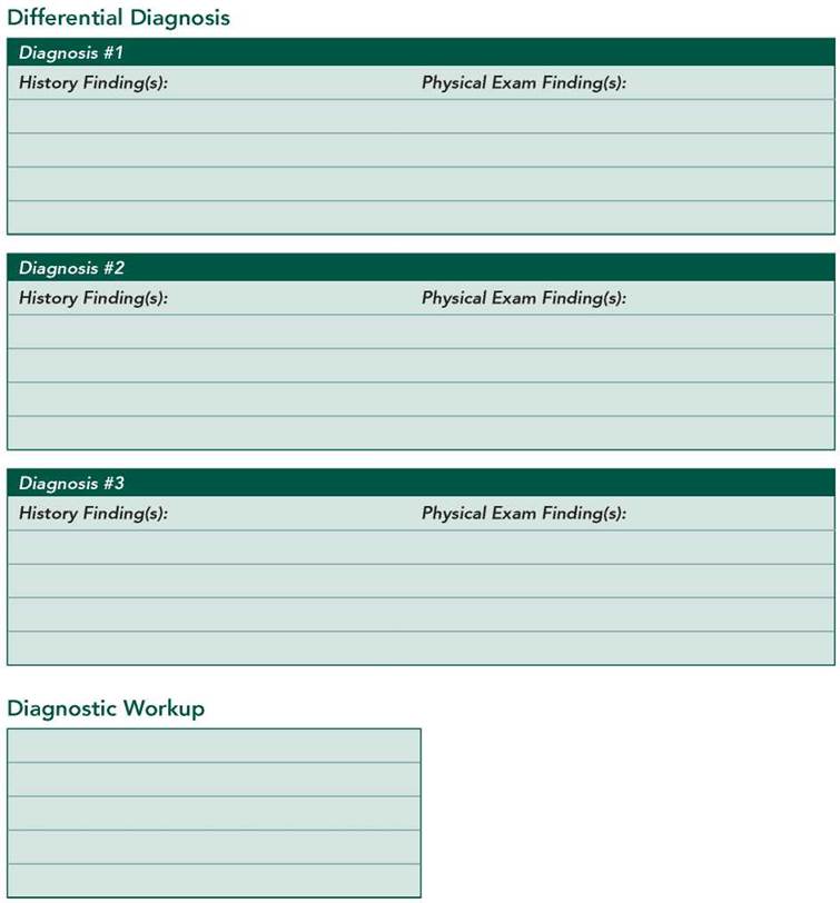
■ Deep venous thrombosis (DVT): DVT is common in the lower limbs and may arise under conditions of stasis, hypercoagulability, and venous endothelial injury. Conditions that result in prolonged immobilization (eg, postsurgery, trauma, sedentary jobs, extended airplane or automobile travel) are predisposing factors.
Other risk factors include advancing age, pregnancy, synthetic estrogens, prior DVT, obesity, malignancy, and thrombophilia. DVT may produce pain and edema of the affected limb or may be asymptomatic. A positive Homans’ sign (pain on dorsiflexion of the ankle) is suggestive of DVT but not diagnostic.
■ Cellulitis: Trauma can lead to cellulitis of the skin and subcutaneous tissue or to myositis of the calf muscle. All the classic signs of inflammation associated with fever (calor, dolor, rubor, tumor) may point to this diagnosis. Regional lymph node enlargement and tenderness are commonly seen. Myositis ossificans may occur as a complication of this disorder, causing hardening of the muscle and pain on contraction. Radiographs may show ossification in the muscle.
■ Rupture of Baker's cyst: Baker’s cysts (also known as popliteal cysts) are seen in the popliteal fossa. Arthritis or a cartilage tear of the knee joint may cause excess synovial fluid to be accumulated, forming a cyst. A ruptured Baker’s cyst may mimic a DVT. Ruptures can present with tightness and swelling behind the knee, pain on knee extension, and stiffness of the calf muscle.
Additional Differential Diagnoses
■ Hematoma: Injuries can cause bleeding intramuscularly (in which no bruising occurs) or intermuscularly (in which bruising is usually present). Patients present with pain, swelling, and restricted movement. The condition may lead to posterior compartment syndrome.
■ Rupture of the gastrocnemius muscle: This presents with sudden pain associated with rupture at the musculotendinous junction of the gastrocnemius muscle, halfway between the knee and the heel. There may be bruising and pain on standing on the tips of the toes. Patients also present with pain on dorsiflexion of the ankle against resistance.
■ Spasm/sprain: Undue strain may cause physical tearing of muscles or tendons, inducing spasm and pain. Ligaments can be ruptured or torn as a result of overstretching or injuries.
Diagnostic Workup
■ Doppler U/S—legs: An initial diagnostic test that is noninvasive and can visualize clots in the veins of the leg.
■ D-dimer: A cross-linked fibrin degradation product that may be increased in DVT. It is usually indicated in cases with a low to intermediate probability of thromboembolism. The negative predictive value of this test is sufficiently high to rule out DVT.
■ Hypercoagulability testing: Several autoantibodies are implicated in thrombophilic states. Proteins C and S deficiency, partial antithrombin deficiency, prothrombin gene mutations, factor V Leiden, hyperhomocysteinemia, antiphospholipid antibody syndrome, and paroxysmal nocturnal hemoglobinuria may all lead to increased coagulability. Hypercoagulability testing should be done on patients with no predisposing factors, recurrent DVT, or a family history of DVT.
■ CBC with differential: To detect infections such as cellulitis.
■ Wound and blood cultures: To work up an infectious etiology of cellulitis.
■ CPK and myoglobin: Both can be elevated in muscle injury (myositis).
■ CT/MRI: CT venography is used to diagnose DVT in conjunction with contrast-enhanced spiral CT to rule out pulmonary embolism. MRI is noninvasive and can detect acute, symptomatic proximal DVTs as well as muscle or tendon rupture.