EQUIPMENT
 Drape
Drape
Skin-marking pencil
Ruler and tape measure
Stethoscope with bell and diaphragm
EXAMINATION
Have patient sit, disrobed to waist.

Thoracic landmarks. A, Anterior thorax. B, Right lateral thorax. C, Posterior thorax.
|
TECHNIQUE |
FINDINGS |
|
CHEST AND LUNGS |
|
|
Inspect front and back of chest |
|
|
See thoracic landmarks. |
|
|
|
|
|
|
EXPECTED:Supernumerary nipples possible (can be clue to other congenital abnormalities, particularly in whites). |
|
|
EXPECTED:Ribs prominent, clavicles prominent superiorly, sternum usually flat and free of abundance of overlying tissue. Chest somewhat asymmetric. Anteroposterior diameter often half of transverse diameter. |
|
UNEXPECTED:Barrel chest, posterior or lateral deviation, pigeon chest, or funnel chest. |
|
|
|
UNEXPECTED:Clubbed fingernails (usually symmetric and painless; may indicate disease, may be hereditary), pursed lips, flared alae nasi. |
|
Assess skin, lips, and nails. |
UNEXPECTED:Superficial venous patterns. Cyanosis or pallor of lips or nails. |
|
|
UNEXPECTED:Malodorous. |
|
Evaluate respirations |
|
|
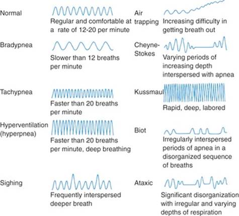
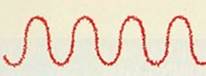
See patterns of respiration in figure below. |
EXPECTED:Breathing easy, regular, without distress. Pattern even. Rate 12 to 20 respirations per minute. Ratio of respirations to heartbeats about 1 : 4. UNEXPECTED:Dyspnea, orthopnea, paroxysmal nocturnal dyspnea, platypnea, tachypnea, hypopnea. Use of accessory muscles, retractions. |
|
Patterns of respiration. The horizontal axis indicates the relative rates of these patterns. The vertical swings of the lines indicate the relative depth of respiration. |
|
|
|
UNEXPECTED:Air trapping, prolonged expiration. |
|
Inspect chest movement with breathing |
|
|
|
EXPECTED:Chest expansion bilaterally symmetric. |
|
UNEXPECTED:Asymmetry. Unilateral or bilateral bulging. Bulging on expiration. |
|
|
Listen to respiration sounds audible without stethoscope |
|
|
EXPECTED:Generally bronchovesicular. |
|
|
UNEXPECTED:Crepitus, stridor, wheezes. |
|
|
Palpate thoracic muscles and skeleton |
|
|
|
EXPECTED:Bilateral symmetry. Some elasticity of rib cage, but sternum and xiphoid relatively inflexible and thoracic spine rigid. |
|
UNEXPECTED:Pulsations, tenderness, bulges, depressions, unusual movement, unusual positions. |
|
|
Stand behind patient. Place palms in light contact with posterolateral surfaces and thumbs along spinal processes at tenth rib, as shown in figure at right. Watch thumb divergence during quiet and deep breathing. Face patient; place thumbs along costal margin and xiphoid process with palms touching anterolateral chest. Watch thumb divergence during quiet and deep breathing. |
EXPECTED:Symmetric expansion. UNEXPECTED:Asymmetric expansion. |
|
Palpating thoracic expansion. The thumbs are at the level of the tenth rib. |
|
|
|
EXPECTED:Nontender sensations. |
|
UNEXPECTED:Crepitus or grating vibration. |
|
|
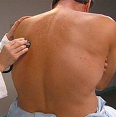
Ask patient to recite numbers or words while systematically palpating chest with palmar surfaces of fingers or ulnar aspect of clenched fist, using firm, light touch. Assess each area, front to back, side to side, lung apices. Compare sides. |
EXPECTED:Great variability; generally, fremitus is more intense with males (lower-pitched voice). UNEXPECTED:Decreased or absent fremitus; increased fremitus (coarser, rougher); or gentle, more tremulous fremitus. Variation between similar positions on right and left thorax. |
|
Note position of trachea |
|
|
Using index finger or thumbs, palpate gently from suprasternal notch along upper edges of each clavicle and in spaces above, to inner borders of sternocleidomastoid muscles. |
EXPECTED:Spaces equal side to side. Trachea midline directly above suprasternal notch. Possible slight deviation to right. |
|
UNEXPECTED:Significant deviation or tug. Pulsations. |
|
|
Perform percussion on chest |
|
|
Percuss as shown in figure below. Compare all areas bilaterally, following a sequence such as shown in figures on p. 103 |
|
|
See table on p. 103 for common tones, intensity, pitch, duration, quality. |
|
|
Method for percussion. |
|
|
Suggested sequence for systematic percussion and auscultation of the thorax. A, Posterior thorax. B, Right lateral thorax. C, Left lateral thorax. D, Anterior thorax. The pleximeter finger or the stethoscope is moved in the numeric sequence suggested; however, other sequences are possible. |
|

Percussion Tones Heard Over the Chest
|
TECHNIQUE |
FINDINGS |
|
Have patient sit with head bent and arms folded in front while percussing posterior thorax, then with arms raised overhead while percussing lateral and anterior chest. Percuss at 4- to 5-cm intervals over intercostal spaces, moving superior to inferior, medial to lateral. The female breast may obscure findings. You or the patient may need to shift the breast, but pay careful attention to modesty. |
EXPECTED:Resonance over all areas of lungs, dull over heart and liver, spleen, areas of thorax. UNEXPECTED:Hyperresonance, dullness, or flatness. |
|
Ask patient to breathe deeply and hold breath. Percuss along scapular line on one side until tone changes from resonant to dull. Mark skin. Allow patient to breathe normally, then repeat on other side. Have patient take several breaths, then exhale as much as possible and hold. On each side, percuss up from mark to change from dull to resonant. Tell patient to resume breathing comfortably. Measure excursion distance. |
EXPECTED:3 to 5 cm (higher on right than left). UNEXPECTED:Limited descent. |
|
Measuring diaphragmatic excursion. Excursion distance is usually 3 to 5 cm. |
|
|
Auscultate chest with stethoscope diaphragm, apex to base |
|
|
Have patient breathe slowly and deeply through mouth. Follow set auscultation sequence, holding stethoscope as shown in figure below. Ask patient to sit upright (1) with head bent and arms folded in front while auscultating posterior thorax, (2) with arms raised overhead while auscultating lateral chest, (3) with arms down and shoulders back while auscultating anterior chest. Listen during inspiration and expiration. Auscultate downward from apex to base at intervals of several centimeters, making side- to-side comparisons. |
EXPECTED:See expected breath sounds in table on p. 106. UNEXPECTED:Amphoric or cavernous breathing. Sounds difficult to hear or absent. Crackles, rhonchi, wheezes, or pleural friction rub, as described in box on p. 107. |
|
Auscultation with a stethoscope. |
|
|
Ask patient to recite numbers or words. |
EXPECTED:Muffled and indistinct sounds. UNEXPECTED:Bronchophony, whispered pectoriloquy, or egophony. |
Characteristics of Expected Breath Sounds
|
Sound |
Characteristics |
Findings |
|
Vesicular |
Heard over most of lung fields; low pitch; soft and short expirations; will be accentuated in a thin person or a child and diminished in overweight or very muscular patient |
|
|
Bronchovesicular |
Heard over main bronchus area and over upper right posterior lung field; medium pitch; expiration equals inspiration |
|
|
Bronchotracheal (tubular) |
Heard only over trachea; high pitch; loud and long expirations, often somewhat longer than inspiration |
|
Modified from Thompson et al, 1997.
Modified from Thompson et al, 1997.
Adventitious Breath Sounds
|
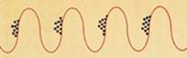
Fine crackles: High-pitched, discrete, discontinuous crackling sounds heard during end of inspiration; not cleared by cough |
|
|
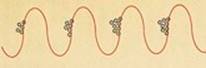
Medium crackles: Lower, more moist sound heard during midstage of inspiration; not cleared by cough |
|
|
Coarse crackles: Loud, bubbly noise heard during inspiration; not cleared by cough |
|
|
Rhonchi (sonorous wheeze): Loud, low, coarse sounds, like a snore, most often heard continuously during inspiration or expiration; coughing may clear sound (usually means mucus accumulation in trachea or large bronchi) |
|
|
Wheeze (sibilant wheeze): Musical noise sounding like a squeak; most often heard continuously during inspiration or expiration; usually louder during expiration |
|
|
Pleural friction rub: Dry rubbing or grating sound, usually caused by inflammation of pleural surfaces; heard during inspiration or expiration; loudest over lower lateral anterior surface |
|
AIDS TO DIFFERENTIAL DIAGNOSIS
|
ABNORMALITY |
DESCRIPTION |
|
Pleural effusion |
Subjective Data:Cough with progressive dyspnea is the typical presenting concern. Pleuritic chest pain occurs with an inflammatory effusion. |
|
Objective Data:Findings on auscultation and percussion vary with the amount of fluid present and with the position of the patient. These include dullness to percussion and tactile fremitus, which are the most useful findings for pleural effusion. When the fluid is mobile it will gravitate to the most dependent position. The affected areas, the breath sounds are muted and the percussion note is often hyperresonant in the area above the perfusion. |
|
|
Lung cancer |
Subjective Data:Cough, wheezing, a variety of patterns of emphysema and atelectasis, pneumonitis, and hemoptysis. Peripheral tumors without airway obstruction may be asymptomatic. |
|
Objective Data:Findings are based on the extent of the tumor and the patterns of its invasion and metastasis. With airway obstruction a postobstructive pneumonia can develop with consolidation. A malignant pleural effusion may develop with corresponding findings. |
|
|
Pneumonia |
Subjective Data:Rapid onset (hours to days) of cough, pleuritic chest pain and dyspnea. Sputum production is common with bacterial infection (see table on p. 110). Chills, fever, rigors, and nonspecific abdominal symptoms of nausea and vomiting may be present. Involvement of the right lower lobe can stimulate the tenth and eleventh thoracic nerves to cause right lower quadrant pain and simulate an abdominal process. |
|
Objective Data:Febrile, tachypneic, and tachycardic. Crackles and rhonchi are common with diminished breath sounds. Egophony, bronchophony, and whisper pectoriloquy. Dullness to percussion occurs over the area of consolidation. |
|
|
Asthma |
Subjective Data:Episodes of paroxysmal dyspnea and cough. Chest pain is common and, with it, a feeling of tightness. Episodes may last for minutes, hours, or days. May be asymptomatic between episodes. |
|
Objective Data:Tachypnea with wheezing on expiration and inspiration. Expiration becomes more prolonged with labored breathing, fatigue, and anxious expression as airway resistance increases. Hypoxemia by pulse oximetry. |
|
|
Chronic bronchitis |
Subjective Data:Dyspnea may be present although not severe. Cough and sputum production are impressive. |
|
Objective Data:Wheezing and crackles. Hyperinflation with decreased breath sounds and a flattened diaphragm. Severe chronic bronchitis may result in right ventricular failure with dependent edema. |
|
|
Emphysema |
Subjective Data:Dyspnea is common even at rest. Cough is infrequent without much production of sputum. |
|
Objective Data:Chest may be barrel shaped, and scattered crackles or wheezes may be heard. Overinflated lungs are hyperresonant on percussion. Inspiration is limited with a prolonged expiratory effort (i.e., longer than 4 or 5 seconds) to expel air. |
Assessing Sputum
|
Cause |
Possible Sputum Characteristics |
|
Bacterial infection |
Yellow, green, rust-colored (blood mixed with yellow sputum), clear, or transparent; purulent; blood streaked; mucoid, viscid |
|
Viral infection |
Mucoid, viscid; blood streaked (not common) |
|
Chronic infectious disease |
All of the above; particularly abundant in early morning; slight, intermittent blood streaking; occasionally large amounts of blood |
|
Carcinoma |
Slight, persistent blood streaking |
|
Infarction |
Blood clotted; large amounts of blood |
|
Tuberculous cavity |
Large amounts of blood |
Pediatric Variations
EXAMINATION
|
TECHNIQUE |
FINDINGS |
|
|
Chest and Lungs |
||
|
Inspect front and back of chest |
||
|
|
EXPECTED:Infant’s chest is expected to measure 2 to 3 cm less than head circumference. |
|
|
Evaluate respirations |
||
|
|
EXPECTED: |
|
|
Age |
Respirations per Minute |
|
|
Newborn |
30-80 |
|
|
1 year |
20-40 |
|
|
3 years |
20-30 |
|
|
6 years |
16-22 |
|
|
10 years |
16-20 |
|
|
17 years |
12-20 |
|
|
Perform direct or indirect percussion on chest |
||
|
|
EXPECTED:Hyperresonance may be heard in children. |
|
|
Auscultate chest with stethoscope diaphragm, apex to base |
||
|
|
EXPECTED:In infants and children, expect transmitted breath sounds throughout chest. Vesicular sound is accentuated in a child. Absent or diminished breath sounds are harder to detect. |
|
SAMPLE DOCUMENTATION
Subjective.A 45-year-old woman complaining of cough and fever for 4 days. Cough is nonproductive, persistent, and worse when she lies down. She feels ill and short of breath. Her chest feels “heavy.” Fever up to 38.3° C (101° F). Taking acetaminophen and nonprescription cough syrup without relief.
Objective.Pulse 104 per minute, temperature 38.2° C, blood pressure 122/82, respirations 32 per minute and somewhat labored; no retractions or stridor. Minimal increase in anteroposterior diameter of chest, without kyphosis or other defect. Trachea in midline without tug. Thoracic expansion symmetric. No friction rubs or tenderness over ribs or other bony prominences. Over posterior left base, diminished tactile fremitus, dull percussion note, and on auscultation, crackles that do not clear with cough, diminished breath sounds. Remaining lung fields are clear and free of adventitious sounds, with resonant percussion tones. Diaphragmatic excursion 3 cm bilaterally.
 Size/shape/symmetry