
The laboratory diagnosis of infection requires the demonstration—either direct or indirect—of viral, bacterial, fungal, or parasitic agents in tissues, fluids, or excreta of the host. The traditional detection methods of microscopy and phenotypic characterization are time-consuming and are increasingly being replaced by nucleic acid probe assays.
MICROSCOPY
• Wet mounts: the simplest method for microscopic evaluation and useful for certain large and/or motile organisms. For example, combining wet mounts with dark-field illumination permits detection of spirochetes in genital lesions or of Borrelia and Leptospira in blood.
– Fungal elements may be identified in skin scrapings with 10% KOH wet mount preparations.
– Some wet mounts use staining to enhance detection and morphologic identification—e.g., india ink for visualization of encapsulated cryptococci in CSF and lactophenol cotton blue for morphologic identification of fungal elements.
• Stains: Staining techniques permit organisms to be seen more clearly.
– Gram’s stain: differentiates between organisms with thick peptidoglycan cell walls (gram-positive) and those with thin peptidoglycan cell walls and outer membranes that can be dissolved with alcohol or acetone (gram-negative).
• This stain is particularly useful for examining sputum samples for PMNs and bacteria. The presence of >10 epithelial cells per low-power field and multiple bacterial types suggests contamination with oral flora.
• In normally sterile fluids (e.g., CSF, pleural and joint fluid), the detection of bacteria suggests an infectious etiology (Fig. 85-1) and correlates with the presence of >104 bacteria/mL.

FIGURE 85-1 Interpretation of Gram’s stain.
• Sensitivity is increased by centrifugation of the sample.
– Acid-fast stains: identify organisms that retain carbol fuchsin dye after acid/organic solvation (e.g., Mycobacterium spp.). Modification of this procedure permits detection of weakly acid-fast organisms (e.g., Nocardia).
– Immunofluorescent stains: utilize antibodies—either labeled directly with a fluorescent compound or detected indirectly by a secondary immunofluorescent antibody—to detect viral antigens (e.g., CMV, HSV, and respiratory viruses) within cultured cells and difficult-to-grow bacteria (e.g., Legionella pneumophila).
MACROSCOPIC ANTIGEN DETECTION
• Latex agglutination assays and EIAs are rapid and inexpensive tests that identify bacteria, viruses, or extracellular bacterial toxins by means of their protein or polysaccharide antigens.
• The assays may be performed either directly on clinical specimens or after growth of the organisms in the laboratory.
CULTURE
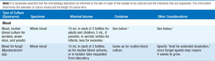
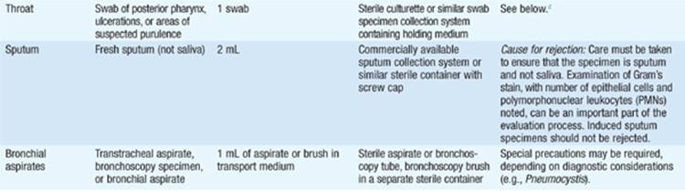
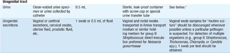
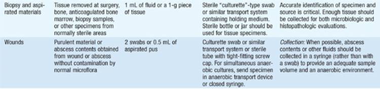
• The success of efforts to culture a specific pathogen often depends on use of appropriate collection and transport procedures in conjunction with a laboratory-processing algorithm suitable for the specimen. Instructions for collection and transport are listed in Table 85-1.
TABLE 85-1 INSTRUCTIONS FOR COLLECTION AND TRANSPORT OF SPECIMENS FOR CULTURE





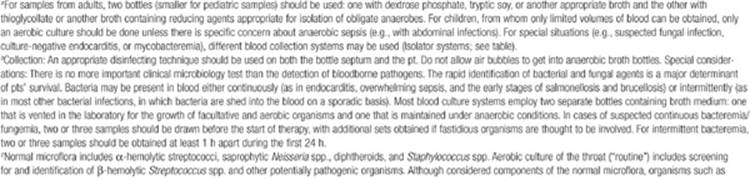
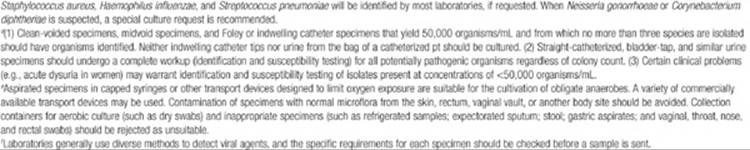
• Bacterial isolation relies on the use of artificial media that support bacterial growth in vitro. Once bacteria are isolated, different methods are used to characterize specific isolates (e.g., phenotyping based on enzymatic and metabolic functions, gas-liquid chromatography, nucleic acid tests).
• Viruses are grown on a monolayer of cultured cells sensitive to infection with the suspected virus. After incubation, cells are examined for cytopathic effects or immunofluorescent studies are performed to detect viral antigens.
SEROLOGY
• Measurement of serum antibody provides an indirect marker for past or current infection with a specific pathogen.
• Quantitative assays detect increases in antibody titers, most often with paired serum samples obtained at illness onset and 10–14 days later (i.e., acute- and convalescent-phase samples). A 4-fold increase is regarded as evidence for acute infection.
• Serology can also be used to document protective levels of antibody, particularly in diseases for which vaccines are available (e.g., rubella, VZV infections).
NUCLEIC ACID PROBES
• Techniques for the detection and quantitation of specific DNA and RNA base sequences in clinical specimens have become powerful tools for the diagnosis of infection and are useful in four settings:
1. To detect and/or quantify specific pathogens in clinical specimens (e.g., Neisseria gonorrhoeae, HIV)
2. To identify organisms that are difficult to grow or identify by conventional methods (e.g., Tropheryma whipplei, Legionella)
3. To determine whether two or more isolates belong to the same clone or strain
4. To predict sensitivity (typically of viruses) to chemotherapeutic agents (e.g., HIV, Mycobacterium tuberculosis)
• The sensitivity and specificity of probe assays for direct detection are comparable to those of more traditional assays, including EIA and culture.
• Amplification strategies (e.g., PCR) enhance the sensitivity of RNA or DNA assays, but false-positive findings can result from even low levels of contamination.
SUSCEPTIBILITY TESTING
• Susceptibility testing allows the clinician to choose the optimal antimicrobial agents and to identify potential infection-control problems (e.g., the level of methicillin-resistant Staphylococcus aureus in a hospital).
• Qualitative measures (e.g., disk/agar diffusion and breakpoint) and quantitative measures [e.g., broth dilution, epsilometer (E-test)] are available.
• Antifungal susceptibility testing is becoming more commonplace; however, testing of some species (e.g., Aspergillus) remains technically difficult and is performed primarily in reference labs.
CONSIDERATIONS FOR DIAGNOSIS OF PARASITIC INFECTIONS
The cornerstone for the diagnosis of parasitic diseases, as for that of many other infections, is the elicitation of a thorough history of the illness and of epidemiologic factors such as travel, recreational activities, and occupation. Table 85-2 summarizes the diagnosis of some common parasitic infections.
TABLE 85-2 DIAGNOSIS OF SOME COMMON PARASITIC INFECTIONS


INTESTINAL PARASITES
• Most helminths and protozoa can be detected by examination of fecal samples; contamination with urine or water should be avoided.
• Fecal samples should be collected before the ingestion of contrast agents and before treatment with antidiarrheal agents or antacids; these substances alter fecal consistency and interfere with microscopic detection of parasites.
• The collection of three samples on alternate days is recommended because of the cyclic shedding of most parasites in the feces; examination of a single sample can be up to 50% less sensitive.
• Analysis of fecal samples entails macroscopic examination for adult worms or tapeworm segments and microscopic examination that includes direct wet mounts, concentration techniques, and application of permanent stains.
• Alternative diagnostic methods such as sampling of duodenal contents (e.g., for Giardia lamblia, Cryptosporidium, and Strongyloides larvae) and the “cellophane tape” method (e.g., for pinworm ova or Taenia saginata) may be required.
BLOOD AND TISSUE PARASITES
• Diagnosis of tissue-invasive parasites requires an understanding of the pathophysiology of the parasite in question (e.g., examination of urine sediment to detect Schistosoma haematobium).
• The laboratory procedures for detection of parasites in other body fluids are similar to those used for examination of feces.
• The parasites most commonly detected in Giemsa-stained blood smears are plasmodia, microfilariae, and African trypanosomes; however, wet mounts may be more sensitive for microfilariae and African trypanosomes, given their motility.
• The timing of blood collection is crucial—e.g., to diagnose Wuchereria bancrofti infection, blood must be drawn near midnight, when the nocturnal microfilariae are active.

For a more detailed discussion, see McAdam AJ, Onderdonk AB: Laboratory Diagnosis of Infectious Diseases, Chap. e22; and Reed SL, Davis CE: Laboratory Diagnosis of Parasitic Diseases, Chap. e25, in HPIM-18.