
Timur S. Durrani, MD, MPH, MBA
Kent R. Olson, MD
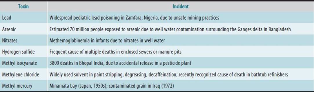
Medical toxicology is a subspecialty focusing on the diagnosis, management, and prevention of poisoning and other adverse health effects due to drugs, occupational and environmental toxic substances, and biological agents. All substances are potentially toxic; it is the dose that makes the poison. Many activities, including occupations and hobbies, can result in exposure to toxic substances. Although occupational exposure to toxic substances is considered to be underreported, it is estimated that 5% of all poison control consultations are occupationally related, suggesting a high rate of chemical exposure in the workplace. Table 16–1 lists selected activities and potential toxic exposures. Historically, many populations have also been recognized to be at risk for toxic environmental exposure (Table 16–2).
Table 16–1. Selected job processes at high risk for specific toxic exposures.
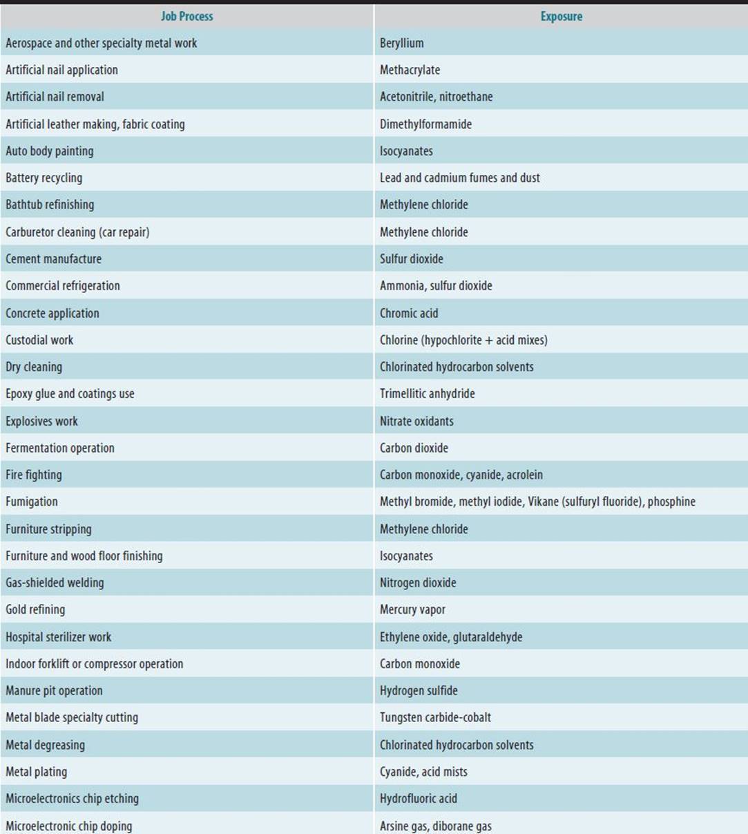
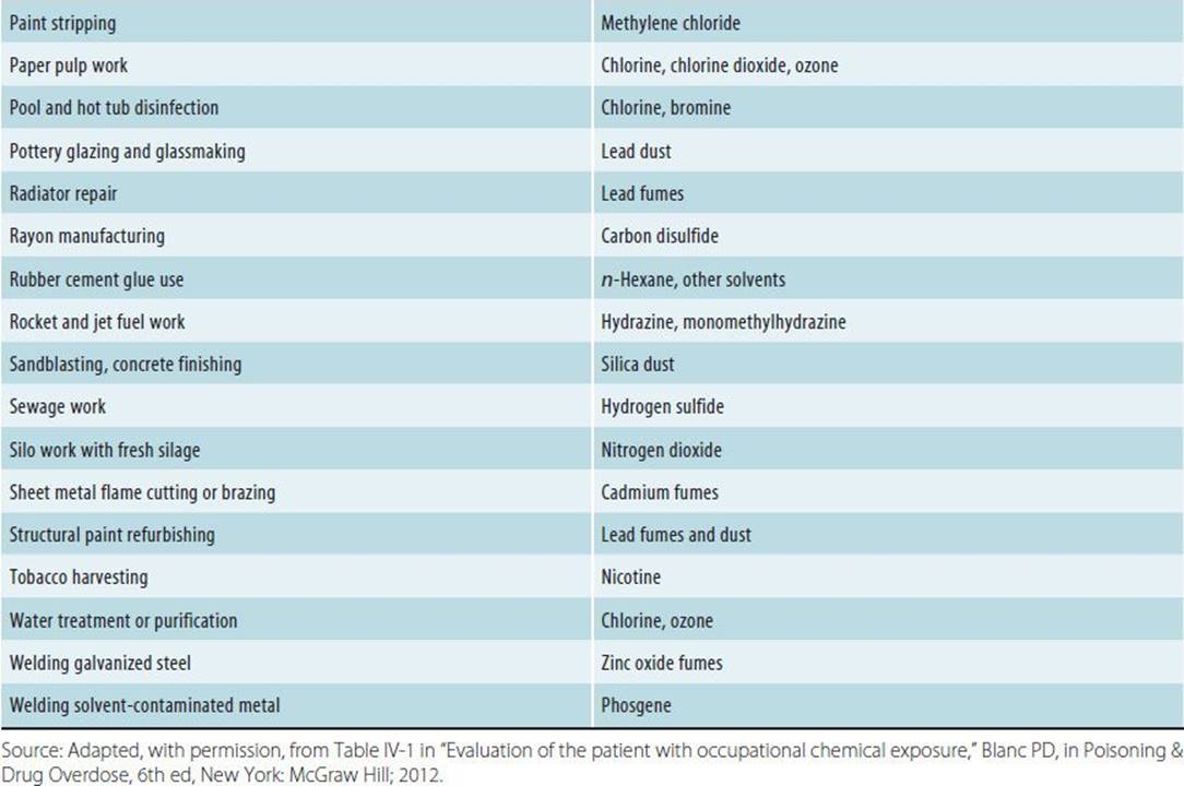
Table 16–2. Examples of toxic exposures affecting public health.
Toxicity does not necessarily equate with hazard. An extremely toxic chemical that is in a sealed container on a shelf has inherent toxicity but presents little or no hazard. However, when the chemical is removed from the shelf and used by a worker without appropriate protection, the hazard may become significant. Thus, the manner of use affects how hazardous the substance will be in the workplace.
TOXIC AGENTS & THEIR EFFECTS
 Classification of Toxic Agents
Toxic agents can be classified by their:
A. Physical State
A metal such as lead may be harmless in solid form, moderately toxic as a dust, and extremely toxic as a fume.
B. Chemical Structure
Chemical structure can determine toxicity. Often one but not another isomer of a compound possesses toxicity. For example, aromatic amines are carcinogenic when substituted in other than the para-positions. The stability of a substance and the presence of impurities, contaminants, or additives can also affect toxicity.
C. Size
Nanoparticles are engineered structures that are less than 100 nm in size. These structures have demonstrated unique physical and chemical properties, which may be independent of the dose. Their small size and unique shape may facilitate absorption, theoretically allowing them direct access to the central nervous system via the olfactory nerve or causing increased fibrosis in the lung similar to asbestos. Due to their unique properties, these materials may display altered biological activity, which may result in positive health effects (eg, antioxidant effects, improved absorption for drug delivery), or negative health effects (eg, unexpected toxicity, oxidative stress), or the results may be mixed. These materials are used in a number of products including computers, food storage containers, clothing, cosmetics, and medications. There is little known about their human toxicity. The measurement of dose and of levels in biologic tissues may be difficult for some nanostructures.
D. Medium or Solution
The medium in which a toxic substance is found in part determines the population exposed and thus to some extent the hazard. Some toxic substances occur in a specific medium—for example, oxides of nitrogen in air (from vehicular exhaust), trihalomethanes in water (from chlorination), and nitrosamines in food (from nitrites). Often, the active chemical in a commercial product is dissolved in a liquid solvent because it is insoluble in water; the solvent may have potential toxicity independent of the active ingredient.
E. Site of Injury
Toxic agents can be described in terms of their effects on target organs (hepatotoxins, nephrotoxins, etc).
F. Mechanism of Action
Simple asphyxiants (inert gases, such as carbon dioxide) act by displacing oxygen in tissue without causing other toxic effects. In contrast, chemical asphyxiants such as carbon monoxide actively interfere with the delivery or utilization of oxygen by combining with hemoglobin to form carboxyhemoglobin, which decreases the oxygen-carrying capacity of the blood and inhibits release of oxygen to tissues.
G. Clinical Effects
1. Onset of effects—Toxic effects can be immediate, as occurs with some irritants that cause direct damage to tissues at the point of initial contact, usually resulting in inflammation; or delayed, as with chemical carcinogens.
2. Reversibility of effects—Whether or not the toxic effects of a substance are reversible depends on the capacity of damaged cells to regenerate or recover. For instance, brain and other nervous system cells have little capacity to regenerate, whereas liver and muscle cells are more likely to regenerate or recover after injury.
Factors Affecting Clinical Response to a Toxic Agent
The following factors affect the dose-response relationship and the clinical response of humans to a toxic agent:
A. Duration, Frequency, and Route of Exposure
The severity of injury is related to the duration, frequency and route of exposure. For example, ethylene glycol is toxic when ingested but poses little threat in the workplace except when sprayed or heated.
B. Environmental Factors
Toxicity is affected by atmospheric pressure, temperature, and humidity. For example, a concentration of carbon monoxide that has little effect at sea level can cause impairment of work capacity at an altitude of 5000 ft. Chemicals are more readily absorbed through skin that is injured or wet with perspiration and has increased blood flow in response to heat and humidity.
C. Individual Factors
Individual factors that determine “susceptibility” include racial and genetic background, age and maturity, gender, body weight, nutrition, lifestyle, immunologic and hormonal status, and presence of disease or stress. These factors are not independent of one another. For instance, genetic factors determine many of the other factors, and poor nutrition can affect immunologic status.
1. While much concern about the effect of age on individual susceptibility has focused on the fetus, the elderly also metabolize many chemicals less efficiently. As the work force ages, this may become an increasing concern.
2. The effect of nutritional deficiency on susceptibility to toxic agents has been of concern in developed countries primarily during war or famine, but it is relevant in developing countries as they industrialize. While toxicologic studies in animals readily demonstrate the effects of nutritional deficiency on susceptibility, the results of these studies are difficult to extrapolate to humans. There is controversy about the role of genetic factors and the development and use of genetic screening tests to identify individuals with increased susceptibility to toxic agents in the workplace. It is questioned whether such tests are accurate and whether job discrimination could result from their use as preemployment screening. Examples of genetic traits that might increase the risk of toxicity from exposure to chemicals or radiation glucose-6-phosphate dehydrogenase (G6PD) deficiency, sickle cell anemia, and α1-antitrypsin deficiency.
G6PD deficiency is an X-linked recessive disorder that primarily affects American black males and persons of Mediterranean descent. Affected individuals are susceptible to hemolysis from many drugs. Some chemicals—notably naphthalene and arsine—can cause hemolysis following overexposure. There is no evidence that G6PD-affected workers exposed to these chemicals are at increased risk. Screening for G6PD deficiency is not supported by solid evidence of its utility.
Similarly, there is no evidence that any of the 7–13% of American blacks with sickle cell trait are at increased risk of hypoxia when working as airplane pilots or of hemolysis when working with hemolytic agents, despite the fact that these “risks” have been cited to justify screening of individuals for these occupations.
Severe α1-antitrypsin deficiency, when present in the rare homozygous genotype, can lead to early emphysema in the absence of environmental agents. The more common heterozygous genotype, which affects 4–9% of the US population, may in combination with other factors place affected individuals at increased risk of developing emphysema from exposure to environmental agents.
TOXICOKINETICS & TOXICODYNAMICS
Toxicokinetics is the study of the movement of toxic substances within the body (ie, their absorption, distribution, metabolism, and excretion) and the relationship between the dose that enters the body and the level of toxic substance found in the blood or other biologic sample. Toxicodynamics is the study of the relationship between the dose that enters the body and the measured response. Simply put, toxicokinetics is the study of the effect of the body on the substance, and toxicodynamics is the study of the effects of the substance on the body. The magnitude of a toxic response is usually related to the concentration of the toxic substance at its site of action.
Bioavailability
The bioavailability of a toxic substance indicates the extent to which the agent reaches its site of action. In some instances, an agent will be inactivated before it reaches the site of action. For example, when cyanide is taken orally, it is absorbed and passes to the liver, where the enzyme rhodanese may detoxify a portion of the ingested cyanide. On the other hand, if the cyanide in the form of gaseous hydrocyanic acid (HCN) is absorbed through the pulmonary circulation, it goes directly to the brain, where it may cause damage due to hypoxia.
Cell Membrane Permeability & Cellular Barriers & Cell Signaling
Absorption, distribution, metabolism, and excretion all involve passage of toxic agents across cell membranes. Permeability is dependent upon a toxic substance’s molecular size and shape, degree of ionization, and relative lipid solubility. The distribution of some toxic agents is altered by unique cellular barriers, for example, the blood-brain barrier, the blood-testis barrier, and the placenta, which may exclude toxic substances.
Bone is an important deep reservoir for many heavy metals (especially lead) and for radioactive materials, and the effects of these materials can persist long after they have left the circulation. This storage property can be used in determining previous exposure and toxic burden. Substances rely on many different cell signaling pathways to induce toxicity. These pathways include G-protein coupled receptors (for muscarinic receptors that are susceptible to organophosphate agonism), ligand gated ion channels (used by nicotine) and intracellular enzymes such as soluble guanylate cyclase (used by nitrovasodilators such as nitrate).
Absorption
The rate of absorption is dependent on the concentration and solubility of the toxic agent. Absorption is enhanced at sites that have increased blood flow or large absorptive surfaces such as the adult lung and gastrointestinal tract.
A. Gastrointestinal Absorption
The amount of absorption through the gastrointestinal tract is usually proportionate to the gastrointestinal surface area and its blood flow and depends on the physical state of the agent. Most toxic substances are absorbed in the small intestine. Therefore, agents that accelerate gastric emptying will increase the absorption rate, while factors that delay gastric emptying will decrease it. Some toxic substances may be affected by gastric juice; for example, the acidity of the stomach may release cyanide products and form hydrogen cyanide gas, which is even more toxic than the cyanide salt.
B. Pulmonary Absorption
The most common route of occupational exposure is pulmonary absorption. Gaseous and volatile toxic substances may be inhaled and absorbed through the pulmonary epithelium and mucous membranes in the respiratory tract. Access to the circulation is rapid because the surface area of the lungs is large and the blood flow is great. The nasal hair, the cough reflex, and the mucociliary barrier help prevent dust particles and fumes from reaching the lung.
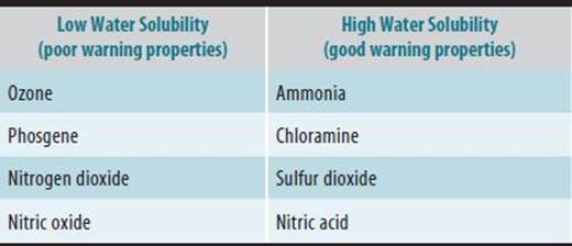
The solubility of gases affects their absorption. Highly water-soluble gases such as ammonia and sulfur dioxide are absorbed in the upper airways and cause marked irritation there. This serves as a warning and may help limit the injury to the lung because the victim leaves the site of exposure. In contrast, noxious gases of lower water solubility such as nitrogen dioxide and phosgene, which have few early warning properties, can reach the bronchioles and alveoli and cause delayed injury (Table 16–3).
Table 16–3. Irritant gases.
C. Percutaneous Absorption
Many toxic substances can pass through intact or broken skin. The amount of skin absorption is generally proportionate to the surface area of contact and to the lipid solubility of the toxic agent. The epidermis acts as a lipid barrier, and the stratum corneum provides a protective barrier against noxious agents. The dermis, however, is freely permeable to many toxic substances. Absorption is enhanced by toxic agents that increase the blood flow to the skin. It is also enhanced by use of occlusive skin coverings (eg, including clothing and industrial gloves) and topical application of fat-solubilizing vehicles. Hydrated skin is more permeable than dry skin. The thick skin on the palms of the hands and the soles of the feet is more resistant to absorption than is the thin skin on the face, neck, and scrotum. Burns, abrasions, dermatitis, and other injuries to the skin may alter its protective properties and allow absorption of larger quantities of the toxic substance.
The pH of the substance can affect the degree of tissue injury and ultimate skin. Highly acidic substances can cause an immediate coagulation-type necrosis that creates an eschar, which tends to self-limit further damage. In contrast, highly alkaline substances cause a liquefactive necrosis with saponification and continued penetration into deeper tissues, resulting in extensive damage.
D. Ocular Absorption
The eye is also a ready site of absorption. When chemicals enter the body through the conjunctiva, they bypass hepatic first-pass elimination and may cause systemic toxicity. Early decontamination of the eye may thus prevent systemic as well as local damage.
Distribution of Toxins in the Body
After absorption, toxic substances are transported to various regions of the body. Some are removed by the lymph, and some insoluble compounds are transported through tissues such as the lung via cells such as macrophages. Most toxic substances enter the bloodstream and are distributed into interstitial and cellular fluids. The pattern of distribution depends on the physiologic and physicochemical properties of the material. The initial phase of distribution usually reflects the cardiac output and regional blood flow. Agents that penetrate membranes poorly are restricted in their distribution, and their potential sites of action are therefore limited. The blood-brain and blood-testis barriers limit the distribution of water-soluble but not lipid-soluble chemicals to these organs. Distribution may also be limited by the binding of toxic substances to plasma proteins. Toxic agents can accumulate in higher concentration in some tissues as a result of pH gradients, binding to special cellular proteins, or partitioning into lipids. Some agents accumulate in tissue reservoirs, and this may serve to prolong the toxic action, for example, lead may be stored for years in bone and may be released later. Some properties allow for substances to be removed by extracorporeal means such as dialysis. Substances with a small volume of distribution, low molecular weight, high water solubility, and low protein binding are more likely to be removed by dialysis.
Metabolism
Before a toxic substance can be excreted, it may require metabolic conversion (biotransformation), for example, to a more water-soluble substance that can be eliminated in the urine. The most common site for biotransformation is the liver, but it can also occur in plasma, lung, or other tissue. Biotransformation may result in either a decrease (detoxification or inactivation) or an increase (activation) in the toxicity of a compound. Differences in the metabolism of toxic substances account for much of the observed differences between individuals and between animal species.
Biotransformation occurs in the liver by hydrolysis, oxidation, reduction, and conjugation. Microsomal cytochrome P450 metabolizing enzymes play a key role in the process by primarily catalyzing the oxidation of toxic substances. The activity of the CYP450 enzyme system can be increased (induced) by many environmental and pharmacologic agents. Individual differences in microsomal enzyme activity and susceptibility to induction are genetically determined and account for the marked variability in bioavailability of many toxic substances. Other factors that regulate key liver enzyme systems are hormones (which account for some gender-dependent differences) and disease states (eg, the presence of hepatitis, cirrhosis, or heart failure). Because the activity of many hepatic metabolizing systems is low in neonates—particularly premature neonates—they may be much more susceptible to toxic substances that are inactivated by liver metabolism. Inefficient metabolizing systems, an altered blood-brain barrier, and inadequate mechanisms of excretion combine to make the fetus and neonate more sensitive to the toxic effects of many agents.
Excretion
A. Pathways and Mechanisms of Excretion
Toxic substances are excreted either unchanged or as metabolites. Excretory organs other than the lungs eliminate polar (water-soluble) compounds more efficiently than they eliminate nonpolar (lipid-soluble) compounds. The kidney is the primary organ of elimination for most polar compounds and metabolites. Excretion of toxic substances in the urine involves glomerular filtration, active secretion, and passive tubular reabsorption. Alkalization or acidification of the urine may dramatically change excretion of some agents. When tubular urine is more alkaline, weak acids are excreted more rapidly because they are ionized and passive tubular reabsorption is decreased. In contrast, when tubular urine is made more acid, excretion of weak acid is reduced.
Many toxic substances metabolized by the liver are excreted first in the bile and later eliminated in the stool. After biliary excretion, some substances are efficiently reabsorbed into the blood, a process known as enterohepatic recirculation. This recirculation can be a cause of repeat exposure and injury. This process can be interrupted by the use of binding agents, such as activated charcoal given in multiple doses. Toxic substances can also be excreted in sweat, saliva, and breast milk, and there may be some minor removal in hair or skin.
B. Clearance
Clearance is the rate at which a toxic agent is excreted, divided by the average concentration of the agent in the plasma. Most toxic substances are eliminated as a function of concentration, ie, a constant fraction of the toxic material is eliminated per unit of time, a process known as “first-order” elimination. If the point of saturation is reached, the body will no longer be able to eliminate a constant fraction of the material but will instead eliminate a constant amount per unit of time, a process known as “zero-order” elimination. Under these circumstances, the clearance becomes quite variable. Note that clearance is a measure not of how many milligrams of toxin is being removed but rather of the volume of fluid that is freed of the toxic agent per unit of time.
C. Volume of Distribution
The volume of distribution is calculated by dividing the amount of the toxic substance in the body (eg, a known dose) by the concentration measured in the blood. This number is not necessarily a physiologic volume; it is an “apparent” volume that reflects the degree of distribution of the toxic agent in tissues. The volume of distribution for most toxic agents depends on its size, pH, protein binding, partition coefficients, and regional differences in blood flow and binding to special tissues.
D. Half-Time and Half-Life
The time it takes for the plasma concentration of a substance to be reduced by 50% is the half-time. For substances that are eliminated according to first-order kinetics, the time it takes to eliminate 50% of the substance is called the half-life. For a substance eliminated by first-order kinetics, about 90% of the amount in the body will be eliminated in 3.5 half-lives after the end of the period of exposure.
TESTS OF TOXIC EFFECTS
Much of our information about the toxic effects of different agents comes from studying various strains and species of animals. Toxic substances frequently cause effects in animals, some immediately after administration and others after a prolonged period. Acute effects are sometimes qualitatively quite different from chronic effects. For example, the acute effect of benzene is central nervous system depression, while its chronic effects are aplastic anemia and leukemia.
Although tests in animals are the most common methods of identifying agents that cause toxicity, the results are difficult to extrapolate to humans, given the disparity among life spans (18–24 months for rodents versus 75 years for humans). In addition, different strains and species of animals may show both qualitative and quantitative differences in the pattern or intensity of response to a toxic agent. Even with the best statistical approaches and the best evidence of toxic responses in animals, there is no certain way of estimating the incidence of toxicity or determining the type of response to a toxic substance in a human population. Furthermore, there is no absolute certainty that safety factors for exposure to a toxic substance based on studies in animals would be valid for humans.
Tests for Acute, Subacute, & Chronic Toxic Effects
Tests for acute effects are usually performed when there are no data available on the potential toxicity of a single exposure or a few exposures to a specific agent. An appropriate route of administration is chosen, and a specific end point (eg, death of the laboratory animal) is selected. The signs and symptoms before death are observed, and the animal is later examined for gross and histologic damage to tissues. In some cases, topical application of an agent is used to test for skin or eye injury.
Tests for subacute or sublethal effects of a specific agent are usually performed during a period of 21–90 days in animals, with the route of administration chosen on the basis of anticipated human exposure. Two different species of rodents are usually involved in each test.
Tests for chronic effects are performed in animals when long-term human exposure to a specific agent is anticipated or a long latency period between exposure and toxicity is expected. Rats and mice are usually exposed from a few weeks of age until their premature death or their sacrifice at the end of the expected lifetime. Short-term tests for genotoxicity, including mutagenicity, are used to prioritize agents for long-term testing or to provide supportive data for the results of long-term testing.
Tests for Teratogenesis & Toxic Effects on Reproductive Organs
Teratologic tests involve exposing pregnant female animals to a specific agent at a critical time during pregnancy and then examining their offspring for malformations. Usually two or three species are used for comparison and controls. In reproductive studies, male and female animals are exposed to an agent and subsequently observed for reproductive failure or success. In cases of successful reproduction, the first- and second-generation offspring are also observed for their ability to reproduce. In cases of unsuccessful reproduction, male animals are often tested for sperm motility, count, and morphology.
TOXICOLOGIC RISK ASSESSMENT
Steps in Risk Assessment
Risk assessment is the characterization of the potential adverse health effects of human exposure to hazardous substances. It can be divided into the following steps:
Step 1. Hazard identification—(a) Description of the population exposed to a substance (population at risk). (b) Determination of the adverse health effects that would be caused by that substance (eg, cancer and birth defects).
Step 2. Dose-response assessment—(a) Collection of epidemiologic and experimental dose-response data on the effects of the substance. (b) Identification of a “critical” dose-response relationship (discussed in detail below). (c) Quantitative expression of the dose-response relationship by mathematical extrapolation from high doses in animals to low doses in humans.
Step 3. Exposure assessment—Estimation of past, present, and future exposure levels of the population at risk and of actual doses received.
Step 4. Risk characterization—Estimation of the incidence of adverse health effects in the population predicted from the dose-response assessment (step 2) as applied to the exposure assessment (step 3).
Uncertainties Inherent in Risk Assessment
There are a number of uncertainties inherent in risk assessment for toxic substances: (1) Human data are frequently lacking or are limited due to inability to detect low-incidence effects. Epidemiologic studies do not demonstrate causation or provide quantitative dose-response data, nor do they account for mixed and multiple exposures, a sufficient latency period for effects to be expressed, and differences between the populations studied. (2) Animal data are often of uncertain relevance to humans. A rational choice of the most appropriate species may not be possible. Toxicokinetic and toxicodynamic data are usually lacking. The route, frequency, and duration of exposure may be different from those of the human population. The doses are usually much higher, and the animals studied are genetically homogeneous and free of exposure to other toxic substances. (3) The mechanisms of action for effects are poorly understood. (4) The exposure of the population at risk may not be quantified, and calculation of doses may not be possible.
Because of these uncertainties, the practice of quantitative risk assessment is sometimes criticized for being “unscientific.” However, since human exposure to toxic substances may result in medical and public health risks, risk assessment often provides the only basis for decisions on how to manage potential risks. Methods for estimation of health risk assessment are discussed in Chapter 50.
DOSE-RESPONSE CURVES
A dose-response relationship exists when changes in dose are followed by consistent changes in response, as shown in dose-response curves. A variety of toxicologic phenomena can be demonstrated by these curves. Figure 16–1shows the intensity of the response to various doses in an individual. Line L depicts a linear response, where there is a linear relationship between dose and toxicity. Line T depicts a threshold response, where a toxic response isn’t elicited until a dose above a particular threshold is met. Line H depicts hormesis, a theoretical dose-response, where a minimal dose results in a protective response, such as increasing detoxification ability, but at a higher dose, results in a toxic response, where by the organ can no longer detoxify and is subsequently harmed. Line S depicts a supralinear dose response, where toxicity is increased in relation to dose, particularly at the lower spectrum.
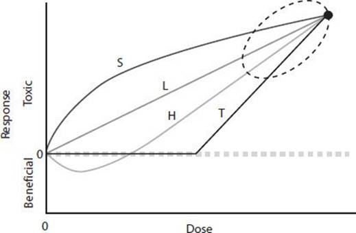
 Figure 16–1. In this figure, dose-response curves are abbreviated as follows: H = hormetic (biphasic); L = linear (no threshold); S = supralinear; T = threshold. The dose-response curves show the range of possible dose response relationships in an individual.
The frequency of a response in a population can be related to dose as a frequency distribution (as in Figure 16–2) or as a cumulative frequency (as in Figures 16–3 and 16–4).
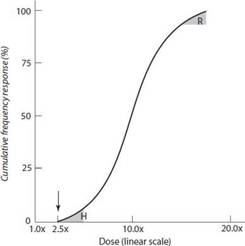
Figure 16–2. The existence of a threshold in this dose response curve is indicated by the arrow. Doses below this point do not produce a response. Individuals who exhibit the response at doses well below the average or the mean are considered hypersusceptible (H), whereas those who respond only to doses well above the average or the mean are considered resistant (R).
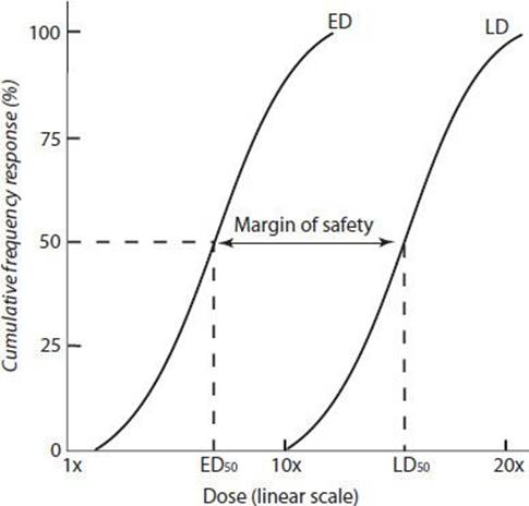
Figure 16–3. Dose-response curves comparing two doses of the same toxic substance. ED = effective dose; LD = lethal dose. The area between the ED and LD is the margin of safety.
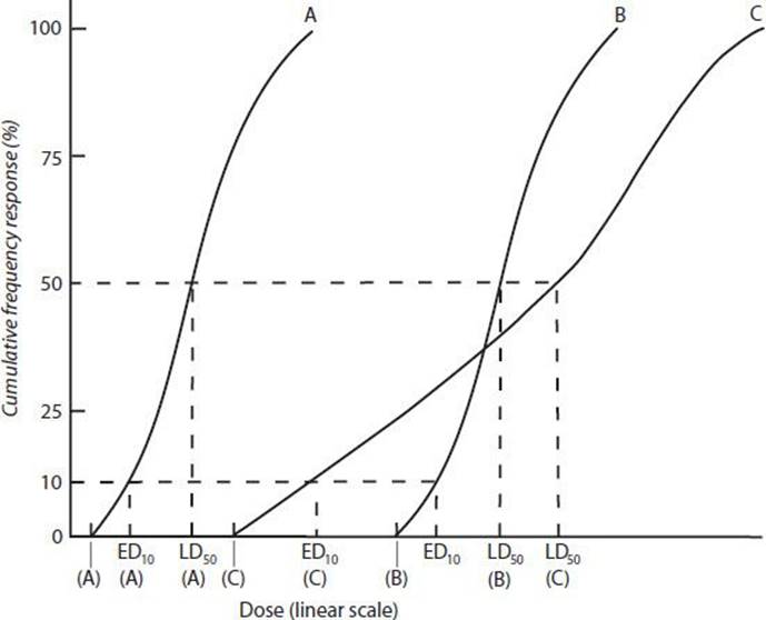
Figure 16–4. Dose-response curves comparing the doses at which the same toxic effect is elicited by three different toxic substances (A, B, and C).
In Figure 16–2, the existence of a threshold is indicated by the arrow at the point where the curve intersects the dose coordinate. Doses below this point do not produce a response. Individuals who exhibit the response at doses well below the average or the mean are considered hypersusceptible (H in Figure 16–2), while those who respond only to doses well above the average or the mean are considered resistant (R in Figure 16–2).
In Figure 16–3, cumulative frequency curves are used to compare two doses of the same toxic substance to the dose that is lethal to 50% of the population (LD50) and the dose that has an effect on 50% (ED50). The ED50 may, for example, represent an effect that is not harmful, such as odor. The ratio between comparable points on the curves (ie, the ratio of LD50 to ED50) will then represent the margin of safety for odor as a warning against a toxic or lethal effect.
In Figure 16–4, cumulative frequency curves are used to compare the doses at which the same toxic effect is elicited by three different toxic substances (A, B, and C). Substance A is clearly the most toxic, because at every dose level a greater percentage of the population exhibits the response to A than to B or C. The LD50, the ED10, and the threshold for A are all lower than the corresponding values for B and C. The comparison between B and C is less clear and demonstrates the need to consider the entire dose-response curves rather than individual points when comparing toxicities. Because the LD50 of B is lower than that of C, at this dose B is more toxic than C. However, because the ED10 of C is lower than that of B, at the lower dose C is more toxic than B. The shape of a dose-response curve is important for assessing the hazard of a toxic substance. A substance that has a low threshold and shallow dose-response curve (such as C) may be more hazardous at low doses, while a substance that has a steep dose-response curve (such as B) may be more hazardous as the dose increases. Adequate assessment of the hazard of a toxic substance requires evaluation of dose-response data over a wide range of doses.
DIAGNOSIS OF TOXIC EFFECTS
In general, the manifestations of acute toxicity due to high-dose exposures will be more overt than those due to chronic toxicity or toxicity associated with low-dose exposures. Table 16–4 lists selected occupational exposures and their symptoms and signs.
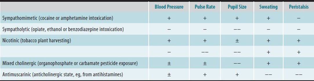
Table 16–4. Occupational/environmental toxic syndromes.
ACUTE EXPOSURE
Recognition of symptoms consistent with toxic exposure is key in evaluating and ultimately ordering tests to confirm acute exposure. Vital signs including blood pressure and pulse rate as well as examination of the pupils, skin, and bowel sounds can assist in the diagnosis (Table 16–4, Toxidromes). For example, a worker who becomes ill after harvesting tobacco may present with an increased blood pressure, heart rate, pupil size, and sweating, due to the toxic effects of nicotine absorbed across the skin. In the setting of an acute poisoning, access to adequate emergency care is essential, including advanced cardiac and respiratory supportive care. Few if any specific toxicologic tests are available on a rapid turnaround basis in the setting of an acute poisoning.
CHRONIC EXPOSURE
In cases of suspected chronic toxin exposure, signs and symptoms may be subtle or even missed if there is a low index of suspicion for a specific toxin. While many validated tests are available for toxin exposure (Chapter 42), these tests are best employed after a through history and physical has been performed, and a specific differential diagnosis is being explored. With chronic exposure to low doses, toxic agents are more likely to cause an increase in the incidence of disorders already present in the population rather than a novel disorder.
A common scenario the clinician is confronted with is a patient who requests testing for various environmental toxins that the patient suspects may be causing a multitude of symptoms. For example, fear of chronic heavy metal poisoning is often invoked. Metals are ubiquitous in the environment, resulting in constant human exposure, including nonessential metals such as lead. Clinical testing is able to detect very low levels of heavy metals, at concentrations lower than those associated with known toxic effects. The practice of administering a chelating agent then testing for urine heavy metals is (postchallenge or postprovocation testing) not supported by strong evidence and results are difficult to interpret. Moreover, administration of chelating agents can increase the elimination of some essential minerals such as iron, copper, and zinc.
Hair analysis has been used in determining exposure to heavy metals including arsenic, methyl mercury, and selenium and drugs of abuse including amphetamines. However, the results of hair testing are difficult to interpret as hair samples are subject to external contamination.
Trace metals analysis can be technically difficult, as specimens are easily contaminated during the collection process, including needles and vacutainers used in collection. Special care should be used in collection and testing of trace metals from body fluid to reduce the risk of contamination.
MANAGEMENT OF TOXIC EFFECTS
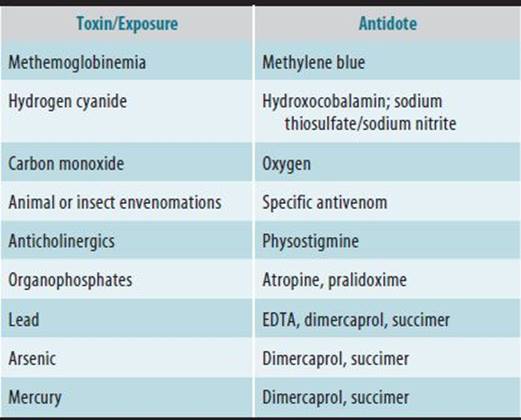
Management of acute toxicity consists of removal from exposure, symptomatic treatment, and supportive care. In cases of life-threatening toxicity, maintenance of cardio-pulmonary function and fluid and electrolyte balance are of high priority. After acute ingestion of a poisonous drug or chemical, measures to limit gastrointestinal absorption by administration of activated charcoal or whole bowel irrigation may be employed. For chemicals on the skin or in the eye, topical decontamination should be carried out, usually with copious water or saline solution. Methods to enhance elimination such as hemodialysis may be effective for a few acute poisonings. A number of specific treatments or antidotes exist for acute poisoning. Chelating agents may help eliminate some metals (eg, lead, arsenic, and mercury), but they are less likely to have an effect on subacute or chronic toxicity. Atropine and pralidoxime can be lifesaving in reversing the acute cholinesterase-inhibiting effects of organophosphate pesticides. Hydroxocobalamin (vitamin B12a) is used as an antidote for cyanide, and methylene blue can be used in patients with methemoglobinemia. Use of oxygen counters the effect and enhances the elimination of carbon monoxide. Table 16–5 lists several common antidotes used in occupational and environmental toxic exposures.
Table 16–5. Common antidotes used in occupational and environmental exposure.
Carcinogens
The International Agency for Research on Cancer (IARC) rates the carcinogenic potential of chemical agents for humans. The agency currently rates over 100 chemical agents as “known” carcinogens. These ratings are based primarily on human and animal data.
Although substances are implicated in the abnormal development of the fetus, less than 10% of birth defects can be attributed to exposure to a toxic substance. The human fetus is most susceptible to teratogenic agents in first 3–8 weeks of gestation. Known workplace teratogens include antineoplastic agents, carbon monoxide, mercury, lead, and tobacco smoke.
RESOURCES
American Associations of Poison Control Centers. www.aapcc.org.
International Agency for Research on Cancer. www.iarc.fr.
The Hospital for Sick Children. www.motherisk.org.
SELF-ASSESSMENT QUESTIONS
Select the one correct answer for each question.
Question 1: Nanoparticles
a. are engineered structures that are less than 100 nm in size
b. have no chemical properties
c. are blocked from the central nervous system
d. are easily measured in biologic tissues
Question 2: Toxicity
a. affects all ages to the same degree
b. is independent of racial and genetic background
c. has no important genetic factors
d. is affected by atmospheric pressure, temperature, and humidity
Question 3: G6PD deficiency
a. primarily affects white males and persons of Mediterranean descent
b. is an X-linked recessive disorder
c. prevents hemolysis from many drugs
d. screening is supported by solid evidence
Question 4: Biotransformation
a. occurs in the liver by hydrolysis, oxidation, reduction, and conjugation
b. is independent of microsomal cytochrome P450
c. can be arrested by many environmental and pharmacologic agents
d. is accelerated in neonates
Question 5: Clearance
a. is the rate at which a toxic agent is excreted, multiplied by the average concentration of the agent in the plasma
b. is a function of concentration in the urine, unrelated to dose
c. is a measure of how many milligrams of toxin is being removed
d. is a measure of the volume of fluid that is freed of the toxic agent per unit of time
Question 6: Hair analysis
a. is used only for evaluating heavy metal exposures
b. is the preferred method for evaluating toxic exposures
c. requires little training in collection and analysis
d. results are difficult to interpret as hair samples are subject to external contamination
Question 7: Management of acute toxicity
a. consists of removal from exposure, symptomatic treatment, and supportive care
b. first identifies the exact toxin so the appropriate antidote can be given
c. follows contact with public health authorities
d. focuses on preventing damage to the central nervous system