
Jeffrey L. Kishiyama, MD
Immune hypersensitivity mechanisms play a part in many disorders of occupational medicine. A basic appreciation of the components and physiology of normal immunity are central to the understanding of the pathophysiology of hypersensitivity diseases of the immune system.
OVERVIEW OF THE IMMUNE RESPONSE
The function of the immune system is to protect the host from invasion by foreign antigens by distinguishing “self” from “nonself” antigens. Such a system is necessary for survival in all living animals. A normal immune response relies on the careful coordination of a complex network of specialized cells, organs, and biological factors necessary for the recognition of pathogens and subsequent elimination of foreign antigens. An abnormal, exaggerated immune response can cause hypersensitivity to foreign antigens, with resultant tissue injury and the expression of a variety of clinical syndromes, which are often seen in the practice of occupational medicine.
Innate & Adaptive Immunity
Living organisms have two levels of response against external invasion: (1) a nonspecific, innate system of natural immunity and (2) an adaptive system, which is acquired and relies on immunologic memory (Figure 17–1). Innate immunity is present from birth, does not require previous antigenic exposure, and is nonspecific in its activity. The skin and mucosal barriers serve as the first line of defense of the innate immune system. Soluble factors, such as proteolytic enzymes, chemoattractants, acute-phase proteins, cytokines and leukocytes, including phagocytes and natural killer cells, provide additional layers of protection. Toll-like receptors (TLR), found on macrophages, mast cells, and immature dendritic cells, recognize conserved patterns found in microbial proteins, DNA, RNA, and lipopolysaccharide (LPS), initiating inflammatory responses prior to adaptive response. Through a series of proteolytic activations, the serum and membrane components of the complement cascade amplify and regulate microbial killing and inflammatory responses. Despite the lack of specificity, innate immunity is largely responsible for protection against a vast array of environmental microorganisms and foreign substances.
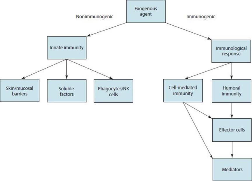
 Figure 17–1. Host response to exogenous agents or exposures. NK, natural killer.
Higher organisms have evolved the adaptive immune system, which is triggered by encounters with foreign agents that have evaded or penetrated the innate immune defenses. The adaptive immune system has specificity for individual foreign antigens and immunologic memory, which allows for an intensified response upon subsequent encounter with the same or closely related agent. Primary adaptive immune responses require clonal expansion, leading to a delayed response to new exposures. Secondary immune responses are more rapid, larger, and more efficient. Stimulation of the adaptive immune system triggers a complex sequence of events initiating the activation of lymphocytes, the production of antigen-specific antibodies (humoral immunity), and effector cells (cellular or cell-mediated immunity), and ultimately, the elimination of the inciting substance. Although adaptive immunity is antigen specific, the repertoire of responses is tremendously diverse, with an estimated 109 antigenic specificities.
Antigens & Immunogens
Foreign substances that can induce an immune response are called antigens or immunogens. Immunogenicity implies that the substance has the ability to react with antigen-binding sites on antibody molecules or T-cell receptors. Complex foreign agents possess distinct and multiple antigenic determinants or “epitopes,” dependent on the peptide sequence and conformational folding of immunogenic proteins. Most immunogens are proteins, although pure carbohydrates may be immunogenic as well. The immune response to a particular immunogen may also depend on the route of exposure to the foreign substance. Blood-borne substances are normally immunoglobulin bound and removed via the reticuloendothelial system. Occupational allergen exposure through respiratory mucosal surfaces can lead to vigorous local production of immunoglobulin, along with recruitment, activation, and proliferation of leukocytes in involved tissues and regional lymphoid tissues.
Cells of the Immune System
A number of effector cells participate in immune defense and hypersensitivity reactions. These include mast cells, basophils, polymorphonuclear neutrophils, eosinophils, macrophages, monocytes, platelets, and lymphocytes. Depending on the type of immune response, many or all play a part. Derived from hematopoietic stem cells, fully-differentiated effector cells have membrane receptors for various chemoattractants and mediators and participate in activation or destruction of target cells.
Lymphocytes are responsible for the initial specific recognition of antigen. They are functionally and phenotypically divided into B- and T lymphocytes. Structurally, B- and T lymphocytes cannot be distinguished visually from each other under the microscope; they can be enumerated by flow cytometric phenotyping or by immunohistochemical methods. Approximately 70–80% of circulating blood lymphocytes are T cells and 10–15% are B cells; the remainder are referred to as natural killer cells (also known as NK cells or null cells).
The thymus-derived cells (T lymphocytes or T cells) are involved in cellular immune responses. B-lymphocytes or B cells are involved in humoral or antibody responses. Precursors of T cells migrate to the thymus, where they develop some of the functional and cell surface characteristics of mature T cells. Through positive and negative selection, clones of autoreactive T cells are eliminated, and mature T cells migrate to the peripheral lymphoid tissues. There they enter the pool of long-lived lymphocytes that recirculate from the blood to the lymph.
T lymphocytes are heterogeneous with respect to their cell surface markers and functional characteristics. Numerous subpopulations of T cells are now recognized. Helper-inducer T cells (CD4) help to amplify B-cell production of immunoglobulin and amplify T-cell (CD8)–mediated cytotoxicity. Activated CD4 T cells regulate immune responses through cell-to-cell contact and by elaboration of soluble factors or cytokines.
Subsets of helper T cells can be identified on the basis of their pattern of cytokine production. T-helper type 1 (TH1) cells produce gamma-interferon (IFN-γ) and tumor necrosis factor-beta (TNF-β), and T-helper type 2 (TH2)cells produce interleukins 4, 5, 9, and 25, among others. Both subsets produce interleukin (IL)-2, IL-3, IL-10, IL-13, and granulocyte-macrophage colony-stimulating factor (GM-CSF). The TH1 and TH2 phenotypes represent diametrically-opposed T-helper immune responses. The TH1 subset of CD4 T cells promotes cellular immune responses to intracellular pathogens and underlies the pathogenesis of delayed-type hypersensitivity. TH2 cells play a central role in immediate hypersensitivity and humoral immune responses, because IL-4/IL-13 promote immunoglobulin (Ig) E production and IL-5 is an eosinophil proliferation and differentiation factor. TH1 and TH2 responses mutually antagonize each other. Although other regulatory mechanisms clearly influence the balance between TH1 and TH2responses, their polarity has led to the “hygiene hypothesis,” a paradigm explaining the immunopathogenesis of atopic diseases.
Cytotoxic or “killer” T cells are generated after mature T cells interact with certain foreign antigens. They are responsible for defense against intracellular pathogens (eg, viruses), tumor immunity, and organ graft rejection. Most killer T cells express the CD8 phenotype, although in certain circumstances, CD4 T cells can be cytotoxic. Cytotoxic T cells may kill their target through osmotic lysis, by secretion of TNF, or by induction of apoptosis, that is, programmed cell death.
Mucosal dendritic cells control the generation of regulatory T cells. TH-17 and T-regulatory cells (TREG) are subsets of T cells, which modulate inflammatory responses through the secretion of regulatory cytokines. TH-17 cells recruit neutrophils to sites of acute inflammation through secretion of IL-17. TREG cells are inhibitory, suppressing activated T effector cells by their secretion of interleukin-10 and TGF-β cellREG cells modulate responses to antigen, thereby regulating homeostasis and tolerance versus inflammation, allergy, and autoimmunity.
B-cell maturation proceeds in antigen-independent and antigen-dependent stages. Antigen-independent development occurs in the marrow where pre-B cells mature into immunoglobulin bearing naive B cell (a cell that has not been exposed to antigen previously). In peripheral lymphoid tissues, antigen-dependent activation produces circulating long-lived memory B cells and plasma cells found predominantly in primary follicles and germinal centers of the lymph nodes and spleen. All mature B cells bear surface immunoglobulin that is their antigen-specific receptor. The major role of B cells is differentiation to antibody-secreting plasma cells. However, B cells may also release cytokines and function as antigen-presenting cells.
Macrophages are involved in the ingestion, processing, and presentation of antigens for interaction with lymphocytes. In addition, they are effector cells for certain types of tumor immunity. Circulating monocytes are recruited to sites of inflammation where they mature into macrophages. Both monocytes and macrophages contain receptors for C3b (activated bound complement) and the Fc portion of IgG and IgE, allowing for activation through antigen-specific and nonspecific immune pathways. Activation of these cells occurs after binding to immune complexes, through exposure to various cytokines and after phagocytosis of antigen or particulates, such as silica and asbestos. They contain proteolytic enzymes and are able to synthesize proinflammatory mediators including cytokines, arachidonic, acid metabolites, and oxidative metabolites. Macrophages constitutively express toll-like receptor 4 (TLR4) which can bind bacterial endotoxin, triggering cytokine release. It is hypothesized that macrophage-derived IL-12 and TNF influences TH1 and TH2 differentiation, thereby affecting the expression of atopy and allergic disease.
NK cells are non-B, non-T lymphocytic cells, which can kill a wide spectrum of target cells. They are recognized by the presence of specific surface antigens (CD16 or CD56). NK cells are capable of binding IgG because of their membrane receptor for the IgG molecule (FcR). Antibody-dependent cell-mediated cytotoxicity (ADCC) occurs when an organism or a cell is coated by antibody and undergoes NK cell–mediated destruction. Alternatively, NK cells can destroy virally infected cells or tumor cells nonspecifically.
Mast cells are basophilic staining cells found chiefly in connective and subcutaneous tissue. They have prominent granules that are the source of many mediators of immediate hypersensitivity and have 30,000–200,000 cell surface membrane receptors for the Fc fragment of IgE. When an allergen molecule cross-links two adjacent mast cell surface–associated IgE antibodies, calcium-dependent cellular activation leads to the release of both preformed and newly generated mediators. Mast cells also have surface receptors for “anaphylatoxins” (activated complement fragments, C3a, C4a, and C5a), cytokines, and neuropeptides, such as substance P. Activation by these non–IgE-mediated mechanisms may contribute to host immunity and provide ties between the immune and neuroendocrine systems. Mast cell–deficient mice display a particular vulnerability to sepsis and rapid death after peritonitis, possibly due to insufficient TNF-α production during bacterial infection. Mast cells also appear in areas of wound healing and in fibrotic lung disease. Experimentally, mast cell–derived mediators promote angiogenesis and fibrogenesis, suggesting their presence in these sites is pathologically relevant.
Neutrophils are granulocytes that phagocytose and destroy foreign antigens and microbial organisms. They are attracted to the site of antigen by chemotactic factors, including plasma-activated complement 5 (C5a), leukotriene B4 (LTB4), granulocyte colony-stimulating factor (G-CSF), GM-CSF, IL-8, and platelet-activating factor (PAF). They possess receptors for the Fc fragment of IgG and IgM antibodies (specific opsonins) and for complement fragment C3b (nonspecific opsonin). Smaller antigens are phagocytosed and destroyed by lysosomal enzymes. Locally released lysosomal enzymes destroy particles too large to be phagocytosed. Neutrophils contain or generate a number of antimicrobial factors, including oxidative metabolites, superoxide, and H2O2; myeloperoxidase, which catalyzes the production of hypochlorite; and proteolytic enzymes, including collagenase, elastase, and cathepsin B. Some or all of these factors may play a part in a number of hypersensitivity reactions, including the type I late asthmatic response, the type II cytotoxic reaction, and type III immune complex disease (see the section “Classification of Immune Hypersensitivity Disorders”).
Eosinophils play both a proactive and a modulating role in inflammation. They are attracted to the site of the antigen-antibody reactions by PAF, C5a, chemokines, histamine, and LTB4. They are important in the defense against parasites. When stimulated, they release numerous inflammatory factors, including major basic protein (MBP), eosinophil-derived neurotoxin, eosinophil cationic protein (ECP), eosinophil peroxidase, lysosomal hydrolases, and LTC4. MBP destroys parasites, impairs ciliary beating, and causes exfoliation of respiratory epithelial cells; it may trigger histamine release from mast cells and basophils. Eosinophil-derived products may play a role in the development of airway hyperreactivity.
Dendritic cells are specialized phagocytic cells, which are abundant near mucosal surfaces that internalize microorganisms and debris, and travel to secondary lymphoid organs and present antigen to circulating naïve T cells. They also display toll-like receptors and upon binding to microorganisms trigger inflammatory cytokine production, bridging innate and adaptive immune responses.
MEDIATORS OF IMMEDIATE HYPERSENSITIVITY
Mediators of immediate hypersensitivity are chemicals generated or released by effector cells following activation. They have various biological activities and normally function in host defense, but they play a pathologic role in immune hypersensitivity. Mediators may exist in a preformed state in the granules of mast cells and basophils, or are newly synthesized at the time of activation of these and some other nucleated cells (Tables 17–1 and 17–2). Increased awareness of the immunologic and physiologic effects of mediators has led to a better understanding of immunopathology and provides potential targets for future pharmacotherapies.
Table 17–1. Mediators of immediate hypersensitivity.
Vasoactive and smooth-muscle constricting mediators
Preformed
Histamine
Generated
Arachidonic acid metabolites (PGD2, LTC4)
PAF
Adenosine
Chemotactic mediators
Eosinophil-directed
Eosinophilic chemotactic factor of anaphylaxis (ECFA)
ECF oligopeptides
PAF
Neutrophil-directed
High-molecular-weight neutrophilic chemotactic factor
LTB4
PAF
Enzymatic mediators
Neutral proteases
Tryptase
Chymase
Lysosomal hydrolases
Other enzymes
Superoxide dismutase
Peroxidase
Table 17–2. Action of mediators in hypersensitivity reactions.
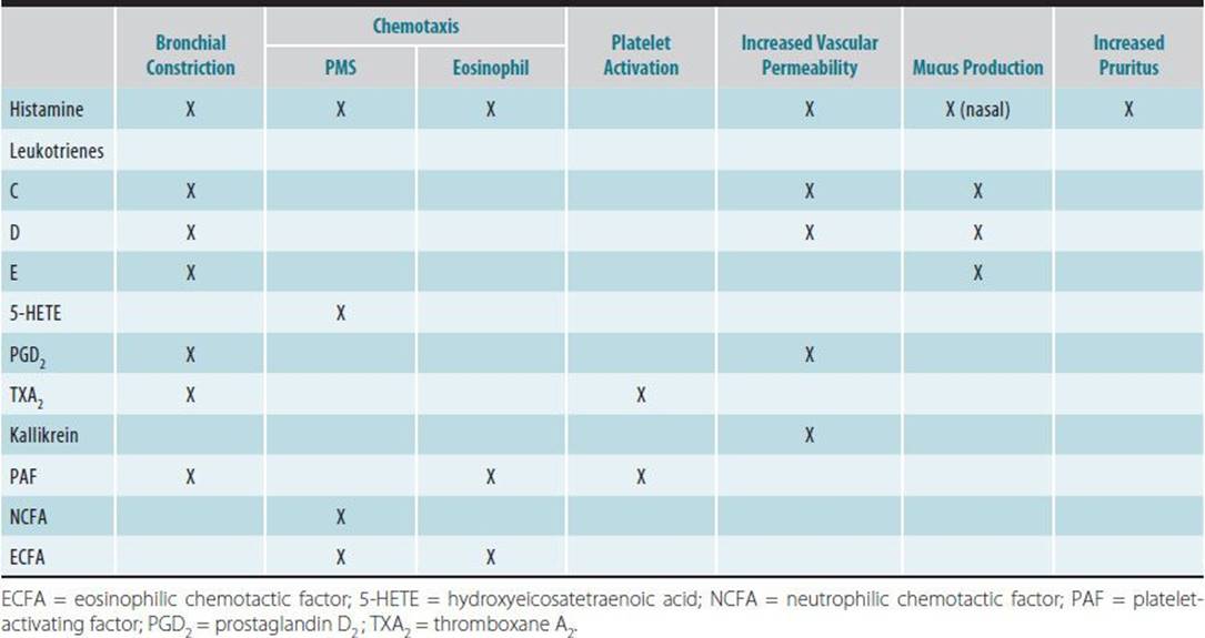
Preformed mediators include histamine, eosinophil and neutrophil chemoattractants, proteoglycans (heparin, chondroitin sulfate), and various proteolytic enzymes. Histamine is a bioactive amine, packaged in dense intracellular granules, that when released binds to membrane-bound H1, H2, and H3 receptors resulting in significant physiologic effects. Binding to H1 receptors causes smooth muscle contraction, vasodilatation, increased vascular permeability, and stimulation of nasal mucous glands. Stimulation of H2 receptors causes enhanced gastric acid secretion, mucus secretion, and leukocyte chemotaxis. Histamine is important in the pathogenesis of allergic rhinitis, allergic asthma, and anaphylaxis.
Newly generated mediators include kinins, platelet-activating factor, and arachidonic acid metabolites, including leukotrienes and prostaglandins. In many immune cells, arachidonic acid, liberated from membrane phospholipid bilayers, is metabolized either by the lipoxygenase pathway to form leukotrienes (LT) or by the cyclooxygenase pathway to form prostaglandins (PG) and thromboxanes A2 and B2(TXA2 and TXB2). LTB4 is a potent chemoattractant for neutrophils. LTC4, LTD4, and LTE4 constitute slow-reacting substance of anaphylaxis, which has bronchial smooth muscle spasmogenic potency 100–1000 times that of histamine and which also causes vascular dilation and vascular permeability.
Almost all nucleated cells generate prostaglandins. The most important members are PGD2, PGE2, PGF2, and PGI2 (prostacyclin). Human mast cells produce large amounts of PGD2, which causes vasodilatation, vascular permeability, and airway constriction. Activated polymorphonuclear neutrophils and macrophages generate PGF2a, a bronchoconstrictor, and PGE2, a bronchodilator. PGI2 causes platelet disaggregation. TXA2 causes platelet aggregation, bronchial constriction, and vasoconstriction.
Macrophages, neutrophils, eosinophils, and mast cells generate PAF, which causes platelet aggregation, vasodilatation, increased vascular permeability, and bronchial smooth-muscle contraction. PAF is the most potent eosinophil chemoattractant described and also plays a role in anaphylaxis. The kinins are vasoactive peptides formed in plasma when kallikrein, released by basophils and mast cells, digests plasma kininogen. Kinins, including bradykinin cause slow, sustained contraction of bronchial and vascular smooth muscle, vascular permeability, secretion of mucus, stimulation of pain fibers contributing to human angioedema and anaphylaxis.
Complement Cascades
The union of antigen with IgG or IgM antibody initiates activation of the classic complement pathway. Complement-fixing sites on these immune complexes are exposed, allowing binding of the first component of the complement sequence, C1q. Other components of the complement sequence are subsequently bound, activated, and cleaved, eventually leading to cell lysis. Important by-products of the classic pathway include activated cleavage products, the anaphylatoxins C3a, C5a, and less-potent C4a. C5a is a potent leukocyte chemotactic factor, and also causes mediator release from mast cells and basophils. C4b and C3b mediate binding of immune complexes to phagocytic cells, facilitating opsonization.
Activation of the complement sequence by the alternative pathway is initiated by a number of agents, including lipopolysaccharides (LPS), trypsin-like molecules, aggregated IgA and IgG, and cobra venom. Activation of the alternative pathway does not require the presence of antigen-antibody complexes, nor does it use the early components of the complement sequence, C1, C4, and C2. Ultimately, as a result of activation of the classic or alternative pathway, activation of the terminal complement sequence occurs, resulting in cell lysis and/or tissue inflammation.
Cytokines
Many immune functions are regulated or mediated by cytokines, which are soluble factors secreted by activated immune cells. Cytokines can be organized functionally into groups according to their major activities: (1) those that promote inflammation and mediate natural immunity, such as IL-1, IL-6, IL-8, TNF, and interferon (IFN)-γ; (2) those that support allergic inflammation, such as IL-4, IL-5, and IL-13; (3) those that control lymphocyte regulatory activity, such as IL-10, IL-12, and IFN-γ; and (4) those that act as hematopoietic growth factors, such as IL-3, IL-7, and GM-CSF (Table 17–3). This complicated network of interacting cytokines functions to modulate cellular function and immunologic responses. Many ongoing research investigations are focused on modulating cytokine responses as a way to control or treat disease processes.
Table 17–3. Major cytokines.

Response to Antigen
The major immunologic responses to antigen include the elimination of antigen through antibody-mediated events (humoral response) and the direct killing of target cells by a subset of T lymphocytes called cytotoxic T lymphocytes (CTLs) (cellular response). The series of events that embody the immune response include antigen processing and presentation, lymphocyte recognition and activation, cellular and/or humoral immune responses, and antigenic destruction or elimination (Figure 17–2).
Immune responses may have both positive and deleterious effects. Tight control of inflammatory mechanisms promotes efficient elimination of foreign substances and prevents uncontrolled lymphocyte activation and unregulated antibody production. Inappropriate activation or dysregulation of the system, however, can perpetuate inflammatory processes leading to tissue damage and organ dysfunction. Inflammation is responsible for hypersensitivity reactions and for many of the clinical effects of autoimmunity.
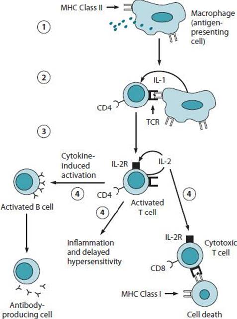
Figure 17–2. The normal immune response. (1) Antigen processing and presentation by antigen-presenting cells. (2) Recognition of antigen-MHC complex by CD4 T lymphocytes induces IL-1 secretion by antigen-presenting cells and subsequent cellular activation. (3) Activated T lymphocytes express IL-2 receptors and secrete IL-2, which upregulate IL-2 receptor expression in an autocrine fashion. (4) Activated CD4 T lymphocytes can stimulate CD8 cytotoxic T lymphocytes to mediate cellular cytotoxicity, B-lymphocyte activation, and differentiation into antibody-producing plasma cells, which mediate humoral immunity or mediate delayed hypersensitivity and other inflammatory reactions.
A. Antigen Processing and Presentation
Most foreign immunogens are not recognized by the immune system in their native form and require capture and processing by specialized antigen-presenting cells. Antigen-presenting cells include macrophages, dendritic cells in lymphoid tissue, Langerhans cells in the skin, Kupffer cells in the liver, microglial cells in the nervous system, and B lymphocytes. Following encounter with immunogens, the antigen-presenting cells internalize the foreign substance by phagocytosis or pinocytosis, modify its parent structure, and display antigenic fragments of the native protein on its surface.
B. T-Lymphocyte Recognition and Activation
The recognition of processed antigen by specialized T lymphocytes known as “helper” T (CD4+) lymphocytes constitutes the critical event in the immune response. The helper T lymphocytes orchestrate the many cells and biological signals that are necessary to carry out the immune response. Helper T lymphocytes recognize processed antigen displayed by antigen-presenting cells only in association with polymorphic cell surface proteins encoded by the major histocompatibility gene complex. MHC genes are highly polymorphic and determine immune responsiveness. They are known as HLA or human leukocyte antigen, with certain HLA genotypes conferring genetic susceptibility to or resistance against a range of specific autoimmune, environmental, and occupational diseases.
Endogenously synthesized viral proteins are processed in association with MHC class I molecules, while exogenous foreign antigens that require an antibody-mediated response are expressed in association with MHC class II structures. All somatic cells express MHC class I, whereas only the specialized antigen-presenting cells can express MHC class II. Helper T lymphocytes expressing the CD4 antigen recognize antigen in the context of MHC class II, while cytotoxic T lymphocytes (CD8+) recognize target cells bearing MHC class I complexed to antigen.
Two signals are required for activation of these T lymphocytes: (1) binding of the antigen-specific T-cell receptor (CD3) to the antigen-MHC complex, and (2) costimulation through CD28(on T cells)-B7(on antigen presenting cell) interactions. These two signals induce the expression of IL-2 receptors on the surface of the CD4+ lymphocytes, as well as the production of various cell growth and differentiation factors (cytokines). Activated CD4+ T helper lymphocytes subsequently trigger the effector cells that mediate the cellular and humoral arms of the immune response.
Cytotoxic T lymphocytes (CTLs) eliminate target cells (virally infected cells, tumor cells, or foreign tissues), constituting the cellular immune response. These “killer” T lymphocytes release substances called cytotoxins, which lead to cytolysis or destruction of infected target cells. CTLs arise from the antigen-driven activation and differentiation of resting mature small lymphocyte precursors. Activated CTLs manufacture a membrane pore-forming protein (perforin or cytolysin), IFN-γ. Killing of target cells by CTLs requires direct cell-to-cell contact and proceeds sequentially by (1) adhesive interactions between CTLs and target cell, (2) activation of CTLs by antigen engagement of CTL receptors, (3) delivery of the lethal hit to target cells by poorly characterized mechanisms, and (4) programmed cell death of target cells.
C. Activation of B Lymphocytes (Humoral Immune Response)
The primary function of mature B lymphocytes is to synthesize antibodies. After proliferation and terminal differentiation, antigen binding to B-cell receptors, that is, surface antibodies, B cells become high-rate antibody-producing cells, called plasma cells. Antibodies are immunoglobulin molecules directed toward specific antigens and mediate humoral immunity. B lymphocytes may also bind and internalize foreign antigen directly, process that antigen, and present it to CD4+ T lymphocytes. A pool of activated B lymphocytes may differentiate to form memory cells, which respond more rapidly and efficiently to subsequent encounters with identical or closely related antigenic structures. These secondary immune responses are more rapid and larger as a consequence of immunologic memory.
D. Antibody Structure and Function
It has been estimated that the repertoire of immunoglobulin antigen specificities in the human body is 107. Immunoglobulins serve a variety of secondary biological roles, including complement fixation, transplacental passive immunization of neonates, and facilitation of phagocytosis (opsonization), all of which participate in host defense against disease (Figure 17–3). Circulating immunoglobulins have both a unique specificity for one particular antigenic structure and diversity to encounter a broad range of antigenic materials. This diversity arises from complex DNA rearrangements and RNA processing within B lymphocytes early in their ontogeny. All immunoglobulin molecules share a four-chain polypeptide structure consisting of two heavy and two light chains. Each chain includes an amino-terminal portion, containing the variable (V) region, and a carboxy-terminal portion containing four or five constant (C) regions. V regions are highly variable structures, which form the antigen-binding site, whereas the C domains support effector functions of the molecules. There are five classes (isotypes) of immunoglobulins, which are defined on the basis of differences in the C region of the heavy chains. IgG is the predominant immunoglobulin in serum. IgG antibodies are strong precipitins, and three subclasses—IgG1, IgG2, and IgG3—can activate complement, qualities contributing to the pathogenesis of serum sickness and certain types of hypersensitivity pneumonitis (eg, bird breeder’s disease).
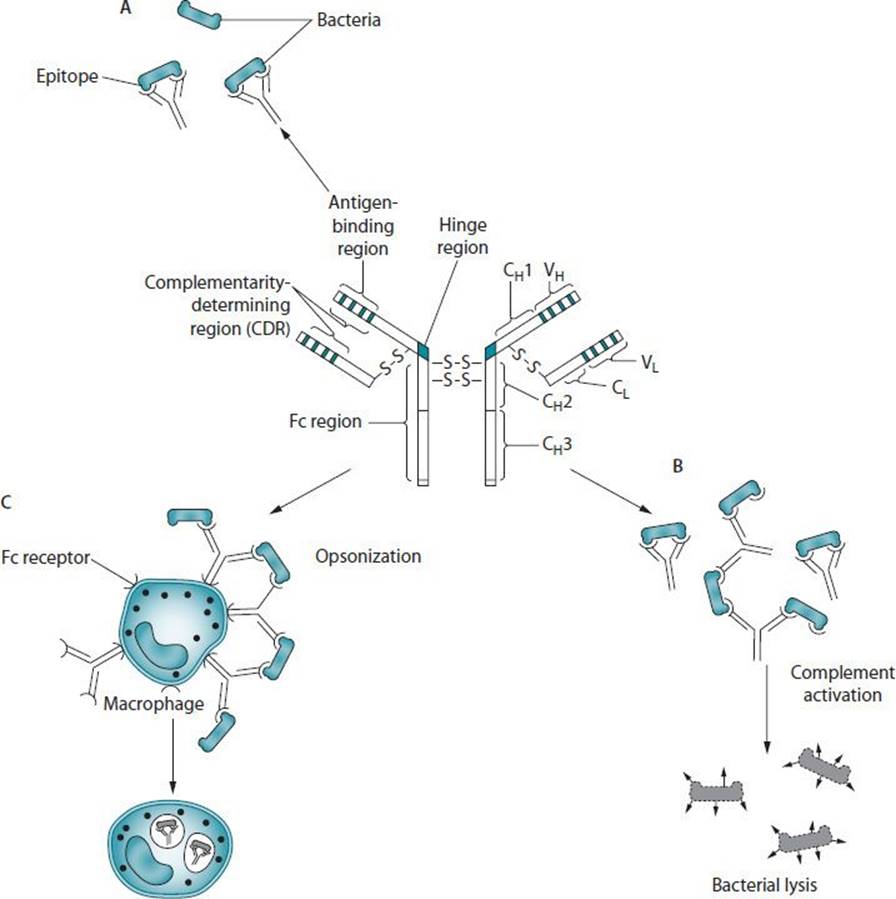
Figure 17–3. Immunoglobulin function. Antibody has multiple functions. The prototypical antibody consists of two heavy (H) and two light (L) chains, each subdivided into constant (CL, CH) and variable (VL, VH) domains. The structure is held together by intra- and interchain disulfide bridges. (A) The complementarity-determining region (CDR) of the antigen-binding portion of the antibody engages the antigenic determinant (epitope) in a lock and key fashion. (B) Antigen-antibody complexes activate complement to produce split complement components that cause bacterial lysis. (C) The Fc portion of antibodies binds to Fc receptors on phagocytes (eg, macrophages, neutrophils) and facilitates uptake of bacteria (opsonization).
IgA is the predominant immunoglobulin on mucous membrane surfaces. It exists predominantly as a monomer in serum and as a dimer or trimer when secreted on mucous membrane surfaces. When the dimer or trimer passes through the epithelial cells to a mucous membrane surface, it acquires a smaller molecule called a secretory piece that stabilizes the molecule and prevents its degradation by proteolytic enzymes. IgA antibodies protect the host from foreign antigens on mucous membrane surfaces, but they do not fix complement by the classic pathway.
IgM is a pentamer that is found almost exclusively in the intravascular compartment. IgM antibodies are potent agglutinins and fix complement. They may mediate the trimellitic anhydride pulmonary anemia syndrome. IgD is a monomeric immunoglobulin. Its biological function is unknown.
IgE is the heaviest immunoglobulin monomer, with a normal concentration in serum varying from 20 to 100 IU, but the concentration may be five times normal or even higher in an atopic individual. The Fc portion of IgE binds to receptors on the surfaces of mast cells and basophils. IgE antibodies play an important role in immediate hypersensitivity reactions such as nasal allergy and allergic asthma in veterinarians, laboratory animal handlers, and enzyme detergent industry workers.
E. Humoral Mechanisms of Antigen Elimination
Antibodies can induce the elimination of foreign antigen through a number of different mechanisms. Binding of antibody to bacterial toxins or foreign agents may neutralize or promote elimination of antigen-antibody “immune complexes” through the reticuloendothelial system. Antibodies can also coat bacterial surfaces, allowing clearance by macrophages in a process known as opsonization. Some classes of antibodies can complex with antigen and activate the complement cascade, which culminates in lysis of the target cell. Finally, the major class of antibody, IgG, can bind to natural killer cells that subsequently complex with target cells and release cytotoxins through antibody-dependent cytotoxicity (ADCC).
CLASSIFICATION OF IMMUNE HYPERSENSITIVITY DISORDERS
Gell and Coombs devised a classification scheme to define the basic immunopathologic mechanisms of hypersensitivity by four distinct types of reactions (types I–IV) (Figure 17–4). Types I–III are all mediated by specific antibodies (Humoral Immune Response), while type IV results from the actions of sensitized T lymphocytes (cell-mediated immune response). All defined mechanisms require an initial exposure to antigen, which induces a primary immune response (sensitization). Subsequent exposure to the same antigen (challenge) following a short lag period (usually at least 1 week) evokes the hypersensitivity response.

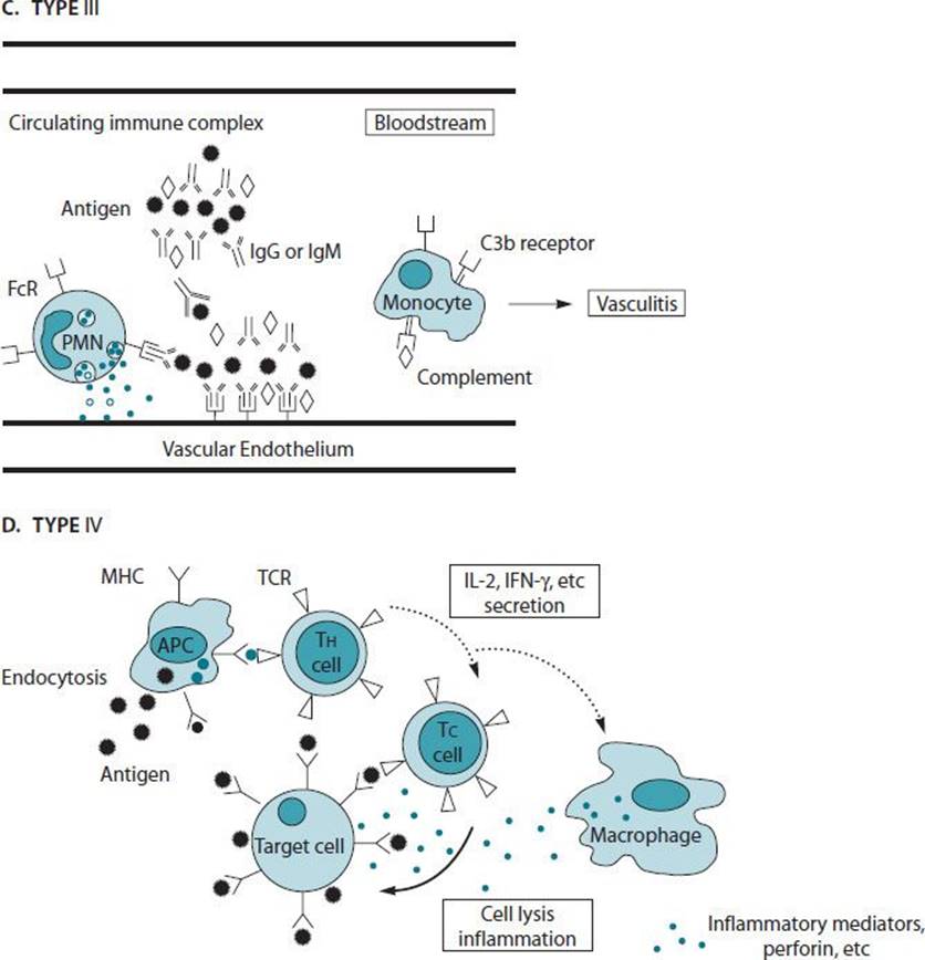
Figure 17–4. Hypersensitivity immune responses. A. Type I reaction. Mast cells and basophils bind IgE via high-affinity Fc receptors (FcεR1). Antigen-binding and cross-linking of FcεR1-IgE complexes induce cellular degranulation and release of inflammatory mediators. B. Type II reaction. IgG or IgM antibodies against tissue or cellular antigens induce complement activation, which results in cell death and tissue injury. C. Type III reaction. Circulating immune complexes composed of soluble antigen and IgG or IgM deposit on vascular endothelium of various tissues, which activates the complement cascade. Polymorphonuclear leukocytes (PMNs) and other phagocytes are attracted to these sites of immune complex deposition via their Fc and C3b receptors and are induced to degranulate and phagocytose the complexes, resulting in local tissue injury and vasculitis. D. Type IV reaction. Via their T-cell receptors, helper T cells recognize target cell antigenic peptides bound to antigen-presenting cells. This T-cell recognition results in the secretion of interleukin 2, interferon-gamma, and other cytokines that are required for the activation of tissue macrophages and cytotoxic T cells. Cytotoxic T cells recognize the same antigen bound to target cells and induce lysis by perforin and other molecules secreted by cytotoxic T cells. ADCC, antibody-dependent cell-mediated cytotoxicity; APC, antigen-presenting cell; Tc, cytotoxic T cell; TCR, T-cell receptor; TH, T-helper cell.
Type I: Anaphylactic or Immediate Hypersensitivity Reactions
These reactions are initiated by the interaction of antigen with specific IgE antibodies bound to mast cells and basophils with the subsequent release of inflammatory mediators. Examples of type I reactions in the practice of occupational medicine include allergic rhinitis and asthma seen in bakers and animal handlers and systemic anaphylaxis in beekeepers and health care workers (latex allergy).
Initial exposure to antigen in a genetically predisposed host leads to the synthesis of antigen-specific IgE by mature B cells, constituting the atopic state. Isotype switching to IgE production requires the cytokine IL-4, along with additional B lymphocyte activation and differentiation factors (IL-5, IL-6, IL-13). In contrast, IFN-γ, a TH1 cytokine, inhibits IL-4–dependent IgE synthesis in humans. It has been hypothesized that the balance of IL-4 responses and those favoring IFN-γ may influence whether atopy develops in an individual.
Helper (CD4+) T lymphocytes play a central role in the induction of normal immune responses. Activated T lymphocytes that release TH2-characteristic cytokines have been found at sites of inflammation in allergic airway disease and are believed to direct the immune response toward allergic inflammation.
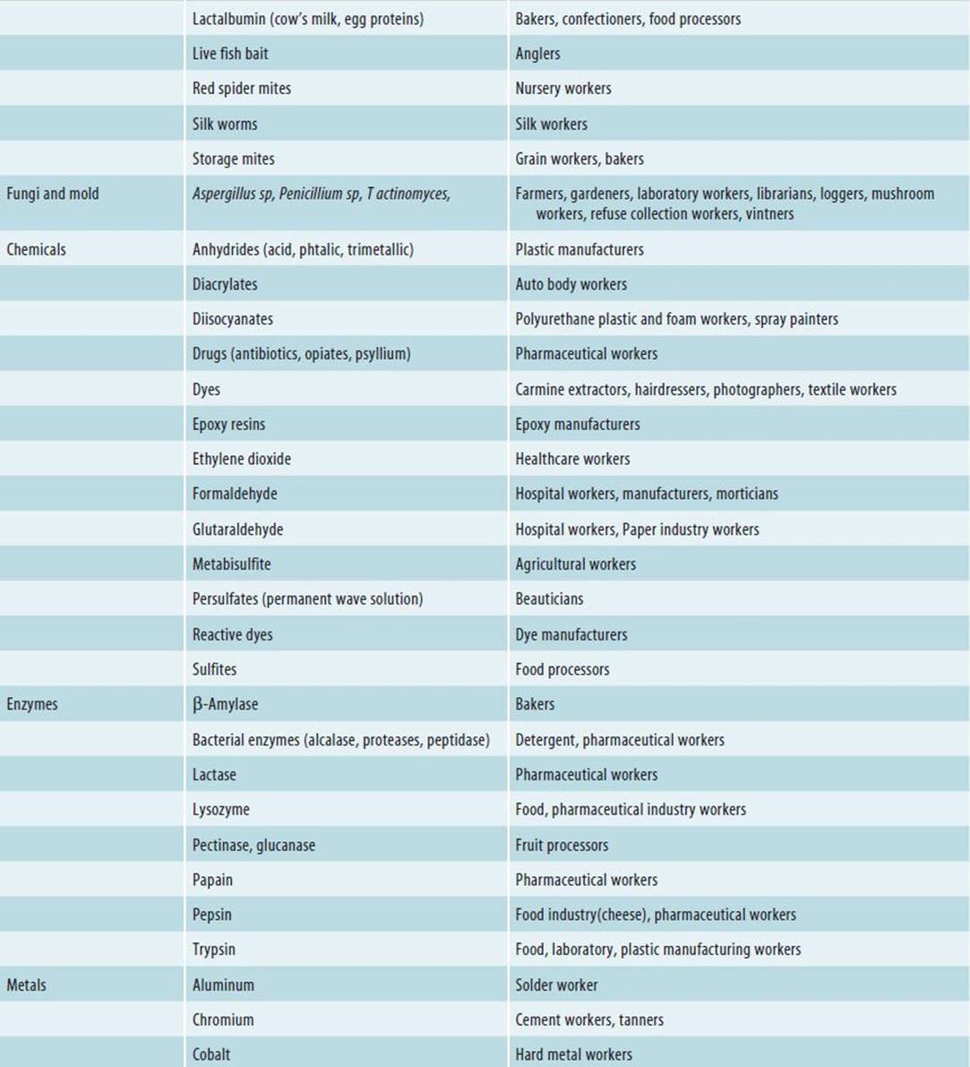
Antigen-specific IgE binds to high-affinity Fc receptors on tissue mast cells and basophils as well as to low-affinity Fc receptors on lymphocytes, macrophages, eosinophils, and platelets, thus sensitizing these cells for future allergen encounters. Upon reexposure to allergen, the sensitized individual can mount an immediate hypersensitivity response. Mast cells, armed with antigen-specific IgE on their surfaces, can bind polyvalent allergen, cross-linking adjacent IgE molecules, thereby activating and degranulating the cell. Table 17–4 includes many occupational causes of immediate hypersensitivity reactions. Common environmental aeroallergens causing IgE-mediated reactions include pollen, house dust mite, mold, and animal dander. Activation of mast cells and basophils induces both the release of preformed mediators from cytoplasmic granules (histamine, chemotactic factors, and enzymes) and the synthesis and release of newly generated mediators (prostaglandins, leukotrienes, and platelet-activating factor). Mast cells and basophils also have the ability to synthesize and release proinflammatory cytokines, growth factors, and regulatory factors, which interact in complex networks.
Table 17–4. Materials causally linked to rhinitis and asthma in the workplace.


The interaction of mediators with specific target organs and cells frequently induces a biphasic response: an early effect on blood vessels, smooth muscle, and secretory glands marked by vascular leakiness, smooth-muscle constriction, mucus hypersecretion, and a late response characterized by mucosal edema and the influx of inflammatory cells. The early-phase response occurs within minutes of an antigen exposure. In allergic rhinoconjunctivitis, the early-phase response is marked grossly by erythema, localized edema, rhinorrhea, and pruritus that result largely from the interaction of histamine with target tissues of the upper airway and conjunctival mucosa. Histologically, the early response is characterized by vasodilatation, edema, and a mild cellular infiltrate of mostly granulocytes.
The late-phase response may either follow the early-phase response (dual response) or occur as an isolated event (isolated late phase). Late-phase reactions begin 2–4 hours after initial antigen exposure, reach maximal activity at 6–12 hours, and usually resolve within 12–24 hours. The late-phase response is characterized grossly by erythema, induration, heat, burning, and itching and microscopically by an influx of primarily eosinophils and mononuclear cells. Eosinophils are frequently sampled from the nasal mucosa of patients with allergic rhinitis and from the sputum of asthmatics. Products of activated eosinophils such as major basic protein and eosinophilic cationic protein are destructive to airway epithelial tissue and are associated with airways hyperreactivity. Epithelial disruption is a feature of patients with both atopic dermatitis and asthma. Immune cells infiltrating tissues in the late response may further elaborate cytokines and histamine-releasing factors that perpetuate the inflammatory response, leading to a sustained hyperresponsiveness and disruption of the target tissue (eg, bronchi, skin, or nasal mucosa). As exposure to allergen persists, onset and resolution of the late-phase response becomes nebulous, as chronic tissue inflammation develops. Late-phase reactivity has been found in atopic disease states, including allergic rhinitis and conjunctivitis, asthma, food-sensitive atopic dermatitis, and anaphylaxis. It is possible that dual asthmatic responses seen in individuals with detergent worker’s asthma, trimellitic anhydride asthma, and baker’s asthma represent clinical examples of bronchial late-phase responses. Targeting the late-phase response with anti-inflammatory therapies, such as topical corticosteroids, remains a cornerstone of the medical management for allergic rhinitis and asthma.
Immediate hypersensitivity can be demonstrated in vivo by a prick or intradermal skin test to specific antigen, or in vitro by the radioallergosorbent test (RAST) or the enzyme-linked immunosorbent assay (ELISA).
Type II: Cytotoxic Reactions
In type II reactions, the target antigen is a cell surface protein or antigenic substance that binds covalently to a cell surface protein (see Figure 17–4). Cell surface–bound antigen-antibody union takes place, facilitating phagocytosis or activating the terminal complement sequence, leading to cell lysis. In transfusion reactions, the antigen is a protein on the cell membrane of the incompatible erythrocyte. The Coombs test is helpful in demonstrating IgG antibody on the surface of red cells and leukocytes. In some type II hypersensitivity disorders, a low-molecular-weight substance such as trimellitic anhydride (TMA), acting as a hapten, attaches to a host cell surface protein that acts as a carrier. In the pulmonary disease-anemia syndrome, occurring in workers repeatedly exposed to high concentrations of volatile TMA, the chemical combines with an erythrocyte membrane protein or pulmonary basement cell membrane protein to form a complete antigen that, in turn, stimulates the formation of anti-TMA-protein–conjugated IgG or IgM antibodies. Rat models of TMA-induced pulmonary disease-anemia demonstrate correlation of elevated antibody titers with intra-alveolar hemorrhage, alveolar septal inflammatory nodules, and evidence of endothelial and epithelial cell injury.
Type III: Immune Complex Reaction
Type III reactions depend on the union of soluble antigen with soluble IgG or IgM antibody and subsequent activation of the complement sequence with end-organ tissue damage (see Figure 17–4). In the Arthus reaction subtype, an immune complex is formed locally; in the second subtype, serum sickness, circulating complexes are deposited in various tissues.
Induration and erythema were noted at the injection site in 2 hours, peaked at 6 hours, and resolved in 12–24 hours. In some instances, necrosis developed at the site. The manifestations of the Arthus reaction are the result of binding of localized but not fixed antigen to circulating antibody, forming an immune complex in situ. This reaction may be operative in hypersensitivity pneumonitis. An example is pigeon breeder’s disease. The antigen, serum dried in pigeon excreta, is inhaled, sensitizing the host and leading to IgG and IgM antibody formation. On subsequent exposure to inhaled antigen, localized alveolar immune complex formation occurs. The complex activates the complement cascade, with opsonin formation, enhancement of phagocytosis, generation of C3a, C4a, and C5a anaphylatoxins, leading to vasodilatation and increased vascular permeability, facilitating the diffusion of other mediators and effector cells to the reaction site. The ingestion of immune complexes by polymorphonuclear causes activation of monocytes and macrophages and stimulates the release of lysosomal enzymes. PAF and thromboxane can also cause platelet aggregation and activation, leading to thrombus formation. In serum sickness, or “immune complex disease,” circulating antigen and IgG antibodies combine, forming immune complexes that in antigen excess form microprecipitates. These are filtered from the circulation at the postcapillary venule in tissues such as skin, kidney, joints, and lungs. Clinical manifestations of serum sickness include generalized urticaria, polyserositis (arthritis, pleuritis, pericarditis), fever, and nephritis.
Examples of circulating immune complex disease include classic serum sickness, which occurs 8–13 days after injection of the foreign serum or administration of drugs, and systemic lupus erythematosus, in which the antigen is host DNA. The presence of antibody in type III reactions may be demonstrated by the Ouchterlony gel diffusion technique.
Type IV: Cellular Immunity
Type IV delayed hypersensitivity reactions are not mediated by antibody; instead, they are mediated primarily by T lymphocytes (cell-mediated immunity). In contrast to type I reactions, which often occur within minutes of antigen challenge dose, type IV reactions require 24–72 hours to appear. Classic examples of type IV immunopathologic changes are the tuberculin skin test reactions and contact dermatitis.
Cytotoxic T lymphocytes (CTLs) that induce necrosis of antigen-bearing cells represent an important immune system function in the elimination of tumor cells, virus-infected cells, or transplanted cells bearing foreign proteins, but inappropriate activation or dysregulation of the system can result in immunologic tissue injury (type IV hypersensitivity). The histologic appearance of T cell–mediated cytotoxicity is characterized by necrosis of affected cells and marked lymphocytic infiltration in affected tissues.
The activated T cells also promote the migration of mononuclear phagocytes into sites of antigen deposition and induce the activation and differentiation of macrophages (see Figure 17–4). Activated macrophages have increased capacity and efficiency for killing microorganisms. Type IV injury may result from the uncontrolled activation of tissue macrophages.
Contact dermatitis is a cutaneous type IV hypersensitivity reaction that usually occurs when low-molecular-weight (MW <500) sensitizers haptenate with dermal proteins, forming a complete antigen. The complete antigen is recognized and bound by sensitized T cells that release cytokines, activating macrophages and promoting the subsequent inflammatory skin reaction. In addition, type IV hypersensitivity may also contribute to the pathogenesis of hypersensitivity pneumonitis. Antigens derived from mold spores or Thermophilic actinomycetes bind with sensitized T lymphocytes to initiate the reaction. Patch testing with standard antigen-impregnated patches is used to demonstrate delayed type contact sensitivity.
NONIMMUNE ACTIVATION OF INFLAMMATORY REACTIONS
Clinical symptoms and inflammatory reactions can also be initiated by nonimmunologic activation of cellular and humoral effector mechanisms. Substances such as plant-derived lectins (concanavalin A from the jack bean, phytohemagglutinins from the red kidney bean, and pokeweed mitogen), gram-negative polysaccharides, pneumococcal polysaccharides, fungal by-products, Epstein-Barr virus, trypsin, papain, silica, zinc oxide, and asbestos act as “pseudoantigens,” nonimmunologically activating lymphocytes, macrophages, mast cells, basophils, and, in some cases, the complement system. Nonspecific activators functioning in concert with specific antigens may play an important role in the induction of immune hypersensitivity reactions observed in many occupational immune disorders. Toxic constituents have been identified in many organic dusts, including endotoxin, mycotoxins, and volatile organic compounds, which have been implicated in occupational syndromes characterized by constitutional symptoms, fevers, and dyspnea. Some of these compounds have been studied in vitro and in vivo, demonstrating nonantigenic release of mediators and proinflammatory effects. Occupational exposure to organic acids, such as plicatic acid (red cedar) and abietic acid (colophony or resin), can cause airway epithelial cell desquamation, activation of complement, induction of bronchial hyperreactivity, and stimulation of afferent C fibers, inducing asthma and airways inflammation through toxic injury and neurogenic mechanisms. In clinical reactions to toxic agents, severity of response is more often related to the magnitude and nature of exposure, rather than individual susceptibility or reactivity.
Reactive airways dysfunction syndrome (RADS) is a syndrome characterized by the acute emergence of bronchial hyperreactivity and symptoms of asthma, after an acute exposure to high levels of respiratory irritants, usually toxic chemicals, smoke, or particulates. Symptoms develop acutely, and in contrast to the hypersensitivity syndromes, there is no latency period. By definition, there is an absence of preexisting pulmonary disease in affected workers and despite the lack of reexposure, the symptoms and bronchial hyperreactivity may become persistent. The pathogenesis is believed to involve respiratory epithelial injury with subsequent neurogenic inflammation and airway remodeling. In 2001, during rescue efforts in New York City terrorist attacks, emergency and recovery workers were exposed to a complex mixture of smoke, particulate matter, and pollutants, which has been associated with RADS in affected individuals.
IMMUNE HYPERSENSITIVITY OCCUPATIONAL DISORDERS
The most common immune hypersensitivity occupational disorders include allergic asthma or rhinoconjunctivitis, hypersensitivity pneumonitis, and allergic contact dermatitis. The reactions are dependent on the host, the duration, the degree and type of sensitization, and the antigen.
Allergic asthma and allergic rhinitis occur when sensitized workers inhale specific antigen. Occupational asthma is probably one of the most prevalent of the immunologically mediated occupational disorders, although knowledge of its incidence is limited by the absence of a uniform definition of the disease, selection bias, underreporting, and differences in prevalence rates in different industries. In the United States and Japan, an estimated 2–15% of newly diagnosed adult asthma is a result of occupational exposure. Variable airflow limitation, bronchial hyperresponsiveness, or both, as a result of conditions in a particular work environment, mark occupational asthma. Histamine and arachidonic acid metabolites contribute to acute bronchoconstriction but chronic airways obstruction also develops as a consequence of edema of bronchial mucosa, hyperplasia of bronchial smooth muscle, hypersecretion of mucus, and airway-remodeling with sub–basement membrane thickening. Both occupational asthma and work-aggravated asthma are associated with wheezing, chest tightness, dyspnea, cough, or some combination of these symptoms. Bronchial hyperreactivity is a hallmark of asthma and is characterized by a heightened sensitivity to both allergenic and nonallergenic stimuli. Nonspecific triggers for asthma include inhalation of cold, dry air, exercise, and exposure to respiratory irritants like exhaust fumes, smoke, particulate matter, and strong odors.
Sensitization may result from a broad array of natural or synthesized chemicals that may appear in a diverse range of materials and processes. The list of documented causal agents has expanded rapidly over the past 5–10 years and now numbers more than 250.
In general, atopic patients are predisposed to sensitization to large-molecular-weight inhalants (proteins) such as animal proteins, pollens, plant proteins, enzymes, mold spores, and house dust. Exposure to high-molecular-weight antigens generally induces classic type I, IgE-mediated hypersensitivity reactions. There is no atopic predisposition to sensitization by low-molecular-weight chemicals such as toluene diisocyanate (TDI), TMA, or platinum salts. Some low-molecular-weight compounds, such as anhydrides and platinum salts, act as haptens and induce specific IgE antibodies by combining with a cell surface or carrier protein, while others, such as isocyanates, do not appear to induce specific IgE antibody.
Several clinical patterns have been observed after respiratory exposure to inhalants, including (1) acute bronchospasm with rapid resolution after removal of exposure; (2) late onset with the development of symptoms 4–6 hours after the exposure (often after a worker has returned home); or (3) acute onset of continuous asthma symptoms without remission between early- and late-phase responses. In general, IgE-mediated reactions occur as isolated early-phase events or biphasic reactions, whereas IgE-independent reactions often occur as isolated late-phase, or atypical, asthmatic reactions.
Surveys among workers in high-risk occupations suggest that the etiologic agent or exposure is the most important risk factor for the development of occupational asthma. The incidence of allergic rhinitis or allergic asthma in workers with exposure to animal proteins is estimated to be between 20% and 30%. The prevalence of occupational asthma in workers exposed to anhydrides is estimated to be 20%, as compared to the National Health Interview Survey estimation of 5% in the general adult population in 1994.
A personal or family history of atopy and concurrent smoking are independent risk factors for IgE-mediated occupational asthma but do not appear to influence IgE-independent processes. Patients may develop occupational asthma early in the course of antigen exposure or may develop symptoms after 10 years of exposure.
Pathologic airway changes characterized by inflammatory cell infiltrates (primarily eosinophils), edema, hypertrophy of smooth muscle, subepithelial fibrosis, and obstruction of airway lumen by exudate or mucus are similar for patients with occupational asthma as for patients with other forms of asthma.
In principle, any agent that causes occupational asthma could also cause allergic rhinitis, or allergic conjunctivitis. The sneezing, rhinorrhea, and nasal pruritus seen in allergic rhinitis are the result of tissue effects from early-phase mediators causing increased vascular permeability, tissue edema, stimulation of efferent C fibers, and mucus glandular secretions. Chronic nasal obstruction appears to be caused by the late-phase reaction with cellular recruitment of mononuclear cells, eosinophils, and production of other inflammatory mediators. A phenomenon of priming has been observed where frequent or chronic exposure to an allergen will lower the threshold for elicitation of symptoms. This appears to be also caused by the accumulation of inflammatory cells in the affected tissues. Recent studies estimate the cumulative prevalence of allergic rhinitis to be 20% in the general US population. Occupational rhinitis can develop through both allergic and irritant mechanisms, and may exist alone or be superimposed on allergic rhinitis caused by environmental pollens, dust mite, animal dander, or molds. Many patients with allergic rhinitis will also suffer from concomitant allergic conjunctivitis, manifest by conjunctival injection, eye pruritus, discharge, or discomfort. Although a non–life-threatening disorder, allergic rhinoconjunctivitis has a measurable impact on quality of life. In some quality-of-life surveys, its impact surpasses that of asthma because of its effect on physical, social, emotional functioning and well being, at home, school, and work. Furthermore, uncontrolled rhinitis may lead to complications such as bacterial sinusitis, cough, and asthma.
Hypersensitivity pneumonitis is a parenchymal pulmonary disease resulting from sensitization and subsequent exposure to a variety of inhalant organic dusts and related occupational antigens. Sensitization to bacterial products, small amounts of serum present in the excreta of animals, Thermophilic actinomycetes (eg, Micropolyspora faeni, Tectibacter vulgaris, and Thermoactinomyces sacchari), fungi, and vegetable proteins have produced hypersensitivity pneumonitis. Examples include pigeon breeder’s disease, farmer’s lung, humidifier lung, malt worker’s lung, mushroom worker’s lung, and bagassosis. Occupational agents demonstrated to induce hypersensitivity pneumonitis include the diisocyanates found in most polyurethane paints, foams, and coatings and epoxies in most plastics. Hypersensitivity pneumonitis caused by plastics appears to have a more insidious course than does the classic farmer’s lung. Prolonged treatment with systemic steroids may be required to clear inflammation and ventilatory impairment. The incidence varies with the type and frequency of antigen exposure and is not age dependent. Sensitization is favored by the alveolar deposition of particulate antigen less than 5 μm in diameter.
With short-term high-level exposure to antigen, the acute disease is characterized by fever, cough, dyspnea, and myalgias, which occur 4–12 hours after heavy exposure and remit within hours to days. The subacute or chronic form of the disease is associated with long-term, low-level antigen exposure and induces an insidious onset of symptoms and eventually an irreversible restrictive ventilatory impairment.
There is evidence both to support and reject the concept that hypersensitivity pneumonitis is a type III reaction. Up to 90% of patients have antigen-specific precipitins in their serum. However, 50% of similarly exposed asymptomatic subjects also have precipitins to the same antigens, which suggests that the precipitins may merely be markers of antigen exposure. Passive transfer of serum from a rabbit with hypersensitivity pneumonitis to a nonsensitized rabbit and subsequent aerosol challenge with antigen has failed to induce the reaction, suggesting that a type III response may not be operative in this species.
Evidence suggests that a type IV or cell-mediated immune reaction to inhaled antigen may play a more predominant role in the development of hypersensitivity pneumonitis. Histopathologic study of the lesions reveals infiltration with neutrophils, lymphocytes, and macrophages; noncaseating granulomas, giant cells, and fibrosis may be present. Granuloma formation favors the diagnosis of cell-mediated immune reaction; however, this may also be induced by nonphagocytosed antigen-antibody complexes. The lymphocytes of sensitized patients release cytokines when exposed to specific antigen. Experimentally, lesions resembling alveolitis can be induced by first sensitizing rabbits using methods favoring a cell-mediated immune response and then challenging with inhaled antigen. Furthermore, when rabbits are passively sensitized by lymphocytes from sensitized rabbits and then challenged, typical lesions consistent with alveolitis develop. These studies favor a type IV response. It is possible that nonimmune activation of effector mechanisms of hypersensitivity may also be operative.
Although the diagnosis is used to rely on finding IgG antibodies against offending agents, efforts now focus on finding an intense alveolar lymphocytosis on bronchoalveolar lavage. Lavage fluid in hypersensitivity pneumonitis is marked by increased numbers of CD8+ T cells, which often help to distinguish the syndrome from sarcoidosis in which CD4+ T cells predominate, and idiopathic pulmonary fibrosis, which is characterized by a neutrophilic infiltration. Serum precipitins can be useful in bird fancier’s disease and farmer’s lung but can be an insensitive test for other etiologies due to the lack of appropriate testing reagents for many antigens. Radiographic imaging and pulmonary function testing can establish interstitial involvement but BAL or histopathology may sometimes be necessary.
Allergic contact dermatitis (ACD) is a type IV, delayed hypersensitivity disorder, caused by a variety of agents in the occupational setting including latex, nickel, formaldehyde, potassium dichromate, thiurams, epoxy resins, mercaptos, parabens, quaternium-15, ethylenediamine, and cobalt (Table 17–5). Rhus or poison oak allergic contact dermatitis is caused by cutaneous exposure to oils from the toxicodendron plants. Acutely, the dermatitis is characterized by erythema and induration with vesicle formation, exudation, and crusting in more advanced stages. Chronic ACD may be associated with fissuring, lichenification, or dyspigmentation. The face and hands are disproportionately affected because of their higher likelihood of exposure. Indeed, the appearance may resemble other forms of dermatitis, such as atopic dermatitis or irritant dermatitis. Overall, irritant dermatitis is four times more common than ACD.
Table 17–5. Occupational agents causing allergic contact dermatitis.
Acrylates
Aldehydes
Anhydrides
Ammonium persulfate
Aromatic amines
Aromatic nitros
Carbamates
Disinfectants
Enzymes
Epoxy resins
Fungicides
Herbicides
Isocyanates
Lanolin
Metals
Phenols
Phthalates
Plant extracts
Pharmaceutical agents
Preservatives
Rubber additives
Solvents
In contrast to agents causing ACD, irritant reactions are not characterized by a sensitization phase and do not result in an antigen-mediated inflammatory response.
ANTIGENS INDUCING OCCUPATIONAL IMMUNE HYPERSENSITIVITY DISORDERS
Antigens inducing occupational immune hypersensitivity disorders may be of animal, vegetable, or chemical origin. Table 17–4 lists and classifies reactions caused by a number of these agents. Immune hypersensitivity reactions occur when a sensitized worker encounters antigens in the work environment.
Animal Products
Occupational exposure to animal products may cause a type I immediate response manifested by symptoms of acute or chronic asthma and rhinitis. Animal danders and excreta, insects, shellfish, and animal enzymes induce IgE antibodies and type I reactions. Cat dander and saliva and dog dander antigens may induce occupational allergies in veterinarians and animal handlers. Mouse urine and rabbit and guinea pig epithelia may sensitize laboratory workers and cause respiratory allergy. Of 5641 workers who were exposed to animals at 137 laboratory animal facilities in Japan, approximately 25% had one or more allergic symptoms related to laboratory animals, most commonly rhinitis. Approximately 70% of workers developed symptoms during their first 3 years of exposure. The presence of atopy, the number of animal species handled, and the time spent in handling correlated significantly with the development of allergy. Bovine epithelial and urinary proteins have been demonstrated to cause asthma and rhinitis in farmers, as defined by immunologic tests and bronchial or nasal provocation. Insects including the red spider mite and other arthropods have induced occupational allergic disease in technicians and pest control workers.
Alcalase, derived from Bacillus subtilis, is used in the manufacture of detergents in Great Britain and was at one time so used in the United States. As a result of daily exposure, atopic workers are particularly predisposed to sensitization and the development of IgE-mediated type I respiratory symptoms.
Vegetable Products
Castor bean, soy bean, and green coffee bean dust are potent antigens for some people, inducing a type I immediate hypersensitivity response manifested as rhinitis and asthma. There are reports of patients living near castor bean processing plants who have developed severe asthma secondary to wind shifts resulting in inhalation of minute amounts of this dust. The inhalation of soybean dust released during the unloading of soybeans into a silo caused outbreaks of asthma in Spain. Installing filters on silos to prevent airborne dissemination of allergenic soybean dust eliminated these outbreaks. It is estimated that 10% of workers handling green coffee beans develop IgE-mediated symptoms, especially rhinoconjunctivitis. The antigenicity of the green coffee bean is destroyed by roasting. Workers who have an adverse immune response to green coffee dust are able to handle the roasted beans without difficulty.
An increasingly common problem and growing public health threat is hypersensitivity to natural rubber latex antigens derived from the commercial rubber tree Hevea brasiliensis. Between 1989 and 1993, the U.S. Food and Drug Administration received more than 1100 reports of injury and 15 deaths associated with latex allergy. Latex is a complex intracellular product, the essential functional unit of which is the rubber particle, a spherical droplet of polyisoprene coated with a layer of protein, lipid, and phospholipid. Numerous rubber proteins are potentially allergenic, although investigators have found several major antigens, which seem particularly effective at generating an IgE response (Table 17–6). Latex antigens coat the surface of a number of common products, including gloves, catheters, and balloons. Latex or “dipped” rubber products (gloves, condoms) are produced through different processes than extruded or injection-molded hard rubber products (eg, tires) and are associated with a much higher risk for hyper-sensitivity reactions. Type I, immediate hypersensitivity reactions to latex may range from mild to life-threatening and include systemic urticaria, rhinitis, conjunctivitis, bronchospasm, and anaphylaxis and may occur following cutaneous, mucosal, or parenteral contact. Aerosol transmission of antigens is a commonly reported route of exposure. Workers at risk of latex allergy include health care workers and rubber industry workers. Since latex can be ubiquitous in certain settings, and the incidence latex hypersensitivity may approach 1% in the general public, it can be difficult to establish the source and onset sensitization. Furthermore, up to 3000-fold differences in allergen content have been noted when various brands of natural latex gloves were examined.
Table 17–6. Major natural rubber latex allergens.

Risk factors for sensitization include increased exposure and atopy. Studies looking at nurses and surgeons have estimated the prevalence of latex hypersensitivity to range between 5% and 17%. The growing numbers of affected workers has been blamed on changes in latex processing and the increased number of exposed individuals because of body fluid exposure precautions in the last two decades. In 1998, Germany banned the use of powdered latex gloves and the incidence of occupational latex allergy has decreased by 80%, demonstrating the impact of exposure reduction measures. Allergic contact dermatitis may also be caused by latex exposure, although these type IV, delayed hypersensitivity reactions are mostly caused by rubber additives, such as thiurams, carbamates, benzothiazoles, thioureas, and amines. These rubber components are added to the plant-derived latex extracts during the manufacturing process as antioxidants, accelerating agents, or dyes. A curious condition, “latex fruit syndrome,” has also been seen in up to 52% of latex allergic patients. Immunologic cross-reactivity between latex allergens and those found in bananas, avocado, kiwi fruit, chestnuts and causes systemic hypersensitivity to these foods.
Five percent of workers in the western US red cedar lumber industry develop asthma after a latent period of exposure that averages about 3–4 years. They exhibit bronchospasm to inhalation challenge with plicatic acid, a low-molecular-weight derivative of red cedar. Skin testing and RAST demonstrate IgE sensitivity in approximately 50% of affected workers, but positive results are also found in unaffected workers. Atopy does not predispose workers to sensitization. It is possible that some cases are caused by IgE-mediated processes. Other wood dusts may also cause occupational asthma, including oak, mahogany, zebrawood, and ash, but many sawmill workers also have significant exposure to molds as well.
Colophony, a pine resin by-product (rosin) that is used as solder flux, causes both immediate and dual respiratory reactions in sensitized workers. The reaction is probably an IgE-mediated hypersensitivity to abietic acid.
In the United States, the most common agent causing occupational dermatitis is the oil from plants of the genus Rhus (poison oak, poison ivy, and poison sumac). Poison oak is found west of the Rocky Mountains; poison ivy and poison sumac are found to the east. The active principle is pentadecylcatechol, a low-molecular-weight substance that binds to one of the skin proteins, forming a complete antigen. Studies reveal that more than 90% of subjects are sensitized on exposure to these antigens. A subject will develop a type IV allergic contact dermatitis reaction 24–72 hours after challenge.
Respiratory symptoms secondary to exposure to flour dust occur in bakers. Potential allergens can be from cereal grain but also from mold spore contaminants, storage mites, egg proteins, or enzymes used in cooking and fermentation. The mean annual incidence of occupational respiratory diseases among bakery workers over a 10-year period in Finland was reported to be 374 per 100,000 workers, compared with a rate of 31 per 100,000 workers in general. Affected workers may exhibit (1) an immediate or (2) an immediate followed by a late-onset reaction; both are probably IgE mediated. There is a direct relation between duration of exposure and the percentage of bakers who exhibit skin test reactivity.
Chemical Agents
Workers in industrial plants may be exposed to a wide variety of chemical agents. Two that have been extensively studied are the isocyanates and anhydrides. In contrast to biological allergens, which are high-molecular-weight sensitizers, these agents are low molecular weight and must haptenate before becoming immunogenic. Isocyanates are used in the manufacture of pesticides, polyurethane foams, and synthetic varnishes. There are many case reports of obstructive airway problems related to TDI. These occur with equal frequency in atopic and nonatopic workers. The mechanism of obstructive airway disease has not been elucidated, but some hypotheses to explain the pathogenesis include the following:
1. An irritant effect. Evidence opposed to this hypothesis includes the latent period observed in many cases and the fact that all workers are not affected.
2. Direct pharmacologic action. In vitro studies demonstrate that TDI acts as a weak beta-adrenergic blocking agents and may also stimulate neuroimmune mechanisms, enhancing substance P and neuropeptide-mediated actions.
3. Airway epithelial cell and innate immune responses. These may mediate cytokine release, oxidative stress reactions and inflammation, independent of IgE. Isocyanate exposure has also been associated with increased chemoattractant release and monocytic and neutrophilic cell accumulation.
4. Immune hypersensitivity response. This is suggested by the insidious onset of symptoms after a latency period of weeks to months, peripheral eosinophilia, and the induction of symptoms in sensitized workers on reexposure to minute quantities of the material. RAST and skin testing with a conjugate of a low-molecular-weight isocyanate with human serum albumin have demonstrated specific IgE antibodies, and in some cases, IgG antibodies; however, because the antibodies can be demonstrated in affected and nonaffected workers, they may better correlate with exposure and not with clinical disease. The ability to mount an immunologic response against isocyanates appears to be genetically determined, as certain HLA genes have been linked to disease susceptibility.
Trimetallic anhydride (TMA) is used in the manufacture of plastics, epoxy resins, and paints. TMA dust or fumes have been associated with four clinical syndromes. In the TMA immediate-type reaction, the patient may have rhinitis, conjunctivitis, or asthma. The reaction requires a latent period of exposure before the onset of symptoms. IgE antibodies to trimellityl-human serum albumin (TMHSA) conjugates have been demonstrated. Although affected workers have no atopic predisposition, this is probably a type I reaction.
The late-reacting systemic syndrome (“TMA flu”) is characterized by cough, occasional wheezing, dyspnea, and systemic symptoms of malaise, chills, myalgia, and arthralgia. These reactions occur 4–6 hours after exposure to TMA. This may be a type III disorder in which immune complexes of IgG antibody and TMA protein conjugates are operative. Repeated exposure and a latent period of weeks to months are required before symptoms develop. IgG antibodies to TMHSA have been demonstrated.
The pulmonary disease–anemia syndrome develops after exposure to TMA fumes. It occurs after repeated high-dose exposure to the volatile fumes of TMA sprayed on heated metal surfaces to prevent corrosion. A Coombs-positive hemolytic anemia and respiratory failure are evident. This is an example of a type II cytotoxic reaction in which antibodies are directed toward TMA bound to erythrocytes and pulmonary basement membrane. High titers of IgG antibody to TMHSA and to a trimellityl-erythrocyte conjugate have been demonstrated.
An irritant respiratory syndrome may occur with a first high-dose exposure to TMA powder and fumes. Patients develop cough and dyspnea. Immune sensitization toward TMA conjugates has not been demonstrated.
Hexahydrophthalic anhydride (HHPA) is a component of some epoxy resin systems. A high fraction of HHPA-exposed workers display nasal symptoms, and some of them have specific serum antibodies. Eleven subjects, who were IgE sensitized against an HHPA-human serum albumin (HSA) conjugate and who reported work-related nasal symptoms, had a significant increase of nasal symptoms and a decrease of nasal inspiratory peak flow after HHPA-HSA nasal provocation. The symptoms were associated with the presence of specific serum IgE and significant increases in eosinophil and neutrophil counts and in levels of tryptase and albumin in nasal lavage fluid, suggesting an IgE-mediated syndrome. Nine subjects who were not sensitized but complained of work-related symptoms and 11 subjects who were not sensitized and had no symptoms displayed no changes in any of these parameters following challenge. Another study reported that risk factors for the development of immunologically mediated respiratory disease caused by HHPA in 57 exposed workers included exposure level and the development of specific IgE or IgG antibodies.
Reactive metallic compounds are found in a wide range of industrial settings. Metallic salts are an important cause of immune hypersensitivity. After poison oak, nickel is the most common cause of contact dermatitis, a type IV reaction. There are also reports of asthma secondary to exposure to fumes of nickel and platinum salts. It is thought that these salts acting as haptens binding with body proteins cause the induction of IgE immune sensitivity and, on subsequent exposure, bronchial asthma or dermatitis. Challenge studies with bronchial provocation by offending metallic substances have demonstrated bronchospasm and bronchial hyperreactivity after exposure to platinum salts, nickel sulfate, cobalt chloride, and vanadium. IgE-mediated hypersensitivity has been found with some but not all of these metallic compounds.
Biological Effects of Diesel Exhaust Particles
A growing body of evidence has documented proinflammatory effects of diesel exhaust particles (DEP) on airway epithelial cells and immune cells, with intriguing data suggesting a promotion of allergic responses with increased IgE production. In vitro studies of DEP demonstrate an adjuvant effect, increasing IL-4 production by cultured B cells, activated by IL-4 and costimulatory factors. Animal models show enhanced airways hyperresponsiveness with eosinophilia and goblet cell hyperplasia, histological hallmarks of allergy, and asthma. While the pathophysiologic mechanisms are still under investigation, numerous epidemiologic studies have shown associations between proximity to car traffic and incidence of cough, asthmatic exacerbations, and bronchitis.
DIAGNOSIS OF HYPERSENSITIVITY DISEASES IN OCCUPATIONAL MEDICINE
From the standpoint of both the patient and the employer, it is important to establish an early diagnosis. Many obstructive pulmonary problems that are reversible with proper early management become fixed disabilities with prolonged exposure to offending agents. Diagnosis of occupational hypersensitivity diseases should include both the diagnosis of the hypersensitivity disease and the establishment of a relationship between the disease and the workplace. The requirements for establishing the relationship to work are generally more stringent for medical situations than for field epidemiologic surveys. Although it is often possible to demonstrate a pattern of symptoms and signs suggesting occupational illness, confirmatory tests for occupational hypersensitivity diseases are generally not available. Occupational hypersensitivity diseases should be suspected in a person exposed at work to agents known to cause occupational disease, although the failure to identify a known agent does not rule out the disorder. An occupational history regarding possible past and current exposures should be obtained, because early exposure to an agent may have induced chronic asthma.
 History & Physical Examination
The initial workup should include a detailed history and physical examination and, when indicated, a chest film, pulmonary function tests, and a blood count.
The type of symptoms, aggravating and relieving factors, their temporal relationship with the work environment, and the effects of vacation and weekends should be noted. A history of improvement of symptoms during weekends and holidays and a worsening on return to work suggests but does not confirm occupational hypersensitivity disease. Late-onset respiratory reactions may not occur until a patient has returned home from work. The personal or family history of atopy (hay fever, allergic asthma, or atopic dermatitis) should be investigated. If there is bronchospasm, it is important to review medications the patient is currently receiving, including beta-blockers, aspirin, and nonsteroidal anti-inflammatory drugs, all of which may induce bronchial asthma; angiotensin-converting enzyme inhibitors may cause cough. The home environment should be reviewed, including any changes that have occurred, the presence of pets and molds, any recent moves, hobbies, and the use of tobacco by the patient or others in the household. Finally, a detailed occupational history should be elicited, including information regarding present and past employment. The assessment should include a detailed history of specific job duties and work processes for both the patient and coworkers. The frequency and intensity of exposures and peak concentrations of potential agents should be assessed. The investigator should review safety data sheets for chemicals in the workplace, industrial hygiene data, and employee health records.
At times it is helpful—with the employer’s permission—to visit the work site.
Physical examination should focus on evaluation of the skin and the upper and lower respiratory tracts, but a full examination should be performed to identify signs of systemic or other medical illness. Evidence of atopy should be sought, including the presence of allergic facies, cobblestoning of the conjunctiva, pale and swollen mucous membranes, posterior pharyngeal lymphoid plaques, expiratory wheezing, and signs of atopic dermatitis. Evidence of clubbing, increased anteroposterior diameter of the chest, and the location and quality of skin rashes should be noted.
Laboratory investigation
The diagnosis of occupational disease should be confirmed by objective data. A complete blood count demonstrating evidence of eosinophilia may aid in the diagnosis of atopy. The presence of eosinophilia on a stained smear of sputum or nasal secretions is consistent with asthma and allergic rhinitis, respectively. Total serum IgE level is often elevated in atopic patients, although this test is neither sensitive nor specific for establishing the diagnosis of atopy.
Baseline posteroanterior and lateral radiographs of the chest should be obtained in patients with pulmonary problems, noting increased anteroposterior chest diameter, flattening of the diaphragms, infiltrates, evidence of bronchiectasis, hyperaeration, and diffuse micronodularity.
Pulmonary function studies before and after bronchodilator administration should be obtained in the case of pulmonary disorders; at a minimum, they should include forced expiratory volume in 1 second (FEV1), forced vital capacity (FVC), forced expiratory flow between 25% and 75% of FVC (FEF25–75), and peak expiratory flow rate (PEFR). Complete pulmonary function tests including lung volumes and diffusion capacity (DLCO) determination may be helpful in ruling out a restrictive component of lung disease. Measurements of blood gases may prove helpful.
Measurement of bronchial hyperresponsiveness to pharmacologic agents including methacholine or histamine is an important step in the diagnostic workup. Bronchoprovocation challenge with pharmacologic agents is helpful in establishing increased nonspecific bronchial hyperreactivity, especially in patients who present without reversible obstructive patterns on their pulmonary function tests or in patients with cough as a sole manifestation of lung disease. In a methacholine challenge test, the subject performs serial spirometric maneuvers after inhaling increasing amounts of methacholine. If bronchial hyperreactivity is present, a detectable decrease in air flows or FEV1 (a 20% decline is standard end point) will occur at low doses of methacholine. Asthmatics are up to 1000 times more sensitive than normal individuals to methacholine bronchoprovocation challenge. The absence of bronchial hyperresponsiveness after a person has worked for 2 weeks under normal working conditions virtually rules out the diagnosis of occupational asthma. The presence of bronchial hyperresponsiveness requires further testing to define the relationship of asthma to the workplace. Sensitization to antigen and pharmacologically induced bronchial hyperresponsiveness are associated with an 80% likelihood of immediate hypersensitivity to antigen in laboratory challenge.
More recently, measurements of FENO (fractional exhaled nitric oxide) have been utilized in bronchial asthma, to quantify airway inflammation. Rapid, reproducible and simple to measure, FENO is a noninvasive biomarker that may be an adjunct to measurements of airflow.
Immunologic Tests
Based on the initial evaluation, specific immunologic tests may be ordered, including immediate allergy skin tests, patch tests, in vitro tests for IgE antibody, Ouchterlony gel diffusion tests, and provocative challenge with specific antigens.
A. Skin Tests
Epicutaneous (prick) and intradermal skin tests are helpful in establishing IgE-mediated sensitivity to a number of inhalant protein antigens, including mold spores, house dust mites, animal danders, feathers, pollens, and extracts of suspected high-molecular-weight antigens in the work environment. Standardized occupational allergens are not commercially available at present. Skin tests are in vivo, rapid, cost-effective, and more sensitive and more specific than currently available in vitro allergen-specific IgE assays. Testing can be accomplished in 30–60 minutes at experienced centers. A positive skin test is marked by a pruritic wheal-and-flare reaction, which peaks at 20 minutes, confirming the presence of antigen-specific IgE bound to skin mast cells. The initial wheal-and-flare reaction may be followed in 4–6 hours by an IgE-mediated late-phase reaction evidenced by erythema, induration, pruritus, and tenderness at the skin test site. Low-molecular-weight materials usually do not give a positive immediate skin test response unless they are linked to a protein carrier such as human serum albumin.
B. Patch Testing
Patch tests are useful in evaluating skin contact sensitivity (type IV delayed hypersensitivity). The test employs standard antigen-impregnated patches applied to the skin. The patches are removed after 48 hours and the test sites are read. A positive reaction consists of erythema, induration, and, in some cases, vesiculation. A follow-up reading is usually done 24–48 hours after the first reading, allowing the technician to discriminate between delayed hypersensitivity and irritant reactions. In addition to antigens available in the standard antigen patch test kit obtained from the American Academy of Dermatology, suspected materials may be used from the work environment.
C. In Vitro Antibody Tests
Radioallergosorbent(RAST) and enzyme-linked immunosorbent(ELISA) are in vitro immunoassays that are used to detect antigen-specific IgE antibody. In these tests, inert particles coated with antigen are incubated with serum. If specific antibody is present, it binds to antigen on the surface of the particles. The complex is washed, incubated with radiolabeled or enzyme-labeled anti-IgE, and then washed again. The amount of anti-IgE measured by radioactivity or enzyme activity determines the amount of bound antigen-specific immunoglobulin. In vitro tests may be valuable in cases when appropriate skin testing reagents are unavailable or in patients with severe dermatographism, on treatment with long-acting antihistamines or tricyclic antidepressants or with an increased risk of systemic anaphylaxis. AlaSTAT (Diagnostic Products Corporation), Immuno-CAP (Pharmacia-UpJohn), and HY-TEC EIA (Hycor) are serologically tested, FDA approved for use in the diagnosis of latex allergy in the United States.
D. Ouchterlony Gel Diffusion Test
This semiquantitative test is used to demonstrate IgG precipitating antibody to a specific antigen. Suspected antigens and the patient’s serum are placed in separate wells cut into a gel-coated plate. The antigen and serum diffuse toward one another. If sufficient specific-antibody is present, precipitin lines composed of antigen-antibody complexes form at some intermediate point. Detection of serum precipitins can confirm a type III hypersensitivity reaction. Precipitating antibodies, specific for avian proteins and fungal antigens, can be found in some cases of hypersensitivity pneumonitis.
E. Inhalation Challenge Tests
These tests are conducted by exposing the worker to the suspected antigen. Caution: Inhalation challenge studies are not without risk. The measureable endpoints are typically a 20% deterioration in airflow. Sensitized patients are susceptible to late-onset asthmatic reactions that may develop up to 12 hours after the initial challenge. These reactions are often refractory to bronchodilator treatment. The severity of underlying lung disease or the inability to omit corticosteroid, antihistamine, and bronchodilators prior to the test may be contraindications for these procedures.
The challenge may be performed in the work environment or in a hospital laboratory situation. The patient probably should be hospitalized and observed for 12–24 hours after a laboratory challenge.
1. Workplace challenge—A workplace challenge may be indicated to establish the association between work-place exposure levels and the provocation of respiratory responses. The patient is instructed to use a hand-held peak-flow meter to monitor and record peak expiratory flow rates four times a day for 2 weeks while at work and for an additional 2 weeks while away from work. There is good correlation with specific inhalation challenge; however, peak-flow diaries can be subject to patient bias and effort. Combining measurement of peak flow with serial measurements of bronchial hyperresponsiveness does not appear to improve sensitivity or specificity. The use of computerized peak-flow meters may improve accuracy but does not correct for patient effort. If initial peak-flow monitoring is suggestive of occupational asthma, a technician may be sent to the workplace to monitor hourly spirometry during the workday.
2. Laboratory inhalation challenge—When there is uncertainty regarding the etiologic relevance of a specific occupational agent and respiratory hazard, an inhalation challenge may be performed. This can be obtained in three ways: (1) After the worker’s condition is stabilized off work, baseline tests are obtained; the worker is placed in a closed environment and asked to transfer suspected antigen dust, mixed in lactose powder, back and forth between two trays. (2) In another variation, the subject, in a hospital setting, is exposed to various volatile agents (eg, solder, varnish) by actually working with the materials. In each of these challenges, pulmonary function tests are obtained immediately before and for several hours after exposure. (3) Aerosol inhalation challenge involves the administration of gradually increasing amounts of aerosolized suspected material while pulmonary function tests are monitored. A 20% or greater fall in FEV1 is considered a positive response. False-negative inhalation challenge test results may occur if the incorrect agent or dose is used, or if the patient has had an extended absence from work and has lost bronchial hyperresponsiveness.
Treatment
The diagnosis of occupational hypersensitivity disease has considerable economic implications for the worker and the worker’s family, the employers, and government agencies. Although pharmacotherapy can help manage symptoms, it is environmental avoidance measures that are the cornerstone of any treatment plan. Safe or threshold levels of exposure are not well known or clearly defined for many agents. Complete removal of the worker from the workplace environment may be ideal but may place considerable economic hardships on all involved. An attempt may be made to retrain workers for other roles within the same company or with another employer, to reduce exposure by improving ventilation or providing a respirator, or to make changes in the workplace to abide by existing laws. Public health agencies should be enlisted to begin surveillance programs when index cases have been identified. Patients who return to the same workplace require close medical monitoring and follow-up. Even after removal from the workplace, patients may continue to have chronic airway disease and require the use of medications. Experience with western red cedar (plicatic acid), TDI, and other low-molecular-weight substances reveals that at least half of the patients will continue to have persistent, even worsening asthma despite removal from the source of exposure. Duration of symptoms greater than 6 months before removal is a strong risk factor for progressive disease even after removal from the workplace. Worker impairment and disability should be evaluated to determine if appropriate compensation is available.
UNPROVEN & CONTROVERSIAL PRACTICES
Many patients complain of malaise and dysesthesia not associated with any measurable or demonstrable organ dysfunction. Practitioners of “clinical ecology” espouse the belief that many of these patients suffer from the controversial “syndrome of multiple chemical sensitivities.” This syndrome is defined by clinical ecologists as an “environmental illness” characterized by recurrent symptoms involving multiple organ systems as a response to exposure to a multitude of unrelated chemical compounds at doses below those generally regarded as safe in the general populace. Moreover, it is accepted by many of these practitioners that no single test of physiologic function correlates with symptoms. Many symptoms are nonspecific. Clinical ecologists have proposed a variety of unsubstantiated “pseudoimmunologic” mechanisms, as the basis for this disorder but none are consistent with a modern understanding of immunologic function. Some patients may also cite a well-publicized but similarly flawed theory of “Candida hypersensitivity” as a cause for multiple nonspecific symptoms. There is no scientifically valid basis underlying this syndrome and it should not be confused with well-established local or systemic infections with C albicans.
Disagreements between the traditional medical community and practitioners of clinical ecology center around the absence of any well-documented, controlled, reproducible studies that demonstrate that the dysesthesia is a result of chemical exposures rather than to a misdiagnosed underlying disease (eg, endocrinopathy, cancer, collagen vascular disease) or an undiagnosed psychiatric disorder. Such misdiagnosis can lead to significant cost in unnecessary diagnostic studies, litigation, morbidity, and even mortality. The issues surrounding this controversial aspect of occupational medicine are fully discussed in Chapter 49. Practitioners of clinical ecology rely on unproven or inappropriate diagnostic tests and therapies (Table 17–7). An unproven test is one that lacks proven validity and has not been subjected to properly designed, placebo-controlled, randomized clinical trials. Unproven procedures are tests or therapies incapable of diagnosing or treating any disease. Some of the tests and therapies are modifications of valid tests and therapies for well-specified existing allergic or immunologic disorders. Inappropriate procedures are those technically capable of diagnosing or treating an illness but not necessarily the symptoms experienced by the patient. Proponents of the inappropriate use of these tests and therapies claim they are valid because “traditional” or “establishment” physicians use them. It is important for physicians specializing in occupational medicine to fully understand the basis and theories behind the unproven or inappropriate procedures and therapies used by many practitioners of clinical ecology so as to be able to inform patients about the validity and utility of the tests and treatments recommended. It is particularly important to recognize that a patient’s perception of symptoms is that patient’s reality, and every effort should be made to generate appropriate and effective diagnostic and treatment plans.
Table 17–7. Unproven and inappropriate tests and therapies.

The California Medical Association Scientific Board Task Force on Clinical Ecology reviewed this subject in 1985, and its conclusion best summarizes the scientific knowledge to date: “No convincing evidence was found that patients treated by clinical ecologists have unique, recognizable syndromes, that the diagnostic tests employed are efficacious and reliable, or that the treatments used are effective.”
REFERENCES
Bernstein DI: Diesel exhaust exposure, wheezing and sneezing. Allergy Asthma Immunol Res 2012;4:178 [PMID: 22754710].
Ghosh RE: Asthma and occupation in the 1958 birth cohort. Thorax 2013;68:365 [PMID: 23339164].
Hamilton RG: Clinical laboratory assessment of immediate-type hypersensitivity. J Allergy Clin Immunol 2010;125:284 [PMID: 20176264].
Peden DB: Advances in environmental and occupational disorders in 2012. J Allergy Clin Immunol 2013;131:668 [PMID: 23384680].
Shah R: Unproved and controversial methods and theories in allergy-immunology. Allergy Asthma Proc 2012;33: S100 [PMID: 23339164].
Turner S: Evaluating interventions aimed at reducing occupational exposure to latex and rubber glove allergens. Occup Environ Med 2012;69:925 [PMID: 23085557]
 SELF-ASSESSMENT QUESTIONS
Select the one correct answer to each question.
Question 1: Lymphocytes are
a. about 70–80% B cells
b. about 10–15% T cells
c. responsible for the initial specific recognition of antigen
d. distinguished visually from each other under the microscope
Question 2: Cytotoxic or “killer” T cells are
a. generated after immature T cells interact with foreign antigens
b. responsible for defense against intracellular pathogens
c. unable to cause programmed cell death
d. do not secrete TNF
Question 3: Macrophages
a. become monocytes after they are recruited to sites of inflammation
b. are unable to synthesize proinflammatory mediators
c. are involved in the ingestion, processing, and presentation of antigens for interaction with lymphocytes
d. inhibit production of bacterial endotoxin
Question 4: Eosinophils.
a. play both a proactive and a modulating role in inflammation
b. are attracted to the site of the antigen-antibody reactions by temperature change
c. play no role in the defense against parasites
d. primarily release prostaglandins
Question 5: Type I anaphylactic or immediate hypersensitivity reactions
a. are initiated by the interaction of antigen with specific IgE antibodies
b. have no subsequent release of inflammatory mediators
c. have a destructive role in inflammation
d. are unrelated to proinflammatory mediators
Question 6: Reactive airways dysfunction syndrome (RADS)
a. symptoms develop slowly
b. is characterized by symptoms of asthma
c. has a long latency period
d. occurs in the presence of preexisting pulmonary disease
Question 7: Allergic contact dermatitis (ACD)
a. usually involves skin covered by clothing
b. is not caused by exposure to latex
c. predominantly results from solvent exposure
d. is a type IV, delayed hypersensitivity disorder
Question 8: Trimetallic anhydride (TMA) flu
a. is characterized by cough, occasional wheezing, and dyspnea
b. is a type IV disorder
c. occurs after low-dose exposure to TMA fumes
d. occurs immediately following exposure to TMA fumes
Question 9: Patch testing
a. is useful in evaluating irritant contact dermatitis
b. is useful in evaluating skin contact sensitivity
c. is read after 24 hours
d. follow-up reading is done 1 week after the first reading
Question 10: Inhalation challenge tests
a. expose the worker to the suspected antigen
b. entail no significant risk
c. should not be performed in the work environment
d. do not require omitting corticosteroids, antihista-mine, and bronchodilators prior to the test