
HEMATOLOGY-ONCOLOGY
PLASMA CELL DYSCRASIAS
MULTIPLE MYELOMA (MM)
Definition and epidemiology (NEJM 2011;364:1046)
• Malignant neoplasm of plasma cells producing a monoclonal Ig = “M protein”
• ~21,700 new cases and ~10,710 deaths/y in U.S. (2012); median age at diagnosis 69 y
• African American:Caucasian ratio  2:1
2:1
Clinical manifestations (CRAB criteria)
• HyperCalcemia due to ↑ osteoclast activity
• Renal disease: multiple mechanisms include toxic effect of filtered light chains → renal failure (cast nephropathy) or type II RTA; amyloidosis or light chain deposition disease → nephrotic syndrome; hypercalcemia, urate nephropathy, type I cryoglobulinemia
• Anemia (normocytic) due to bone marrow involvement and autoimmune Ab
• Bone pain due to ↑ osteoclast activity → lytic lesions, pathologic fx
• Recurrent infxns due to relative hypogammaglob. (clonal plasma cells suppress nl Ig)
• Neurologic: cord compression; POEMS (polyneuropathy, organomegaly, endocrinopathy, M protein, skin changes) syndrome
• Hyperviscosity: usually when IgM >4 g/dL, IgG >5 g/dL, or IgA >7 g/dL
• Coagulopathy: inhibition of or Ab against clotting factor; Ab-coated platelets
• Amyloidosis (see “Amyloidosis”)
Diagnostic and staging evaluation
• Symptomatic MM criteria (all 3 must be met)
1) M protein in serum or urine (no specific level required)
2) bone marrow clonal plasmacytosis (≥10%) or presence of a plasmacytoma
3) myeloma-related organ or tissue impairment (ROTI) = lytic bone lesions, Ca >11.5 g/dL, Cr >2 mg/dL, or Hb <10
• Variants
smoldering MM: M protein >3 g/dL and/or plasmacytosis >10%, but asx & no ROTI risk of prog.: M protein concen., subtype & free light chain ratio (NEJM 2007;356:2582)
solitary bone plasmacytoma: 1 lytic lesion w/o M protein, plasmacytosis, or other ROTI
extramedullary (nonosseous) plasmacytoma: usually upper respiratory tract
plasma cell leukemia: plasma cell count >2000/µL in peripheral blood
nonsecretory MM (~2% of MM Pts): no M protein, but marrow plasmacytosis & ROTI
• Ddx of M component: MM, MGUS (see below), CLL, lymphoma, cirrhosis, sarcoidosis, RA
• Peripheral smear → rouleaux (see insert); ✓ Ca, alb, Cr; ↓ anion gap, ↑ globulin, ↑ ESR
• Protein electrophoresis and immunofixation
serum protein electrophoresis (SPEP): quantitates M component;  in ~80% of Pts
urine protein electrophoresis (UPEP): detects the ~20% of Pts who secrete only light chains ( = Bence Jones proteins), which are filtered rapidly from the blood
immunofixation: shows component is monoclonal and identifies Ig type → IgG (50%), IgA (20%), IgD (2%), IgM (0.5%), light chain only (20%), nonsecretors (<5%)
serum-free light chain assay: important test for dx and follow-up of response to Rx
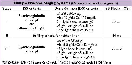
• β2-microglobulin and LDH levels reflect tumor burden
• BM bx cytogenetics: normal karyotype better than abnl. Standard risk = hyperdiploidy or t(11;14); high risk = hypodiploidy, del. 17p13 (~10% of Pts), t(4;14) & t(4;16)
• Gene mutations include TP53, NRAS, KRAS, BRAF & NK-kB pathway (Nature 2011;471:467)
• Skeletal survey (plain radiographs) to identify lytic bone lesions and areas at risk for pathologic fracture; bone scan is not useful for detecting lytic lesions
Treatment (NEJM 2011;364:1046; Am J Hematol 2012;87:79)
• Not indicated for smoldering MM or asx stage I disease
• Decisions generally dictated by risk stratification and transplant eligibility
• Active agents include: bortezomib (V), dexamethasone (D), prednisone (P), lenalidomide (R), thalidomide (T), melphalan (M), cyclophosphamide (C), doxorubicin, carfilzomib (Cz)
• Induction Rx regimens w/ best response rate incl. those w/ proteasome inhib (V, Cz) & immunomod (R), but many 2- or 3-drug options used based on comorbidities and risk. Proteasome inhib containing regimens incl. MPV, RVD, VCD & CzRD.
• If not transplant eligible: induction chemo ↑ survival, not curative; consider maint chemo
• If transplant eligible: induction chemo (eg, RVD, VCD, RD, VTD; Lancet 2010;376:2075) then high-dose chemo + auto-HSCT. Not curative, but ↑ survival c/w chemo (NEJM 2009;360: 2645). Timing of HSCT (upfront vs. relapse) under study. Offer if <70 y w/ good perf. status & no prohibitive comorbidities. Maint Rx w/ R or V until progression or intolerance. Role of tandem auto-HSCT & allo-HSCT remains controversial (NEJM 2003;349:2495).
• Local radiation for solitary or extramedullary plasmacytoma
• Adjunctive Rx
bone: bisphosphonates (JCO 2007;25:2464); XRT for sx bony lesions
renal: avoid NSAIDs & IV contrast; consider plasmapheresis for acute renal failure
hyperviscosity syndrome: plasmapheresis; infxns: consider IVIg for recurrent infections
• Common toxicities of Rx: melphalan → myelosuppression; lenalidomide → low plts & thromboembolism; bortezomib → periph. neuropathy; steroids → hyperglycemia, infxn
MONOCLONAL GAMMOPATHY OF UNCERTAIN SIGNIFICANCE (MGUS)
Definition and epidemiology (NEJM 2006;355:2765)
• M protein <3 g/dL, no urinary Bence Jones proteins, marrow plasmacytosis <10%, no ROTI
• Prevalence ~3% in population >50 y of age, ~5% in population >70 y of age, and
7.5% in population >85 y of age (NEJM 2006;354:1362)
Management
• ✓ CBC, Ca, Cr, SPEP, serum free light chains, UPEP w/ immunofixation (to exclude MM)
• Close observation: repeat SPEP in 6 mo, then yearly thereafter if stable
Prognosis (NEJM 2002:346:564)
• ~1%/y or ~25% lifetime risk → MM, WM, amyloidosis, or malign. lymphoproliferative dis.
• Abnormal serum-free light chain ratio: ↑ risk of progression to MM (Blood 2005;105:812)
WALDENSTRÖM’S MACROGLOBULINEMIA (WM)
Definition (Blood 2009;114:2375)
• B-cell neoplasm (lymphoplasmacytic lymphoma) that secretes monoclonal IgM
• MYD88 (NF-кB pathway) L265P somatic mutation found in 91% of Pts w/ WM and could be used to distinguish WM from MM (NEJM 2012;367:826)
• No evidence of bone lesions (IgM M component + lytic bone lesions = “IgM myeloma”)
Clinical manifestations
• Fatigue from anemia is most common sx
• Tumor infiltration: BM (cytopenias), hepatomegaly, splenomegaly, lymphadenopathy
• Circulating monoclonal IgM
hyperviscosity syndrome (~15%)
neurologic: blurred vision (“sausage” retinal veins on funduscopy), HA, dizziness, Δ MS
cardiopulmonary: congestive heart failure, pulmonary infiltrates
type I cryoglobulinemia → Raynaud’s phenomenon
platelet dysfxn → mucosal bleeding
• IgM deposition (skin, intestine, kidney); amyloidosis and glomerulopathy
• Autoantibody activity of IgM
chronic AIHA (prominent rouleaux; 10% Coombs’ = AIHA)
peripheral neuropathy: may be due to IgM against myelin-associated glycoprotein
Diagnostic evaluation
• SPEP + immunofixation with IgM >3 g/dL; 24-h urine for UPEP (only 20% have UPEP)
• Bone marrow biopsy: ↑ plasmacytoid lymphocytes; β2-microglobulin for prognostic eval
• Relative serum viscosity: defined as ratio of viscosity of serum to H2O (nl ratio 1.8) hyperviscosity syndrome when relative serum viscosity >5–6
Treatment
• Hyperviscosity: plasmapheresis
• Symptoms (eg, progressive anemia): rituximab ± chemotherapy (eg, cyclophosphamide, chlorambucil, fludarabine, cladribine, bendamustine) or bortezomib
• Thalidomide, alemtuzumab, everolimus, ibrutinib & auto-HSCT are investigational Rx