
Definition
• Secondary lung carcinomas from primary GI tract organs
Clinical features
Epidemiology
• Lung metastases could derive from any site of GI tract but are only rarely the initial manifestation of GI malignancies (5% at presentation)
• Disseminated GI malignancies, however, commonly involve lungs with frequency of metastases at autopsy: colon (45%), liver (37%-70%), stomach and esophagus (30%), pancreas (20%-40%)
Presentation
• Colorectal carcinoma classically presents with asymptomatic solitary nodule (up to 40% of all solitary lung metastases), which could be cavitating and calcified
• Gastric carcinomas present with dyspnea due to lymphangitic carcinomatosis or pleural effusions
• Pancreatic cancers commonly present with asymptomatic miliary nodules
• Pulmonary hypertension with tumor emboli and associated lung infarcts are associated with hepatocellular and pancreatic carcinomas, causing pain, hemoptysis, and severe dyspnea
Prognosis and treatment
• Prognosis is dismal in disseminated disease
• Metastasectomy in limited disease; chemotherapy and radiation for widespread disease
Pathology
Gross
• Colorectal carcinoma: well-circumscribed solitary or multiple variable size nodules with central necrosis, cavitation, and calcifications
• Colorectal mucin-secreting tumors: wet, slimy, glistening, yellow-tan cut surface
• Gastric carcinomas: characteristic features are thickening of intervascular bundles, interlobular septae, and pleura, hilar, or mediastinal lymphadenopathy
• Pancreatic cancer: multiple well-circumscribed variably sized nodules
• Tumor emboli of pancreatic and hepatocellular carcinomas display solid, gritty, and myxoid surface and are commonly associated with infarcted lung areas
Histology
• Colorectal carcinoma: cribriform pattern with nuclear pseudostratification and “dirty” necrosis (abundant necrotic debris)
• Pancreatic carcinoma: lepidic growth with cytological atypia, frequently admixed with cribriform areas, necrosis, lymphangitic invasion
• Gastric carcinoma: signet-ring or acinar features
• Hepatocellular carcinoma: trabecular or solid architecture with large polygonal cells
Immunopathology/special stains
• TTF-1 is virtually negative in GI metastases
• The most useful positive and negative immunohistochemical markers for GI metastatic carcinomas are listed in Table 10 in the Appendix
Main differential diagnoses
• Primary lung carcinoma: TTF-1 positive
• Interstitial lung disease, lymphangitic lymphoma or lymphangitic sarcoidosis: absence of cell atypia
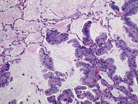
Fig 1 Metastatic carcinomas: gastrointestinal tract. Classic lepidic growth pattern of well-differentiated pancreatic carcinoma mimicking primary adenocarcinoma.

Fig 2 Metastatic carcinomas: gastrointestinal tract. High-grade pancreatic carcinoma can also grow in lepidic fashion, as shown in this image.

Fig 3 Metastatic carcinomas: gastrointestinal tract. Tumor embolus of metastatic carcinoma of the pancreas.

Fig 4 Metastatic carcinomas: gastrointestinal tract. Discohesive pancreatic cancer tumor cells imitating macrophages and megakaryocytes (A), strongly express CK19 (B).

Fig 5 Metastatic carcinomas: gastrointestinal tract. Lymphangitic spreading of signet-ring metastatic carcinoma from pancreas.

Fig 6 Metastatic carcinomas: gastrointestinal tract. Mucicarmine staining is helpful in cases of poorly differentiated metastatic pancreatic cancer for highlighting focal mucin production, as illustrated here.
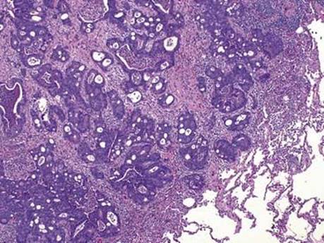
Fig 7 Metastatic carcinomas: gastrointestinal tract. Classic look of colonic metastatic cancer with “dirty” necrosis within glands and cribriform structures.

Fig 8 Metastatic carcinomas: gastrointestinal tract. CK7 is negative in metastatic colonic carcinoma but demonstrates strong reactivity with residual alveolar epithelium.

Fig 9 Metastatic carcinomas: gastrointestinal tract. CK20 positivity is present in more than 90% of colonic adenocarcinomas (shown); however, it is also expressed in more than half of mucinous and primary lung adenocarcinomas.

Fig 10 Metastatic carcinomas: gastrointestinal tract. CDX2 nuclear staining is highly specific for colonic carcinomas.

Fig 11 Metastatic carcinomas: gastrointestinal tract. Hepatocellular carcinoma metastasis exhibiting large polygonal eosinophilic cells arranged in solid sheets.

Fig 12 Metastatic carcinomas: gastrointestinal tract. Hepar1 immunostaining of previous case shows strong cytoplasmic granular reactivity.