
Definition
• A chronic lung disease that develops in some preterm infants who survived hyaline membrane disease
Pathogenesis
• Multiple factors play important roles in BPD:
• Lung immaturity and surfactant deficiency
• Barotrauma due to high pressure of oxygen delivery (not seen with current neonatal management)
• Oxygen toxicity (much less important with surfactant therapy and current management)
• Inflammation
• Pulmonary edema
• Nutritional deficiency
• Presurfactant therapy era: immature lungs with surfactant deficiency are more susceptible to barotrauma, which results in necrotizing bronchiolitis and alveolar septal injury
• Postsurfactant therapy: arrest of further lung development after premature birth leads to fewer alveoli that show compensatory dilatation. Fibrosis is minimal to mild
Clinical features
Epidemiology
• Highest incidence in most immature neonates (<27 weeks’ gestation) with low birth weight (<800 g)
• Occurred in 20% of newborns receiving ventilation before surfactant era
• Male infants tend to have more severe disease
• The use of surfactant therapy in preterm neonates with hyaline membrane disease has significantly decreased the incidence of BPD as well as the mortality rate
Presentation
• Early symptoms are those of hyaline membrane disease, requiring oxygen treatment and assisted ventilation
• Infants who then develop BPD have tachypnea, chest retraction, coughing, paroxysmal respiration, wheezing, and rhonchi; they continue to be oxygen dependent 28 days after birth
• Functional deficits include abnormal gas exchange, increased dead space, decreased compliance, increased work of breathing, and ventilation-perfusion mismatch, as well as mild to moderate pulmonary hypertension
• Radiology: diffuse lung haziness, consolidation, bubble-like cystic space; may be mediastinal shift and mass effect due to lobar overinflation
Prognosis and treatment
• Prevention is most important
• Symptomatic treatment includes diuretics, bronchodilators, vasodilators, and corticosteroids
• Infants with severe BPD are at high risk of pulmonary morbidity and mortality during the first 2 years of life
• Since the introduction of surfactant therapies, BPD is less severe and survival has been greatly improved
Pathology
Gross
• Pleural surface of severe BPD is irregular with a knobby or cobblestone appearance; there is shallow or deep fissure formation
Histology
• Nonsurfactant–treated
• Early phases of BPD in infants show diffuse alveolar damage (DAD) with necrotizing bronchiolitis and hyaline membranes followed by repair and organization
• Late stages show variegated pattern with some lobules with alveolar fibrosis and collapse, while adjacent lobules show overdistention and frequent interstitial air accumulation
• Lung tissue supplied by bronchioles with necrotizing bronchiolitis is relatively protected from further injury
• Since surfactant replacement therapy, the most common histological picture seen in BPD is decreased alveolarization and simplification of lung architecture; there is only minimal to mild alveolar septal fibrosis
• Superimposed infection and pulmonary hypertension (often with cor pulmonale) are usually present at autopsy
Main differential diagnoses
• Early stages with DAD
• Late stages: emphysema, fibrosing interstitial pneumonia, mild pulmonary hypertension or chronic constrictive bronchiolitis
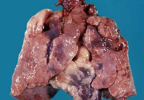
Fig 1 Bronchopulmonary dysplasia. Gross picture of the heart and lung from an infant with BPD. Note the presence of deep fissures and variegated appearance of pleura with dark purple indented areas and pale pink nodular areas; the heart shows right ventricular hypertrophy with prominent epicardial fat deposition.

Fig 2 Bronchopulmonary dysplasia. Septal fibrosis of BPD: H&E (A) and trichrome stain (B) showing blue fibrotic area.
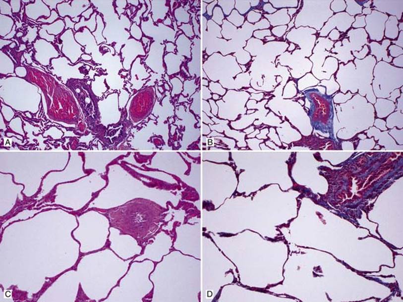
Fig 3 Bronchopulmonary dysplasia. With surfactant therapy, lung with mild BPD showing uniformly dilated acini with thin alveolar septa and only focal interstitial fibrosis: low (A) and high (C) powers of H&E stain; low (B) and high (D) powers of trichrome stain highlighting focal fibrosis.