
Definition
• A low-grade malignant thymoma with similar histological appearance to normal thymus with scattered tumor epithelial cells intermixed with abundant background lymphocytes
Clinical features
Epidemiology
• Relatively rare; accounts for <20% of all thymoma
• Mean age, <50 years
• No gender predilection
Presentation
• About half of cases are completely encapsulated (stage I), one fourth of cases invade the mediastinal fat (stage II), a small percentage of cases invade pleura, pericardium, great vessels, or adjacent organs (stage III); metastases are very rare
Prognosis and treatment
• Surgical resection
• Clinically considered to be a tumor with low-grade malignant potential; 10-year survival rate: >90%
Pathology
Gross
• Well-defined or encapsulated grayish mass with thick fibrous capsule and septa
• Cystic spaces or focal hemorrhagic and necrotic areas may be present
Histology
• Histologically resembles normal thymus with organotypic lobulated architecture separated by fibrous bands
• Composed of expanded normal thymic cortexlike areas with a few small scattered neoplastic epithelial cells surrounded by dense population of nonneoplastic immature T lymphocytes (lymphocyte-rich thymoma)
• Sparse foci of paler areas resembling the thymic medulla are also seen containing loosely packed lymphocytes; Hassall corpuscles may be present but are usually fewer than in normal thymic medulla
• Tingible body macrophages may be present throughout the lesion with a starry-sky appearance
• Features that distinguish from normal noninvoluted thymus include:
• Excessive expansion of cortical areas
• Smaller foci of medullary differentiation with fewer Hassall corpuscles
• Thicker capsule and fibrous septa
Immunopathology/special stains
• Scattered epithelial cells are positive for epithelial markers and negative for CD5
• Cortical T lymphocytes are immature T cells with CD1a, CD4, CD8, CD99, TdT, and CD5 positivity and a high Ki-67 index (approximately 90%)
• Medullary lymphocytes are mature T cells with CD3+, CD5+, CD1a–, CD99–, and TdT– immunophenotype and significantly low proliferation
Main differential diagnoses
• Normal noninvoluted thymus: see earlier chapter
• Thymic lymphoid hyperplasia:
• Absence of a mass
• Normal cortical and medullary distinction with well-formed germinal centers
• B2 thymoma:
• Many more epithelial cells forming aggregates and much fewer intermixed lymphocytes
• Tumor cells are larger in size and have prominent nucleoli
• T-lymphoblastic lymphoma:
• Usually presents as anterior mediastinal mass
• Affects teenagers and young adults
• Additional lymphadenopathy or other organ involvement
• Proliferation of monomorphic lymphocytes with septal and capsular infiltration
• Negative for cytokeratin, except rare entrapped epithelial cells
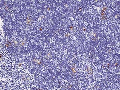
Fig 1 Type B1 thymoma. Keratin AE1/AE3 shows cytoplasmic positivity in the scattered epithelioid cells of this B1 thymoma.
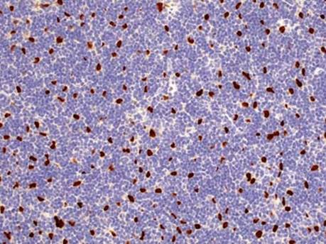
Fig 2 Type B1 thymoma. Stain for p63 shows strong nuclear positivity in epithelioid tumor cells.

Fig 3 Type B1 thymoma. H&E stained section shows a predominance of lymphocytes within which are a few scattered epithelioid tumor cells: low power (A), medium power (B), and high power (C).