
Definition
• Rare pediatric neoplasm of neural crest origin arising from the sympathetic ganglia; with differentiation between neuroblastoma and ganglioneuroma
Pathogenesis
• Thought to arise from differentiation of neuroblastoma
Clinical features
Epidemiology
• Epidemiological factors are similar to neuroblastoma
• Children are usually older than those affected by neuroblastoma but most are still younger than 10 years; rarely, it may be seen in adults
• Female to male ratio is about equal
• Common locations include adrenal medulla, mediastinum, and abdominal cavity
Presentation
• Patients may be asymptomatic or, on the other end of the spectrum, may present with widespread metastatic disease
• Tumor may grow to impinge on other thoracic structures or grow into the spinal canal in a “dumbbell” fashion, producing pain and/or paresthesias
• Similar to neuroblastoma, patients may exhibit Horner’s syndrome and may have increased vasoactive substances (homovanillic acid and vanillylmandelic acid) in their urine
Prognosis and treatment
• Prognosis is thought to be better than neuroblastoma; however, cases are treated similarly due to malignant potential
Pathology
Gross
• Gross and microscopic appearance is heterogeneous based on the degree of differentiation
• The tumor may be encapsulated and have a homogenous cut surface or demonstrate areas of hemorrhage and necrosis
Histology
• Proliferation of neuroblast-like cells akin to neuroblastoma but accompanied by a variable proportion of ganglion cells and intermediate cells; the Schwannian stroma accounts for >50% of the tumor
• Tumors may be classified as poorly or well-differentiated based on degree of differentiation
• Two histological subtypes are recognized: intermixed and nodular
• The nodular type contains nodules of neuroblastic cells with demarcation from large expanses of ganglion cells in a Schwannian fusiform stroma; the latter may be at the periphery of the lesion
• The intermixed type has nests of neuroblastic cells within a ganglioneuromatous stroma
• The nodular form, especially that with high MKI, has a worse prognosis than the intermixed type
Immunopathology/special stains
• Not contributory
Main differential diagnoses
• Neuroblastoma: the ganglioneuromatous component is less than 50%
• Ganglioneuroma: no neuroblastic component; Schwannian stroma
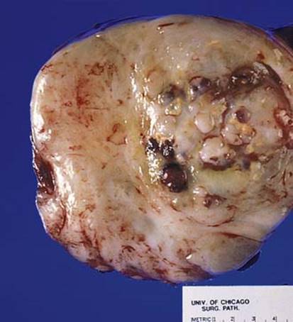
Fig 1 Mediastinal ganglioneuroblastoma. Cut surface of gross specimen shows nodules of hemorrhagic tumor (corresponding to neuroblastoma) embedded in a pale gray-tan tumor (corresponding to ganglioneuroma).
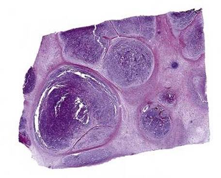
Fig 2 Mediastinal ganglioneuroblastoma. Low-power scanning view of nodular subtype. Note nodules of darker neuroblastic cells surrounded by areas of lighter staining stroma.
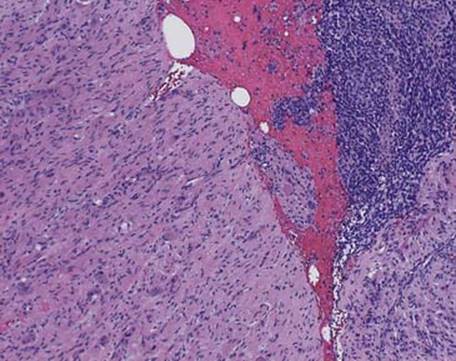
Fig 3 Mediastinal ganglioneuroblastoma. Medium power showing two distinctly different cell populations, one with small, round, blue cells and the other with larger cells with more voluminous cytoplasm (arrows) in a Schwannian stroma.
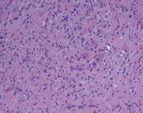
Fig 4 Mediastinal ganglioneuroblastoma. Higher power of the pale area demonstrates scattered ganglion cells in a Schwannian-rich stroma.
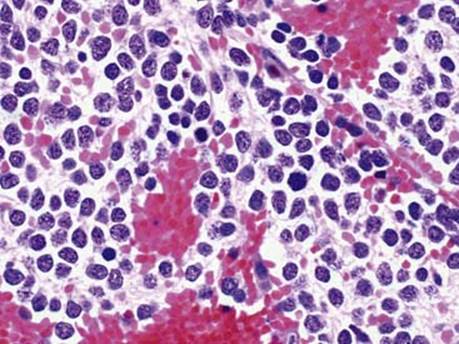
Fig 5 Mediastinal ganglioneuroblastoma. High power of the blue area shows immature neuroblastic cells.
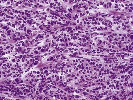
Fig 6 Mediastinal ganglioneuroblastoma. Frozen section diagnosis of neuroblastic component can be made when rosettes and neuropil can be identified, as shown here.
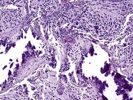
Fig 7 Mediastinal ganglioneuroblastoma. Post therapy, neuroblastoma often shows ganglionic maturation and calcifications as seen in this case. Without 50% Schwannian stroma, this should not be called ganglioneuroblastoma.
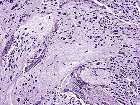
Fig 8 Mediastinal ganglioneuroblastoma. Post therapy neuroblastoma with atypical maturing neuroblasts. Note large areas of neuropil, which are seen presumably where there were more neuroblasts before therapy. There is very little Schwannian stroma.