
Definition
• Infection of the endocardium caused by a variety of organisms, typically on valvular surfaces
Clinical features
Epidemiology
• Approximately 10,000 to 15,000 cases per year in the United States
• Risk factors include intravenous drug abuse, prosthetic heart valves, structural abnormalities of the heart valves, congenital heart disease, chronic hemodialysis, and HIV infection
• In the past, rheumatic heart disease was a common predisposing condition, particularly in young adults
• Today, approximately 50% of cases are adults older than 60 years
• Majority of cases are caused by Staphylococcus and Streptococcus species
• Acute endocarditis is most commonly caused by Staphylococcus aureus
• Subacute endocarditis is most commonly caused by Streptococcus viridans
• Other causes of endocarditis include Staphylococcus epidermidis (especially in patients with prosthetic valves), fastidious gram-negative bacilli (e.g., Haemophilus species), enterococci, and fungi
Presentation
• Aortic and mitral valves are most common, but any valve may be involved
• Acute endocarditis presents with nonspecific but often dramatic symptoms, including fever, chills, fatigue, and congestive heart failure
• Right-sided acute endocarditis is associated with intravenous drug abuse and may also present with pneumonia and/or empyema
• Subacute endocarditis has an insidious onset of vague constitutional symptoms, including low-grade fever, night sweats, fatigue, weakness, weight loss, and back pain
• Subacute endocarditis may be preceded by a dental procedure; gingivitis is a risk factor
• Although diagnosis can be made definitively on pathology, clinical diagnosis is typically made by positive blood culture, evidence of endocardial lesion on echocardiogram, and minor criteria such as fever, vascular phenomena, and predisposing conditions
Prognosis and treatment
• Severe cases of acute endocarditis are treated empirically with broad-spectrum antibiotics (e.g., vancomycin) after blood cultures are drawn
• Antibiotic therapy is then tailored to the specific organism grown on blood culture
• Surgical repair may be necessary in cases with severe valve dysfunction, especially in cases of endocarditis involving a prosthetic valve
• Subacute endocarditis is typically caused by Streptococcus viridans, which is susceptible to penicillin and cephalosporins
• Cardiac complications are seen in up to half of cases of endocarditis and include heart failure secondary to valvular insufficiency, perivalvular abscesses, pericarditis, and aortic dissection
• Septic embolic events are a common complication of left-sided endocarditis, but the risk is reduced by prompt initiation of antimicrobial therapy and/or surgery
• Acute endocarditis has a mortality rate of approximately 20%, although fungal endocarditis is associated with an even higher mortality rate
• Prognosis is good in subacute endocarditis; the cure rate approaches 100%
Pathology
Histology
• Acute endocarditis is characterized by a neutrophilic exudate with fibrin, platelets, and bacterial organisms
• Subacute endocarditis shows granulation tissue with histiocytes and multinucleated giant cells, chronic inflammation, and variable acute inflammation
Immunopathology/special stains
• Immunohistochemical stains for specific organisms (e.g., Bartonella, Coxiella, Chlamydia) may be helpful in identifying the causative organism in some cases
• Special stains for microorganisms may be helpful in identifying bacteria and fungi
Main differential diagnoses
• Nonbacterial thrombotic endocarditis
• Vegetation on the valve surface consisting of degenerating platelets and fibrin without inflammation or organisms
• Can also be associated with an embolic event; in fact, it may be seen with a stroke
• Associated with underlying malignancy and/or hypercoagulable state in the majority of cases
• Symptoms of acute infection (e.g., fever, chills) are not typically present
• Managed with anticoagulation, typically heparin
• Libman-Sacks (verrucous) endocarditis
• Typically seen in patients with systemic lupus erythematosus
• Noninfectious vegetation on atrial and ventricular surfaces of valves
• Vegetation composed of fibrin, mononuclear cells, and fibroblasts
• Usually asymptomatic but, rarely, may produce systemic emboli
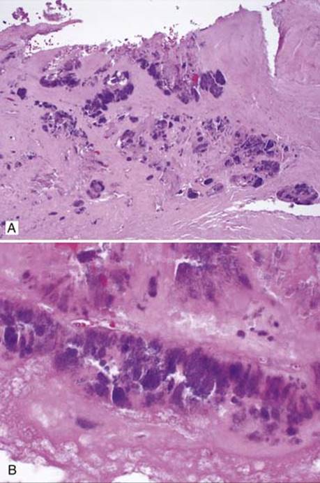
Fig 1 Infectious endocarditis. Bacterial endocarditis. Numerous bacterial colonies are present both at the surface and within fibrin, low power (A) and high power (B).

Fig 2 Infectious endocarditis. Granulation tissue, fibrin, and a marked acute inflammatory infiltrate are present.
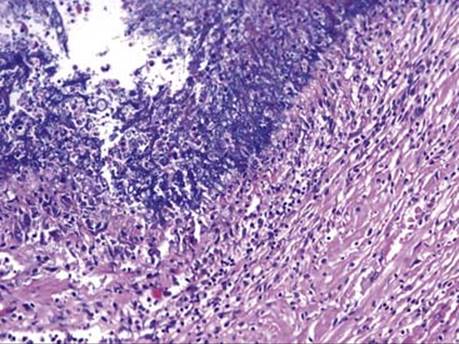
Fig 3 Infectious endocarditis. Fungal endocarditis. Fungal hyphae have destroyed valvular tissue and are growing toward underlying myocardium (seen on the bottom right).

Fig 4 Infectious endocarditis. Fungal endocarditis with numerous yeasts and pseudohyphae consistent with Candida species, H&E (A) and GMS (B) stains.