
Definition
• Acute rheumatic fever: immune-mediated, nonsuppurative, multisystem disease that occurs as a complication of group A streptococcal pharyngitis
• Acute RHD: pancarditis that occurs secondary to molecular mimicry with cross-reactivity of antibodies against group A streptococcal antigens and self antigens in the heart
• Chronic RHD: progressive and permanent deformity of cardiac valves secondary to organized inflammation and fibrosis caused by single or multiple episodes of acute RHD
Clinical features
Epidemiology
• Acute rheumatic fever
• Occurs most often in children ages 5 to 15 years
• Prevalence: worldwide 5/1000; developing countries 24/1000
• Affects up to 3% of patients with group A streptococcus pharyngitis
• Approximately 50% of patients with acute rheumatic fever develop acute carditis
• Chronic RHD
• Significant chronic valve disease occurs in less than 10% of patients after an acute episode
• About 60% of patients with chronic disease have a clinical history of acute rheumatic fever
Presentation
• Acute rheumatic fever
• Seen 2 to 6 weeks after pharyngitis
• Clinical features:
– Must have at least two major or one major and two minor criteria to make the diagnosis
– Major criteria:
– Migratory polyarthritis
– Carditis
– Erythema marginatum
– Subcutaneous nodules
– Sydenham chorea
– Minor criteria:
– Fever
– Arthralgia
– Elevated acute-phase reactants
– Reversible prolongation of PR interval
• Acute RHD: clinical features of acute carditis may include pericardial friction rub, tachycardia, arrythmias, new heart murmur (especially mitral regurgitation), and congestive heart failure
• Chronic RHD: patients may have mitral stenosis or regurgitation, aortic stenosis, atrial fibrillation, infective endocarditis, thromboembolism, and cardiac hemolytic anemia
Prognosis and treatment
• Acute rheumatic fever
• Prognosis is variable. Immediate fatality occurs in 1% to 2%
• Prone to reactivation with subsequent pharyngeal infections leading to cumulative valvular damage (20% have a second occurrence within 5 years)
• Symptomatic treatment may include salicylates, corticosteroids, antibiotics
• Chronic
• Long-term prognosis is highly variable
• Patient may require valve replacement
Pathology
Gross
• The most common specimen is a surgically removed valve from patients undergoing valve replacement due to chronic RHD
• Endocarditis:
• Valves involved: 75% to 80% mitral, 30% aortic (usually in combination with mitral), 5% pulmonic or tricuspid
• Acute: small vegetations along the lines of closure of the valve leaflets
• Chronic:
– Shortening and fusion of chordae tendinae
– Fibrous bridging and calcification of valve leaflets results in “fish mouth” deformity
– MacCallum patch: area of subendocardial thickening in the left atria above the posterior leaflet of the mitral valve
• Myocarditis:
• Acute:
– Dilated ventricles and a floppy heart
– Areas of softening and pallor
• Chronic: focal or diffuse fibrosis in a random distribution
• Pericarditis in acute RHD:
• Serous: serous effusion
• Fibrinous: “bread and butter” pericarditis with fibrinous exudate on visceral and parietal pericardium
Histology
• Endocarditis:
• Acute
– Vegetations are composed of fibrin and mononuclear cells
– Inflamed and edematous valve leaflets
• Chronic:
– Fibrosis and calcification
– Neovascularization
• Myocarditis:
• Acute:
– Aschoff bodies: pathognomonic for acute RHD. Granulomatous lesion with central fibrinoid necrosis surrounded by a collection of lymphocytes, plasma cells, macrophages, and Aschoff giant cells. Most commonly located in the endocardium of the atria and the perivascular spaces of the ventricular myocardium
– Anitschkow cell: macrophages within Aschoff body with a central wavy band of chromatin (in cross-section referred to as owl eyes; in longitudinal section, called caterpillar cells)
• Chronic:
– Patchy fibrosis especially in perivascular areas
• Pericarditis:
• Layers of fibrin and neutrophils overlying reactive mesothelium; subepithelial mixed inflammatory infiltrate of neutrophils, lymphocytes, plasma cells, and macrophages
Immunopathology/special stains
• Not contributory
Main differential diagnoses
• Endocarditis
• Infective endocarditis: large irregular vegetations composed of fibrin, inflammatory cells, and microorganisms, on valve cusp and extending onto chordae
• Libman-Sacks endocarditis: occurs in patients with systemic lupus erythematosus (SLE). Small warty vegetations on atrial or ventricular surface of the mitral and tricuspid valves. Vegetations consist of eosinophilic fibrinous material with remnants of damaged nuclei called hematoxylin bodies
• Nonbacterial thrombotic endocarditis: occurs mainly in patients with cancer, sepsis, and hypercoagulable states. Small vegetations consist of loosely attached thrombi
• Myocarditis
• Lymphocytic myocarditis: diffuse infiltrate of lymphocytes and plasma cells with focal myocyte necrosis
• Giant cell myocarditis: prominent eosinophilic infiltrate; absence of well-formed granulomas
• Infectious myocarditis: can be secondary to viral, bacterial, fungal, or parasitic infection. May have lymphocytic, neutrophilic, or eosinophilic infiltrates and visible microorganisms
• Pericarditis
• Fibrinous pericarditis: uremia; myocardial infarction
• Serous pericarditis: viral infections; SLE

Fig 1 Acute rheumatic fever and rheumatic heart disease. Gross appearance of acute rheumatic endocarditis. Small vegetations are present along the line of valve closure.

Fig 2 Acute rheumatic fever and rheumatic heart disease. Acute rheumatic endocarditis of the mitral valve showing an inflammatory infiltrate and fibrin vegetation.

Fig 3 Acute rheumatic fever and rheumatic heart disease. Aschoff body in acute rheumatic myocarditis. A wavy band of fibrinoid necrosis is surrounded by a mixed inflammatory infiltrate.
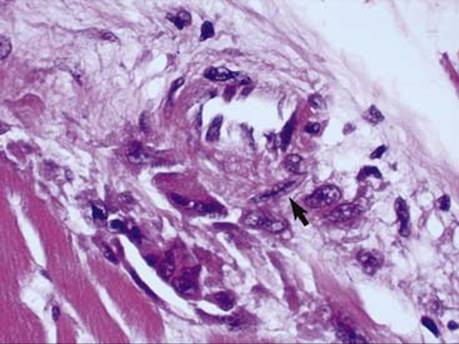
Fig 4 Acute rheumatic fever and rheumatic heart disease. “Caterpillar cell” (arrow) seen in acute rheumatic myocarditis.
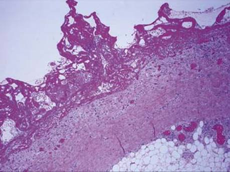
Fig 5 Acute rheumatic fever and rheumatic heart disease. Fibrinous pericarditis. Mounds of eosinophilic, amorphous fibrin overlying pericardium with congested capillaries and an inflammatory infiltrate.
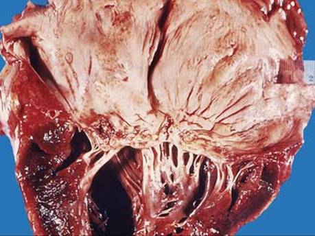
Fig 6 Acute rheumatic fever and rheumatic heart disease. Chronic RHD with a dilated left atrium, thickened endocardium, deformed mitral valve with thickened and fused cusps, and thick, fused chordae tendinae.
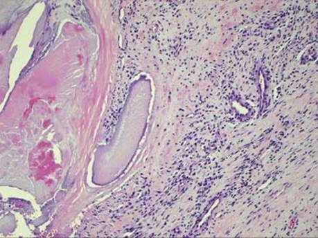
Fig 7 Acute rheumatic fever and rheumatic heart disease. Chronic endocarditis characterized by calcification and neovascularization of the valve.

Fig 8 Acute rheumatic fever and rheumatic heart disease. Mitral valve, surgically removed for valve replacement, with the classic “fish mouth” deformity of chronic rheumatic endocarditis.
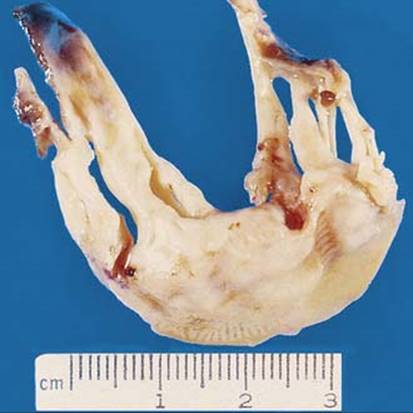
Fig 9 Acute rheumatic fever and rheumatic heart disease. Surgically removed mitral valve with thickened and fused chordae tendinae.