
Definition
• An inflammatory process of the myocardium characterized by a predominantly lymphocytic infiltrate within myocardial fibers with injury to cardiac myocytes, in patients with no other underlying cardiac dysfunction or with low cardiac risk
Pathogenesis
• Approximately 50% of the time, lymphocytic myocarditis is classified as idiopathic
• Well-established causes include viral (enterovirus, coxsackie B, adenovirus, influenza, cytomegalovirus, poliomyelitis), rickettsial (Rocky Mountain spotted fever, Q fever), bacterial (diphtheria, tuberculosis), fungal (candidiasis), protozoal (Chagas disease), and helminthic (trichinosis)
• Other causes include drugs (doxorubicin, anthracyclines), bites/stings (scorpion and snake venoms), and physical agents (heatstroke, radiation)
Clinical features
Epidemiology
• The true incidence is difficult to determine as most cases are believed to be subclinical
• One study of military recruits estimated the incidence at 17 per 100,000 population annually
• Histopathologically defined myocarditis (lymphocytic myocarditis), on the other hand, has been found in about 1% of unselected routine autopsies
Presentation
• Lymphocytic myocarditis is frequently asymptomatic or may have a subclinical course that can later progress to dilated cardiomyopathy
• Symptomatic patients may show nonspecific symptoms of chest pain, fever, sweats, chills, and dyspnea
• In cases with a viral etiology, patients may present with a history of recent (within 1 to 2 weeks) flulike syndrome
• Palpitations, syncope, or sudden cardiac death may develop because of underlying ventricular arrhythmias or atrioventricular block
Prognosis and treatment
• Most patients recover spontaneously with no known sequela
• A small percentage of patients, mostly children and young adults, present with fulminant disease onset and may progress rapidly to sudden death
• Treatment of myocarditis includes supportive therapy for symptoms of acute heart failure and withdrawal of the offending agent, if applicable
Pathology
Gross
• Cardiac hypertrophy may be present with associated ventricular dilation
• The affected myocardium may appear pale, sometimes with hemorrhagic foci
Histology
• A focal or diffuse lymphocytic infiltrate (mostly T-cell type) within myocardial fibers is often admixed with histiocytes
• Myocyte necrosis is typically present
• Interstitial fibrosis of varying degree is commonly seen
• Dallas criteria for diagnosis of myocarditis require presence of both an inflammatory infiltrate and myocyte necrosis or degeneration
• Presence of edema should not be used as a criterion
• The Dallas criteria include a “borderline” category for cases with sparse lymphocytic infiltrate and no associated myocyte damage
Immunopathology/special stains
• CD3 positivity will confirm a T-cell phenotype
Main differential diagnoses
• Giant cell myocarditis
• Characterized by diffuse infiltration of myocardium by a heterogeneous infiltrate composed of abundant lymphocytes, eosinophils, plasma cells, and scattered prominent giant cells
• Occasional poorly formed granulomas may be seen
• Appears clinically distinct with a more fulminant course
• Eosinophilic myocarditis
• Mixed inflammatory cell infiltrate consisting of many eosinophils with histiocytes, lymphocytes, and plasma cells
• Myocardial necrosis is not common and interstitial fibrosis is typically absent or minimal

Fig 1 Lymphocytic myocarditis. Diffuse interstitial infiltrate of inflammatory cells.
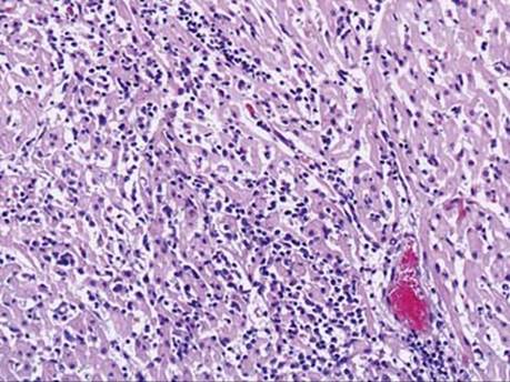
Fig 2 Lymphocytic myocarditis. Infiltrate is composed predominantly of lymphocytes.

Fig 3 Lymphocytic myocarditis. Myocyte damage is present in association with lymphocytic infiltrate.
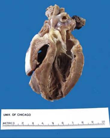
Fig 4 Lymphocytic myocarditis. 13-month-old who died after only a four-day history of respiratory distress and arrhythmias due to myocarditis. The heart at autopsy is enlarged and dilated.
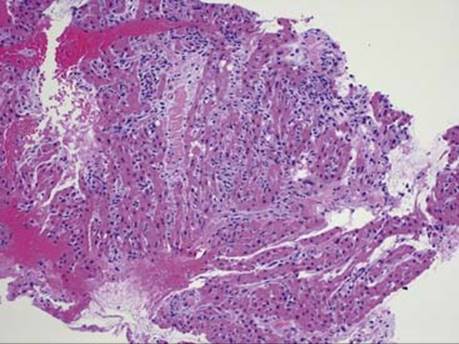
Fig 5 Lymphocytic myocarditis. Although the biopsy from this 1-year-old contained only one fragment of endomyocardium, the amount of inflammation present is enough to make a diagnosis of myocarditis.

Fig 6 Lymphocytic myocarditis. This endomyocardial biopsy (A and B) has only a few lymphocytes without associated myocyte necrosis, which are not enough to make a diagnosis of myocarditis.