
Definition
• Acute rejection that occurs due to formation of antibodies or presensitization of patient to the graft
Clinical features
Epidemiology
• Varies by individual medical centers; approximately 10% to 20% incidence
Presentation
• Occurrence ranges from a few hours to months or years after transplantation
• Hemodynamic compromise such as hypotension, shock, decreased cardiac output
Prognosis and treatment
• Treatment varies and may include corticosteroids, plasmapheresis, and antilymphocyte antibodies (antithymocyte globulin)
• Most patients recover; however, risk of chronic rejection and death is increased
Pathology
Histology
• Endothelial swelling, interstitial edema, and a minimal cellular inflammatory infiltrate
• Intravascular thrombi
• Mild neutrophilic infiltration around capillaries and hemorrhage
• H&E findings are nonspecific; immunofluorescence (IF) or immunohistochemical (IHC) staining is required for diagnosis
Immunopathology/special stains
• IF demonstration of diffuse positivity for C4d and C3d in all capillary endothelium
• Demonstration of strong and diffuse IHC staining for C4d and/or C3d
Main differential diagnosis
• Mild cellular rejection (diffuse type): C4d and C3d stains are negative

Fig 1 Antibody-mediated rejection of the heart. Positive IF for C4d with strong endothelial staining.
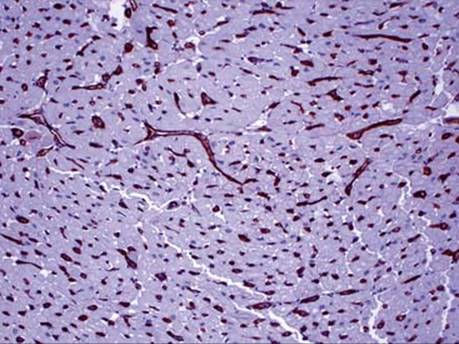
Fig 2 Antibody-mediated rejection of the heart. Strong diffuse uniform endothelial staining with C4d that is seen in positive cases.

Fig 3 Antibody-mediated rejection of the heart. Nonspecific serum and interstitial staining that is frequently seen in C4d IHC stain and should not be interpreted as positive.