
Definition
• Pneumonia caused by aspiration of large amounts of oral/gastric contents
Clinical features
Epidemiology
• Gross aspiration most commonly happens in obtunded or intubated patients who are incapable of protecting their airway
Presentation
• Depends on the amount and type of aspirated material and time course of aspiration
• Can present as bronchiolitis, bronchopneumonia, necrotizing pneumonia, lipoid pneumonia, empyema, or a mixture of these entities
• Common finding at autopsy in severely ill patients who died of other causes
Prognosis and treatment
• Bacterial pneumonia is treated with appropriate antibiotics
• Often associated with development of a lung abscess, which demonstrates a mixture of aerobic and anaerobic bacteria on culture
• Massive aspiration of gastric acid and subsequent bacterial superinfection has a high mortality rate
Pathology
Histology
• Acute inflammatory cells are present within airways and adjacent alveoli
• In necrotizing pneumonia, there is destruction and replacement of lung parenchyma with acute inflammation and necrosis
• Recognition of superficial squames, vegetable material, or skeletal muscle in the alveolar spaces confirms aspiration
• Abscess formation with walling off of infection by granulation tissue and fibrosis
• Occasionally, foreign body giant cell granulomas to aspirated material; lipid-laden macrophages may be present indicating prior episode(s) of aspiration
• Massive aspiration of gastric acid (Mendelson’s syndrome) causes diffuse alveolar damage, necrosis of alveolar lining cells, hemorrhage, and pulmonary edema
Immunopathology/special stains
• Not contributory
Main differential diagnoses
• Bacterial pneumonia without aspiration
• Granulomatous infections: fungal and acid-fast stains should be performed on multiple slides
• Inhalation talcosis: polarizable crystals easily identified
• Sarcoidosis: well-formed granulomas with bronchovascular distribution; no necrosis
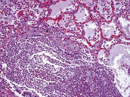
Fig 1 Aspiration pneumonia. There is accumulation of neutrophils within the bronchiole with extension into adjacent alveoli, which also contain edema fluid.
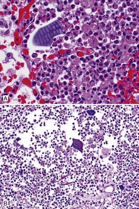
Fig 2 Aspiration pneumonia. Necrotic inflammatory cells with aspirated vegetable material (A) and foreign material, bacterial colonies, and squames (B) in alveolar spaces.