
Definition
• Infection by bacillus Mycobacterium tuberculosis (MTB)
Pathogenesis
• Inhalation of 1-5 μm airborne droplets containing MTB leads to primary disease, which may be reactivated from latency to cause secondary tuberculosis
• Virulence attributed to cell envelope components: mycolic acid, peptidoglycans, lipoarabinomannan (LAM), and mycobacterial entry protein (Mcep)
Clinical features
Epidemiology
• One third of the world’s population is infected with MTB, and active tuberculosis develops in 5% to 10% of those infected
• Males more often affected than females
• In the United States, disease manifestation is mainly in the elderly, homeless, incarcerated, and immunocompromised individuals (Hispanics, blacks, and Asians have higher infection rate than whites)
Presentation
• Presenting signs and symptoms range from asymptomatic infection to a cough, fever, weight loss, anorexia, fatigue, chest pain, and night sweats
• May include hemoptysis, purulent sputum production, and clubbing
Prognosis and treatment
• Long-term multidrug regimens are warranted with excellent prognosis when treatment guidelines are strictly followed
• Current vaccines are ineffective
• Poorer outcomes in infection with multidrug resistant strains of MTB
• The World Health Organization (WHO) estimated 1.7 million deaths due to tuberculosis in 2009 (mainly in Africa)
• Mortality exceedingly rare in the United States
Pathology
Gross
• Usually seen as a necrotizing consolidative process predominantly in lung apices. May be seen as a Ghon lesion (1-2 cm, round, white-gray pulmonary nodule with central necrosis), a Ghon complex (Ghon lesion plus hilar lymphadenopathy), or Ranke complex (fibrosis and calcification of the Ghon complex via cell-mediated immunity)
Histology
• Both necrotizing (caseating) and nonnecrotizing granulomas can be seen in lung parenchyma and/or lymph nodes. MTB (4-μm beaded rods) can be demonstrated using acid-fast stains (Ziehl-Neelsen), although a negative acid-fast stain does not rule out tuberculosis
• Bordering epithelioid cells often form Langerhans-type multinucleated giant cells
Main differential diagnoses
• Nontuberculous mycobacterial infections
• Sarcoidosis
• Wegener granulomatosis
• Nocardiosis

Fig 1 Tuberculosis. Gross photograph of cut surface of lung with caseating granulomas. Note apical emphysema.

Fig 2 Tuberculosis. Frozen section of lung mass shows necrotizing granuloma. Note: with this histological picture, the cryostat used for frozen section needs to be decontaminated.
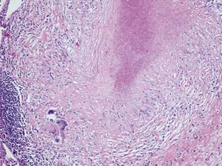
Fig 3 Tuberculosis. Low-power view of necrotizing granuloma characteristic of tuberculosis.
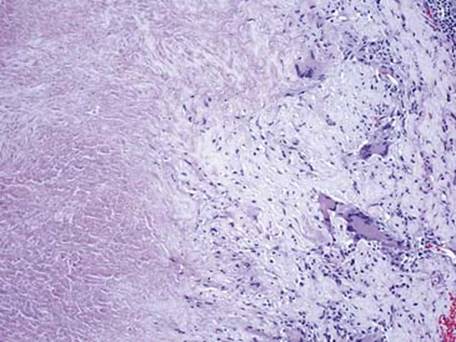
Fig 4 Tuberculosis. High-power view showing irregular necrosis within granuloma, often seen in tuberculosis.

Fig 5 Tuberculosis. Acid-fast stain (Ziehl-Neelsen) shows few acid-fast bacilli photographed here under oil immersion.