
David M. Cline
EPIDEMIOLOGY
 Soft tissue upper extremity injuries account for about 35% of the wounds and lacerations evaluated in the ED.
PATHOPHYSIOLOGY
Injuries may be classified as closed crush, simple lacerations, open crush with partial amputation, and complete amputation.
CLINICAL FEATURES
History should include occupation and hand dominance.
Examination of all arm and hand injuries includes inspection at rest, evaluation of motor, nerve and tendon functions, evaluation of sensory nerve function, and evaluation of perfusion.
Examine in active motion and resistance to passive motion (see Tables 14-1 and 14–2).
Examine all wounds for evidence of potential artery, nerve, tendon, bone injuries, and the presence of foreign bodies, debris, or bacterial contamination.
TABLE 14-1 Motor Testing of the Peripheral Nerves of the Upper Extremity

TABLE 14-2 Sensory Testing of Peripheral Nerves in the Upper Extremity

DIAGNOSIS AND DIFFERENTIAL
A bloodless field is needed to achieve adequate visualization.
If a proximal tourniquet is needed, a Penrose drain can be used for distal finger injures and a manual blood pressure cuff for more proximal injures.
Once adequate visualization is obtained, examine the wound for foreign bodies and tendon and joint capsule injuries.
Examine the hand and arm in the position of injury to avoid missing deep structure injuries that may have moved out of the field of view when examined in a neutral position.
Obtain anteroposterior and lateral radiographs if bony injuries, retained radiopaque foreign bodies, or joint penetration are suspected.
EMERGENCY DEPARTMENT CARE AND DISPOSITION
All wounds require scrupulous cleaning and irrigation after adequate anesthesia.
Provide tetanus prophylaxis as indicated (see Chapter 18).
Consult a plastic or hand surgeon for complex or extensive injuries, injuries requiring skin grafting, injuries requiring technically demanding skills or when the hand is vital to patient’s career (eg, a professional musician).
Additional care instructions of specific injuries are as follows:
FOREARM AND WRIST LACERATIONS
Injury over the wrist raises the possibility of a suicide attempt. Question the patient about intent and a history of depression.
Injuries that involve more than one parallel laceration, classic for suicide attempts, may require horizontal mattress sutures to cross all lacerations to prevent compromising the vascular supply of the island of skin located between incisions (Fig. 14-1).
Examine tendons and distal nerves individually (see Tables 14-3 and 14-4).

FIG. 14-1. Horizontal mattress sutures for multiple parallel lacerations.
PALM LACERATIONS
Injuries to the palm may require a regional anesthetic, for example, a median or ulnar nerve block.
If no deep injury is suspected, close the wound. Pay particular attention to re-opposing the skin creases accurately.
Avoid using deep “bites” with the needle because this risks injury to the underlying tendons or tendon sheaths. Interrupted horizontal mattress sutures (see Chapter 12) with 5–0 monofilament suture are recommended.
Refer patients with deep injuries between the car-pometacarpal joints and the distal creases of the wrist (“no-mans’ land”) to a specialist for exploration and repair.
TABLE 14-3 Extensor Compartments in the Forearm

DORSAL HAND LACERATIONS
Lacerations over the metacarpophalangeal joint suggest a closed fist injury and require special care.
Polymicrobial infections are the rule; Staphylococcus aureus, Streptococcus sp., Corynebacterium sp., and Eikenella corrodens are the most common bacteria.
Obtain radiographs to rule out fracture or embedded teeth.
Irrigate thoroughly and débride as needed.
Infected wounds require IV antibiotics (ampicillin-sulbactam 3 grams every 6 hours), consultation to a hand surgeon, and admission.
Patients with noninfected, minor closed fist injuries can be treated as outpatients with immobilization in position of function (do not close), amoxicillin-clavulanic acid 875 milligrams PO twice daily (22.5 milligrams/kg per dose two times daily in children), and follow-up in 24–48 hours with strict return instructions in the event of erythema, drainage, or increased pain.
The pliable skin and extensive movements of the hand may hide tendon injuries.
Repair skin using 5–0 nonabsorbable sutures.
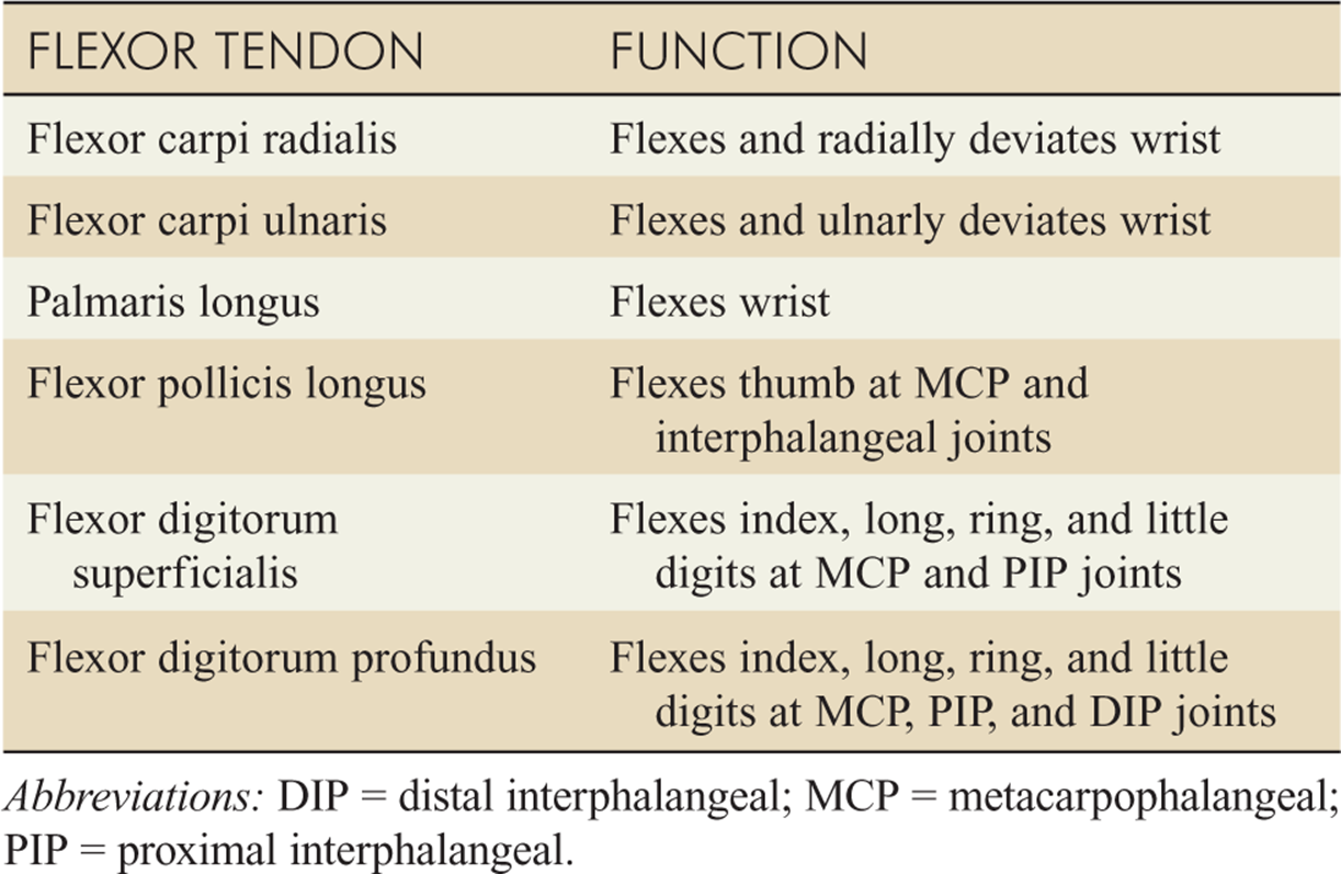
TABLE 14-4 Flexor Tendons in the Forearm
EXTENSOR TENDON LACERATIONS
Experienced emergency physicians may repair (nonbite) extensor tendon injuries over the dorsum of the hand, with the exception of the tendons to the thumb.
Discuss all tendon injuries with a hand specialist for preferred technique and to arrange follow-up.
Use a figure-of-eight stitch, tied on the side of the lacerated tendon, using a 4–0 (5–0 for smaller tendons) nonabsorbable suture material such as polypropylene (Fig. 14-2). Close the skin with nonabsorbable suture and splint the limb.
Lacerations to the extensor tendons over the distal interphalangeal joint may produce a mallet deformity, and if not repaired may result in a swan neck deformity; whereas lacerations over the proximal interphalangeal joint may produce a boutonniere deformity.
Open tendon lacerations require operative repair; closed tendon injuries are either splinted in extension for up to 6 weeks or until operative repair. Refer to a hand surgeon.

FIG. 14-2. Extensor tendon laceration repair with a figure-of-eight stitch.
FLEXOR TENDON LACERATIONS
Refer all flexor tendon injuries to a hand specialist.
Some hand surgeons prefer to repair these injuries within 12 to 24 hours while others delay repair.
If repair is delayed, clean the wound, repair the skin, splint the limb in a position of function, and arrange follow-up within 2 to 3 days with a hand surgeon.
FINGER AND FINGER TIP INJURIES
Most finger lacerations are straightforward and can be repaired by using 5–0 nonabsorbable suture materials.
Suspect digital nerve injuries when static two-point discrimination is distinctively greater on one side of the volar pad than on the other, or when it is greater than 10 mm (normal defined as <6 mm).
Successful repair of fingertip injuries requires knowledge of anatomy (Fig. 14-3) and an understanding of techniques of reconstruction.
Distal fingertip amputations with skin or pulp loss only are best managed conservatively, with serial dressing change only, especially in children.
In cases with larger areas of skin loss (>1 cm2), a skin graft using the severed tip itself or skin harvested from the hypothenar eminence may be required.
Complications of the skin graft technique include decreased sensation of the fingertip, tenderness at the injury and graft site, poor cosmetic result, and hyper-pigmentation in dark-skinned patients.
Injuries with exposed bone are not amenable to skin grafting. Most of these injuries require specialist advice. If less than 0.5 mm of bone is exposed and the wound defect is small, the bone may be trimmed back and the wound left to heal by secondary intention. Injuries to the thumb or index finger with exposed bone nearly always require specialist attention.
Subungual hematomas require decompression by simple trephination of the nail plate. Use of heated paper clip delays healing. Use of nail drill, scalpel, or 18-gauge needle is recommended.
Simple trephination produces an excellent result in patients with subungual hematoma regardless of size, injury mechanism, or presence of simple fracture.
Injuries to the nail bed require careful repair to reduce scar formation. They are associated with fractures of the distal phalanx in 50% of cases.
Remove the nail if there is extensive crush injury, associated nail avulsion or surrounding nail fold disruption, or a displaced distal phalanx fracture on radiograph.
Repair with 6–0 or 7–0 absorbable sutures. If the nail matrix is displaced from its anatomic position at the sulcus, the matrix should be carefully replaced and sewn in place with mattress sutures (Fig. 14-4).
Alternatively, after nail bed repair, tissue adhesive can be used (dripping it onto the perionychium and into nail fold) to secure the nail, avoiding suturing it in place.
Apply mild downward pressure on the nail until the adhesive sets.
If there is extensive injury to the nail bed with avulsed tissue, consult a hand specialist.
In children with fractures of the distal phalanx, the nail plate may come to lie on the eponychium. After careful cleaning and adequate anesthesia, replace the nail plate under the proximal nail fold.

FIG. 14-3. Anatomy of the perionychium.
RING TOURNIQUET SYNDROME
Ring removal is required in all injured fingers. Swelling may require that the ring be cut off. If slower techniques are appropriate, simple lubrication may suffice.
The string technique is an alternative method (Fig. 14-5).
a. String, umbilical tape, or 0-gauge silk may be used.
b. The string is passed under the ring and then wrapped firmly around the finger from proximal to distal.
c. The proximal end of the string is then gently pulled, and the ring advances down the finger.

FIG. 14-4 A and B. Technique for repair of an avulsion of the germinal matrix using three horizontal mattress sutures.
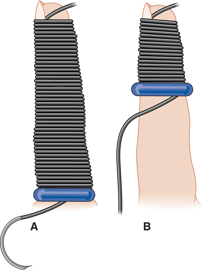
FIG. 14-5. String technique for ring removal. A. Completely wrapped. B. Unwrapping with ring advancing off with the string.
For further reading in Emergency Medicine: A Comprehensive Study Guide, 7th ed., see Chapter 44, “Injuries to the arm, hand, fingertip and nail” by Moira Davenport.