
Types of Abnormal Movements
Tremor
Postural Tremor
Asterixis
Intention (Kinetic) Tremor
Rest Tremor
Chorea
Hemiballismus
Dystonia & Athetosis
Factors Influencing Dystonia
Etiology
Myoclonus
Generalized Myoclonus
Segmental Myoclonus
Propriospinal Myoclonus
Treatment
Tics
Classification
Bradykinesia & Hypokinesia
Clinical Evaluation of Patients
History
Age at Onset
Mode of Onset
Course
Medical History
Examination
Investigative Studies
Blood & Urine Tests
Electrophysiologic Tests
Imaging
Genetic Studies
Psychologic Evaluation
Selected Movement Disorders
Familial or Benign Essential Tremor
Pathogenesis
Clinical Findings
Treatment
Parkinsonism
Etiology
Pathology
Pathogenesis
Clinical Findings
Differential Diagnosis
Treatment
Lewy Body Disease
Clinical Findings
Differential Diagnosis
Treatment
Progressive Supranuclear Palsy
Pathogenesis
Clinical Findings
Differential Diagnosis
Treatment
Prognosis
Corticobasal Degeneration
Clinical Findings
Differential Diagnosis
Treatment
Prognosis
Huntington Disease
Epidemiology
Genetics
Pathology
Clinical Findings
Differential Diagnosis
Treatment & Prognosis
Prevention
Dentatorubral-Pallidoluysian Atrophy
Sydenham Chorea & PANDAS
Primary Generalized Torsion Dystonia
Pathogenesis
Clinical Findings
Differential Diagnosis
Treatment
Course & Prognosis
Dopa-Responsive Dystonia
Dystonia-Parkinsonism
Myoclonic Dystonia
Focal Torsion Dystonia
Heredodegenerative Dystonia
Psychogenic Dystonia
Paroxysmal Dyskinesias
Paroxysmal Dystonic Choreoathetosis (Nonkinesigenic Dyskinesia)
Paroxysmal Kinesigenic Choreoathetosis
Paroxysmal Exercise-Induced Dyskinesias
Wilson Disease
Pathogenesis
Clinical Findings
Differential Diagnosis
Investigative Studies
Treatment
Drug-Induced Movement Disorders
Parkinsonism
Acute Dystonia or Dyskinesia
Akathisia
Tardive Dyskinesia
Other Tardive Syndromes
Neuroleptic Malignant Syndrome
Other Drug-Induced Movement Disorders
Gilles de la Tourette Syndrome
Pathogenesis
Clinical Findings
Differential Diagnosis
Complications
Treatment
Acquired Hepatocerebral Degeneration
Restless Legs Syndrome
References
Movement disorders (sometimes called extrapyramidal disorders) impair the regulation of voluntary motor activity without directly affecting strength, sensation, or cerebellar function. They include hyperkinetic disorders associated with abnormal, involuntary movements and hypokinetic disorders characterized by poverty of movement. Movement disorders result from dysfunction of deep subcortical gray matter structures termed the basal ganglia.Although there is no universally accepted anatomic definition of the basal ganglia, for clinical purposes they may be considered to comprise the caudate nucleus, putamen, globus pallidus (GPi), subthalamic nucleus, and substantia nigra. The putamen and the globus pallidus are collectively termed the lentiform nucleus; the combination of lentiform nucleus and caudate nucleus is designated the corpus striatum.
The basic circuitry of the basal ganglia consists of three interacting neuronal loops (Figure 11-1). The first is a corticocortical loop that passes from the cerebral cortex, through the caudate and putamen, the internal segment of the globus pallidus, and the thalamus and then back to the cerebral cortex. The second is a nigrostriatal loop connecting the substantia nigra with the caudate and putamen. The third, a striatopallidal loop, projects from the caudate and putamen to the external segment of the globus pallidus, then to the subthalamic nucleus, and finally to the internal segment of the globus pallidus. In some movement disorders (eg, Parkinson disease), a discrete site of pathology within these pathways can be identified; in other cases (eg, essential tremor), the precise anatomic abnormality is unknown.

Figure 11-1. Basic neuronal circuitry of the basal ganglia.
TYPES OF ABNORMAL MOVEMENTS
Categorizing an abnormal movement is generally the first step toward arriving at the neurologic diagnosis. Abnormal movements can be classified as tremor, chorea, athetosis or dystonia, ballismus, myoclonus, or tics. Such movements can arise in a variety of contexts, such as in degenerative disorders or with structural lesions. In many disorders, abnormal movements are the sole clinical features.
TREMOR
A tremor is a rhythmic oscillatory movement best characterized by its relationship to voluntary motor activity, that is, according to whether it occurs at rest, during maintenance of a particular posture, or during movement. The major causes of tremor are listed in Table 11-1. Tremor is enhanced by emotional stress and disappears during sleep. Tremor that occurs when the limb is at rest is generally referred to as static tremor or rest tremor. If present during sustained posture, it is called a postural tremor; although this tremor may continue during movement, movement does not increase its severity. When present during movement but not at rest, it is generally called an intention or kinetic tremor. Both postural and intention tremors are also called action tremors.

Table 11-1. Causes of tremor.
POSTURAL TREMOR
Physiologic Tremor
An 8- to 12-Hz tremor of the outstretched hands is a normal finding. Its physiologic basis is uncertain.
Enhanced Physiologic Tremor
Physiologic tremor may be enhanced by fear or anxiety. A more conspicuous postural tremor may also be found after excessive physical activity or sleep deprivation. It can complicate treatment with certain drugs (notably lithium, tricyclic antidepressants, sodium valproate, and bronchodilators) and is often conspicuous in patients with alcoholism or in alcohol or drug withdrawal states. It is common in thyrotoxicosis, and it can also result from poisoning with a number of substances, including mercury, lead, arsenic, and carbon monoxide. There is no specific medical therapy.
Other Causes
The most common type of abnormal postural tremor is benign essential tremor, which often has a familial basis. Postural tremor may also be conspicuous in patients with Wilson disease or cerebellar disorders. A postural tremor of the hands that is indistinguishable from essential tremor may occur in patients with dystonia. The designation dystonic tremor refers to a postural or intention tremor that occurs in a part of the body already affected by dystonia. It is a part of the dystonia and is most prominent when an attempt is made to oppose the dystonic posturing.
ASTERIXIS
Asterixis may be associated with postural tremor, but is itself more properly considered a form of myoclonus (see later discussion) than of tremor. It is seen most commonly in patients with metabolic encephalopathy such as occurs with hepatic or renal failure.
To detect asterixis, the examiner asks the patient to hold the arms outstretched with fingers and wrists extended. Episodic cessation of muscular activity causes sudden flexion at the wrists followed by a return to extension, so that the hands flap in a regular or, more often, an irregular rhythm. A similar phenomenon may be demonstrable at the ankles. The asterixis resolves with clearing of the metabolic encephalopathy.
INTENTION (KINETIC) TREMOR
Intention or kinetic tremor occurs during activity. If the patient is asked to touch his or her nose with a finger, for example, the arm exhibits tremor during movement, often more marked as the target is reached. This form of tremor is sometimes mistaken for limb ataxia, but the latter has no rhythmic oscillatory component.
Intention tremor results from a lesion affecting the superior cerebellar peduncle. Because it is often very coarse, it can lead to severe functional disability. No satisfactory medical treatment exists, but stereotactic surgery of the contralateral ventrolateral nucleus of the thalamus or high-frequency thalamic stimulation through an implanted device is sometimes helpful when patients are severely incapacitated.
Intention tremor can also occur—together with other signs of cerebellar involvement—as a manifestation of toxicity of certain sedative or anticonvulsant drugs (eg, phenytoin) or alcohol; it is seen in patients with Wilson disease.
REST TREMOR
Parkinsonism
Rest tremor usually has a frequency of 4 to 6 Hz and is characteristic of parkinsonism whether the disorder is idiopathic or secondary (ie, postencephalitic, toxic, or drug-induced in origin). The rate of the tremor, its relationship to activity, and the presence of rigidity or hypokinesia usually distinguish the tremor of parkinsonism from other forms of tremor. Tremor in the hands may appear as a “pill-rolling” maneuver—rhythmic, opposing circular movements of the thumb and index finger. There may be alternating flexion and extension of the fingers or hand or alternating pronation and supination of the forearm; in the feet, rhythmic alternating flexion and extension are common. Parkinsonism is discussed in more detail later.
Other Causes
Less common causes of rest tremor include Wilson disease and poisoning with heavy metals such as mercury.
CHOREA
The word chorea denotes rapid irregular muscle jerks that occur involuntarily and unpredictably in different parts of the body. In florid cases, the often forceful involuntary movements of the limbs and head and the accompanying facial grimacing and tongue movements are unmistakable. Voluntary movements may be distorted by the superimposed involuntary ones. In mild cases, however, patients may exhibit no more than a persistent restlessness and clumsiness. Power is generally full, but there may be difficulty in maintaining muscular contraction such that, for example, handgrip is relaxed intermittently (milkmaid grasp). The gait becomes irregular and unsteady, with the patient suddenly dipping or lurching to one side or the other (dancing gait). Speech often becomes irregular in volume and tempo and may be explosive in character. In some patients, athetotic movements or dystonic posturing (see later) may also be prominent. Chorea disappears during sleep.
The pathologic basis of chorea is unclear, but in some cases it is associated with cell loss in the caudate nucleus and putamen, and it can be provoked by dopaminergic agonist drugs. Causes of chorea are shown in Table 11-2 and are discussed later in this chapter. When chorea is due to a treatable medical disorder, such as polycythemia vera or thyrotoxicosis, adequate treatment of the primary disorder abolishes the dyskinesia.
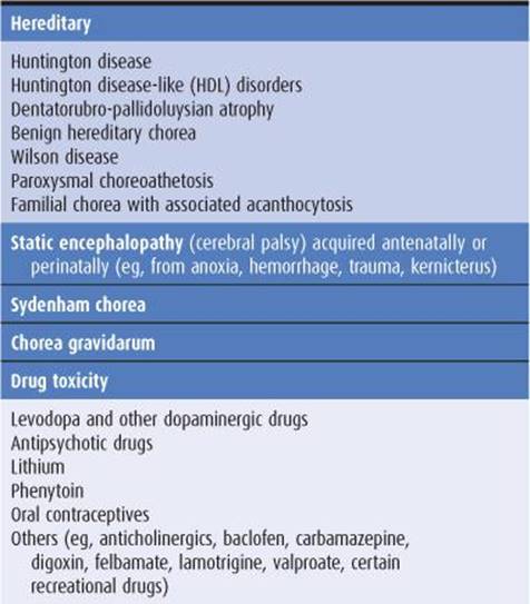
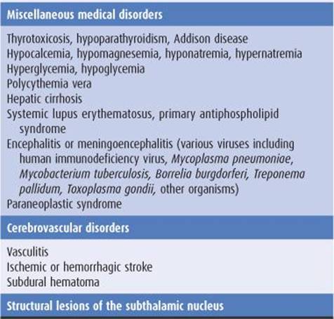
Table 11-2. Causes of chorea.
HEMIBALLISMUS
Hemiballismus is unilateral chorea that is especially violent because the proximal muscles of the limbs are involved. It is due most often to vascular disease in the contralateral subthalamic nucleus and commonly resolves spontaneously in the weeks after its onset. It is sometimes due to other types of structural disease; in the past, it was an occasional complication of thalamotomy. Pharmacologic treatment is similar to that for chorea (discussed later).
DYSTONIA & ATHETOSIS
The term athetosis generally denotes abnormal movements that are slow, sinuous, and writhing in character. When the movements are so sustained that they are better regarded as abnormal postures, the term dystonia is used, and many now use the terms interchangeably. In dystonia, excessive or inappropriate contraction of muscles (often agonists and antagonists) leads to sustained abnormal postures of the affected region of the body. The abnormal movements and postures may be generalized or restricted in distribution, such as to the neck (torticollis), hand and forearm (writer’s cramp), or mouth (oromandibular dystonia). With restricted dystonias, one or more of the limbs may be affected (segmental dystonia), or the disturbance may be restricted to localized muscle groups (focal dystonia). Generalized dystonia may be idiopathic or secondary (Table 11-3).
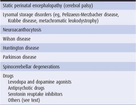
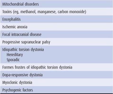
Table 11-3. Causes of dystonia and athetosis.
FACTORS INFLUENCING DYSTONIA
The abnormal movements are not present during sleep. They are generally enhanced by emotional stress and by voluntary activity. In some cases, abnormal movements or postures occur only during voluntary activity and sometimes only during specific activities such as writing, speaking, or chewing.
ETIOLOGY
Table 11-3 lists some of the conditions in which these movement disorders are encountered. Perinatal anoxia, birth trauma, and kernicterus from hyperbilirubinemia are the most common causes. In these circumstances, abnormal movements usually develop before the age of 5 years. Careful questioning usually discloses a history of abnormal early development and often of seizures. Examination may reveal signs of cognitive dysfunction or a pyramidal deficit in addition to the movement disorder.
Dystonic movements and postures are the cardinal features of idiopathic torsion dystonia (discussed later). Torsion dystonia may also occur as a manifestation of Wilson disease or Huntington disease or as a sequela of encephalitis.
Acute dystonic posturing may result from treatment with dopamine receptor antagonist drugs (discussed later).
Lateralized dystonia may occasionally relate to focal intracranial disease, but the clinical context in which it occurs usually identifies the underlying cause.
MYOCLONUS
Myoclonic jerks are sudden, rapid, twitchlike muscle contractions. They can be classified according to their distribution, relationship to precipitating stimuli, or etiology. Generalized myoclonus has a widespread distribution, whereas focal or segmental myoclonus is restricted to a particular part of the body. Myoclonus can be spontaneous, or it can be brought on by sensory stimulation, arousal, or the initiation of movement (action myoclonus). Myoclonus may occur as a normal phenomenon (physiologic myoclonus) in healthy persons, as an isolated abnormality (essential myoclonus), or as a manifestation of epilepsy (epileptic myoclonus). It can also occur as a feature of a variety of degenerative, infectious, and metabolic disorders (symptomatic myoclonus) affecting the cerebral cortex, brainstem, or spinal cord. Myoclonus is sometimes manifest not by a sudden twitchlike muscle contraction but by a sudden loss of muscle activity (negative myoclonus). This is best seen in asterixis: The patient tries to maintain the wrists hyperextended in the outstretched arms, but the sudden intermittent transient relaxation of the extensor muscles followed by return of the contraction leads to a “flapping tremor.”
GENERALIZED MYOCLONUS
The causes of generalized myoclonus are summarized in Table 11-4. Physiologic myoclonus includes the myoclonus that occurs upon falling asleep or awakening (nocturnal myoclonus), as well as hiccup.Essential myoclonus is a benign condition that occurs in the absence of other neurologic abnormalities and is sometimes inherited. Epileptic myoclonus may be impossible to differentiate clinically from nonepileptic forms. It may be possible to distinguish the two types electrophysiologically, however, by the duration of the electromyographic burst associated with the jerking, by demonstrating an electroencephalographic (EEG) correlate with a consistent temporal relationship to the jerks, or by determining whether muscles involved in the same jerk are activated synchronously.
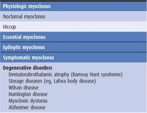
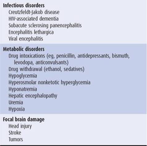
Table 11-4. Causes of generalized myoclonus.
SEGMENTAL MYOCLONUS
Segmental myoclonus can arise from lesions affecting the cerebral cortex, brainstem, or spinal cord. For example, involvement of the dentatorubroolivary pathway by stroke, multiple sclerosis, tumors, or other disorders can produce palatal myoclonus, which may be associated with an audible click or synchronous movements of ocular, facial, or other bulbar muscles. Rhythmic vertical oscillation of the soft palate occurs that is best regarded as a tremor. Segmental myoclonus can result from many of the same disturbances that produce symptomatic generalized myoclonus (Table 11-4). Metabolic disorders such as hyperosmolar nonketotic hyperglycemia can cause epilepsia partialis continua, in which a repetitive focal epileptic discharge occurs from the contralateral sensorimotor cortex and leads to segmental myoclonus. Segmental myoclonus is usually unaffected by external stimuli and persists during sleep.
PROPRIOSPINAL MYOCLONUS
Propriospinal myoclonus arises in the spinal cord and then spreads up and down the cord, leading to a brief bodily contraction. Its myoclonic basis is often not recognized. Electromyographic surface recordings may be necessary to show the spread of muscle activity in an orderly sequence and may help to localize the site of origin of the myoclonus. The underlying spinal lesion may be revealed by imaging in some cases.
TREATMENT
Although myoclonus can be difficult to treat, it sometimes responds to anticonvulsant drugs such as valproic acid 250 to 500 mg orally three times daily or to benzodiazepines such as clonazepam 0.5 mg orally three times daily, gradually increased to as much as 12 mg/d. Postanoxic action myoclonus has been found to be remarkably responsive to 5-hydroxytryptophan, the metabolic precursor of the neurotransmitter 5-hydroxytryptamine (serotonin). The dosage of 5-hydroxytryptophan is increased gradually to a maximum of 1 to 1.5 mg/d orally and may be combined with carbidopa (maximum, 400 mg/d orally) to inhibit metabolism in peripheral tissues.
TICS
Tics are sudden, recurrent, quick, coordinated abnormal movements that can usually be imitated without difficulty. The same movement occurs again and again and can be suppressed voluntarily for short periods, although doing so may cause anxiety. Tics tend to worsen with stress, diminish during voluntary activity or mental concentration, and disappear during sleep.
CLASSIFICATION
Tics can be classified into four groups depending on whether they are simple or multiple and transient or chronic.
1. Transient simple tics are very common in children, usually terminate spontaneously within 1 year (often within a few weeks), and generally require no treatment.
2. Chronic simple tics can develop at any age but often begin in childhood, and treatment is unnecessary in most cases. The benign nature of the disorder must be explained to the patient.
3. Persistent simple or multiple tics of childhood or adolescence generally begin before the age of 15 years. There may be single or multiple motor tics, and often vocal tics, but complete remission occurs by the end of adolescence.
4. The syndrome of chronic multiple motor and vocal tics is generally referred to as Gilles de la Tourette syndrome, after the French physician who was one of the first to describe its clinical features. It is discussed in detail later. Tics also may occur with levodopa or amphetamine use and after chronic neuroleptic use (tardive tic), after head trauma or viral encephalitis, and in autistic children. They can occur in association with degenerative disorders of the basal ganglia, such as Huntington disease, and are well described in neuroacanthocytosis, when they may have a self-mutilating character.
BRADYKINESIA & HYPOKINESIA
Bradykinesia (slowed movement) and hypokinesia or akinesia (poverty or lack of movement) are major features of parkinsonism and may be more disabling than the presence of abnormal movements. Manifestations include a fixity of facial expression (the so-called masked facies, with reduced blinking, widened palpebral fissures, and an apparently impassive appearance) and a paucity of spontaneous movement of the limbs (eg, a reduced arm swing on walking). Some patients have “freezing,” by which is meant a difficulty in starting or maintaining a rhythmic repetitive activity such as walking. Such symptoms are difficult for patients to describe; they are often erroneously attributed to weakness.
These phenomena are tested clinically by, for example, asking the patient to make repetitive alternating movements of each extremity in turn. This can involve the repetitive tapping of the thumb on the terminal interphalangeal joint of the index or third finger or on the pad of these digits, repetitive pronation and supination of the raised arm (as if screwing a light bulb into the ceiling), repetitive opening and closing of the fist of each hand, and repetitive tapping the foot on the floor while the heel is maintained on the ground. A progressive reduction in amplitude or speed of the movements, irregularity in rhythm, or arrests in movement are indicative of abnormality. Activity should be continued until at least 15 repetitions have occurred, and sometimes for longer. It is important to distinguish any abnormalities from the slowness of movement without fatiguing and decrement that may occur in patients with pyramidal or cerebellar dysfunction (often with an irregular rhythm in the latter context). The inexpressive face of depressed patients may simulate the masked facies of parkinsonism and should be distinguished by the lack of other extrapyramidal findings and the abnormal affect.
CLINICAL EVALUATION OF PATIENTS
HISTORY
AGE AT ONSET
The age at onset of a movement disorder may suggest the underlying cause. For example, onset in infancy or early childhood suggests birth trauma, kernicterus, cerebral anoxia, or an inherited disorder; abnormal facial movements developing in childhood are more likely to represent tics than involuntary movements of another sort; and tremor presenting in early adult life is more likely to be of the benign essential variety than due to Parkinson disease.
The age at onset can also influence the prognosis. In idiopathic torsion dystonia, for example, progression to severe disability is much more common when symptoms develop in childhood than when they develop in later life. Conversely, tardive dyskinesia is more likely to be permanent and irreversible when it develops in the elderly than when it develops in the adolescent years.
MODE OF ONSET
Abrupt onset of dystonic posturing in a child or young adult should raise the possibility of a drug-induced reaction; a more gradual onset of dystonic movements and postures in an adolescent suggests the possibility of a chronic disorder such as idiopathic torsion dystonia or Wilson disease. Similarly, the abrupt onset of severe chorea or ballismus suggests a vascular cause, and abrupt onset of severe parkinsonism suggests a neurotoxic cause; more gradual, insidious onset suggests a degenerative process.
COURSE
The manner in which the disorder progresses from its onset may also be helpful diagnostically. For example, Sydenham chorea usually resolves within about 6 months after onset and therefore should not be confused with other varieties of chorea that occur in childhood.
MEDICAL HISTORY
Drug History
It is important to obtain an accurate account of all drugs that have been taken by the patient over the years, because many of the movement disorders are iatrogenic. The phenothiazine and butyrophenone drugs may lead to the development of abnormal movements either while patients are taking them or after their use has been discontinued, and the dyskinesia may be irreversible. These drugs and the dyskinesias associated with their use are discussed later in this chapter.
Reversible dyskinesia may develop in patients taking certain other drugs, including oral contraceptives, levodopa, and phenytoin. Several drugs, especially lithium, tricyclic antidepressants, valproic acid, and bronchodilators, can cause tremor. Serotonin reuptake inhibitors have been associated with a number of movement disorders including parkinsonism, akathisia, chorea, dystonia, and bruxism.
General Medical History
1. Chorea may be symptomatic of the disease in patients with a history of rheumatic fever, thyroid disease, systemic lupus erythematosus, polycythemia, hypoparathyroidism, or cirrhosis of the liver.
2. Movement disorders, including tremor, chorea, hemiballismus, dystonia, and myoclonus, have been described in patients with acquired immunodeficiency syndrome (AIDS). Opportunistic infections such as cerebral toxoplasmosis appear to be the cause in some cases, but infection with human immunodeficiency virus type 1 (HIV-1) may also have a direct pathogenetic role.
3. A history of birth trauma or perinatal distress may suggest the cause of a movement disorder that develops during childhood.
4. Encephalitis lethargica is no longer encountered clinically; it was epidemic in the 1920s, however, and was often followed by a wide variety of movement disorders, including parkinsonism. It is therefore important to inquire about this disease when elderly patients are being evaluated.
Family History
Some movement disorders have an inherited basis (Tables 11-5 and 11-6), and it is essential that a complete family history is obtained, supplemented if possible by personal scrutiny of close relatives. Any possibility of consanguinity should be noted.


Table 11-5. Selected hereditary nonparkinsonian movement disorders.


Table 11-6. Genes implicated in hereditary parkinsonism.
EXAMINATION
Clinical examination will indicate the nature of the abnormal movements and motor deficit, the extent of neurologic involvement, and the presence of coexisting disease; these in turn may suggest the diagnosis.
The mental status examination may suggest psychiatric disease, raising the possibility that the abnormal movements are related to the psychiatric disorder or to its treatment with psychoactive drugs—or that the patient has a disorder characterized by both abnormal movements and behavioral disturbances, such as Huntington disease or Wilson disease.
Focal motor or sensory deficits raise the possibility of a structural space-occupying lesion, as does papilledema. Kayser-Fleischer rings suggest Wilson disease. Signs of vascular, hepatic, or metabolic disease may suggest other causes for a movement disorder, such as acquired hepato-cerebral degeneration or vasculitis.
INVESTIGATIVE STUDIES
Several investigations may be of diagnostic help.
BLOOD & URINE TESTS
1. Serum and urine copper and serum ceruloplasmin levels are important in diagnosing Wilson disease.
2. Complete blood count and sedimentation rate are helpful in excluding polycythemia, vasculitis, or systemic lupus erythematosus, any of which can occasionally lead to a movement disorder. A wet film of the blood may reveal circulating acanthocytes.
3. Blood chemistries may reveal hepatic dysfunction related to Wilson disease or acquired hepatocerebral degeneration; hyperthyroidism or hypocalcemia as a cause of chorea; or a variety of metabolic disorders associated with myoclonus.
4. Serologic tests are helpful for diagnosing movement disorders caused by systemic lupus erythematosus or lupus anticoagulant syndrome. Neurosyphilis and HIV-1 infection can be manifested clinically in a variety of ways and should always be excluded by appropriate serologic tests in patients with neurologic disease of uncertain etiology.
ELECTROPHYSIOLOGIC TESTS
An EEG is sometimes helpful in diagnosing patients with myoclonus and in distinguishing paroxysmal dyskinesias from seizures; otherwise, it is of limited usefulness. Electromyography and somatosensory evoked potentials may help to determine the level of neural involvement in myoclonus.
IMAGING
Radiologic studies are occasionally helpful in evaluating patients with movement disorders. In some patients, intracranial calcification may be found by skull x-rays or computed tomography (CT) scans; the significance of this finding, however, is not clear. CT scans or magnetic resonance imaging (MRI) may also reveal a tumor or other lesion associated with focal dyskinesia or dystonia or with symptomatic myoclonus, caudate atrophy due to Huntington disease, or basal ganglia abnormalities associated with Wilson disease. Positron emission tomography (PET) using 18F-dopa can monitor the loss of nigrostriatal projections in Parkinson disease and may be helpful diagnostically in patients with incomplete parkinsonian syndromes, but is not widely available. Dopamine transporter imaging using beta-CIT single-photon emission computed tomography (SPECT) can also be used for this purpose.
GENETIC STUDIES
Recombinant DNA technology has been used to generate probes for genes that determine certain inheritable movement disorders, such as Huntington disease and Wilson disease. Genetic markers are therefore of diagnostic value in such disorders (Tables 11-5 and 11-6). Their use may be limited, however, by the genetic heterogeneity of some diseases, imprecise gene localization by certain probes, ethical concerns about adverse psychologic reactions to the presymptomatic diagnosis of fatal disorders, and the potential for misuse of such information by prospective employers, insurance companies, and government agencies.
PSYCHOLOGIC EVALUATION
Cognitive and affective disturbances can be documented and characterized by neuropsychologic evaluation. This may be helpful in diagnosing certain disorders such as Huntington disease or diffuse Lewy body dementia. Some movement disorders, such as Gilles de la Tourette syndrome, are associated with behavioral abnormalities such as attention deficit disorder and obsessive-compulsive disorder. The findings also may be important in guiding decisions regarding invasive interventions such as deep brain stimulation. Such a therapeutic intervention is contraindicated in patients with atypical parkinsonian syndromes or in classic Parkinson disease when significant dementia or major depression is also present.
SELECTED MOVEMENT DISORDERS
The more common and well-defined diseases or syndromes characterized by abnormal movements are discussed here with the principles of their treatment.
FAMILIAL OR BENIGN ESSENTIAL TREMOR
PATHOGENESIS
A postural tremor may be prominent in otherwise normal subjects. Although the pathophysiologic basis of this disorder is uncertain, it often has a familial basis with an auto-somal dominant mode of inheritance. At least three gene loci have been implicated; in some cases (ETM1), the disorder is related to a polymorphism in the D3 dopamine receptor gene (DRD3).
CLINICAL FINDINGS
Symptoms may develop in the teenage or early adult years but often do not appear until later. The tremor typically involves one or both hands or the head and voice, whereas the legs tend to be spared. Examination usually reveals no other gross abnormalities, but some patients may have ataxia or personality disturbances. Although the tremor may become more conspicuous with time, it generally leads to little disability other than cosmetic and social embarrassment. In occasional cases, tremor interferes with the ability to perform fine or delicate tasks with the hands; handwriting is sometimes severely impaired. Speech is affected when the laryngeal muscles are involved. Patients commonly report that a small quantity of alcohol provides remarkable but transient relief; the mechanism is not known.
TREATMENT
If treatment is warranted, propranolol, 40 to 120 mg orally twice daily, can be prescribed, but it will need to be taken for an indefinite period. Other beta-blockers, such as atenolol and sotalol, have also been used. If tremor is particularly disabling under certain predictable circumstances, it can be treated with a single oral dose of 40 to 120 mg of propranolol taken in anticipation of the precipitating circumstances.
Primidone has also been effective, but patients with essential tremor are often very sensitive to this drug, so that it must be introduced more gradually than when it is used to treat epilepsy. Patients are therefore started on 50 mg/d, and the daily dose is increased by 50 mg every 2 weeks until benefit occurs or side effects limit further increments. A dose of 100 or 150 mg three times a day is often effective. There is no evidence that high doses (exceeding 750 mg daily) provide any added benefit.
Occasional patients respond to alprazolam, up to 3 mg/d in divided doses. Some patients reportedly benefit from gabapentin (1,200 mg/d), topiramate (400 mg/d), zonis-amide (up to 200 mg daily), or intramuscular injections of botulinum toxin. Anecdotal reports of benefit from mirtazapine were not confirmed in a double-blind study, which found no effect on the tremor in most patients.
Some patients have disabling tremor that is unresponsive to pharmacologic measures. Thalamotomy may be helpful, but a significant morbidity is associated with bilateral procedures. High-frequency thalamic stimulation by an implanted electrode is an effective alternative to thalamotomy and has a lower morbidity. It may be particularly useful for treatment of the unoperated side in patients who have already undergone unilateral thalamotomy. Benefit is maintained over the years in most patients with severe disability.
PARKINSONISM
Parkinsonism occurs in all ethnic groups; in the United States and western Europe it has a prevalence of 1 to 2 per 1,000 population, with an approximately equal sex distribution. The disorder becomes increasingly common with advancing age. It is characterized by tremor, hypokinesia, rigidity, and abnormal gait and posture.
ETIOLOGY
Idiopathic
The most common variety of parkinsonism occurs without obvious cause; this idiopathic form is called Parkinson disease or paralysis agitans when there are no atypical features, it is not secondary to some known cause, and there is a sustained response to treatment with dopaminergic medication. A preclinical phase extending back for several years before the development of the motor deficit is now recognized, during which hyposmia, constipation, anxiety, depression, and rapid-eye-movement (REM) sleep behavior disorder may be present. This is important to recognize in studies seeking predictive biomarkers and in attempts to identify protective factors.
Encephalitis Lethargica
In the first half of the 20th century, parkinsonism often developed in patients with a history of von Economo encephalitis. Because this type of infection is not now encountered, cases of postencephalitic parkinsonism are becoming increasingly rare.
Drug- or Toxin-Induced Parkinsonism
1. Therapeutic drugs—Many drugs, such as phenothiazines, butyrophenones, metoclopramide, reserpine, and tetrabenazine, can cause a reversible parkinsonian syndrome (see later). This is important to recognize because it is usually reversible by withdrawing the offending medication, although symptoms and signs may take many months to resolve.
2. Toxic substances—Environmental toxins such as manganese dust or carbon disulfide can lead to parkinsonism; manganese used in the home manufacture of methcathinone appears to have been responsible for parkinsonism in intravenous users of this illegal stimulant. The disorder may also appear as a sequela of severe carbon monoxide poisoning or exposure to fumes during welding. Experimental studies suggest that pesticide exposure is associated with the development of parkinsonism as well.
3. MPTP (1-methyl-4-phenyl-1,2,5,6-tetrahydropyri-dine)—A drug-induced form of parkinsonism has been described in individuals who synthesized and self-administered a meperidine analogue, MPTP. This compound is metabolized to a toxin that selectively destroys dopaminergic neurons in the substantia nigra and adrenergic neurons in the locus ceruleus and induces a severe form of parkinsonism in humans and in subhuman primates. The ability of this drug to reproduce neurochemical, pathologic, and clinical features of Parkinson disease suggests that an environmental toxin could be responsible for the idiopathic disorder. MPTP-induced parkinsonism has been used as a model to assist in the development of new drugs for treatment of this disease.
Vascular Parkinsonism
Multiple subcortical white-matter infarcts may lead to symptoms and signs suggestive of parkinsonism, usually accompanied by brisk tendon reflexes and extensor plantar responses. Tremor is often relatively inconspicuous and, in some patients, abnormalities of gait are especially evident (“lower-body parkinsonism”). The MRI findings help to suggest or support the diagnosis, and management is focused on preventing stroke. The response to antiparkinsonian medication is usually disappointing.
Post-traumatic Parkinsonism
Boxers and those engaged in certain other sports, such as football, may develop a syndrome of dementia (dementia pugilistica), behavioral and psychiatric disturbances, parkinsonism, and pyramidal and cerebellar deficits from recurrent head trauma leading to a chronic traumatic encephalopathy. There is no satisfactory treatment.
Familial & Genetic Parkinsonism
Rarely, parkinsonism occurs on a familial basis. Autosomal dominant parkinsonism may result from mutations of one of several genes, including α-synuclein (SNCA), leucine-rich repeat kinase 2 (LRRK2), and ubiquitin carboxyl-terminal esterase L1 (UCHL1) (Table 11-6). Mutations in parkin (PARK2) and DJ1 cause early-onset, autosomal recessive, and sporadic juvenile-onset parkinsonism. Several other genes or chromosomal regions have been implicated in familial forms of the disease or as susceptibility factors, including the gene for beta glucosidase (GBA), the enzyme deficient in the lysosomal storage disorder Gaucher disease.
Parkinsonism Associated with Other Neurologic Diseases
Parkinsonism that occurs in association with symptoms and signs of other neurologic disorders is considered briefly in the later section on differential diagnosis.
PATHOLOGY
Idiopathic parkinsonism (Parkinson disease) is a proteinopathy characterized by the misfolding and aggregation of α-synuclein. It is thus also referred to as a synucleinopathy. Histopathologic examination at advanced stages shows loss of pigmentation and cells in the substantia nigra and other brainstem centers, cell loss in the globus pallidus and putamen, and filamentous eosinophilic intraneural inclusion granules (Lewy bodies) containing α-synuclein in the basal ganglia, brainstem, spinal cord, and sympathetic ganglia. The distribution of Lewy bodies is more widespread than originally appreciated, with early involvement of the lower brainstem (eg, dorsal motor nucleus of the vagus [X] nerve), olfactory bulb, and enteric nervous system, and subsequent spread to the locus ceruleus, substantia nigra, transentorhinal cortex, hippocampus, and neocortex. Lewy bodies are not seen in postencephalitic parkinsonism; instead there may be nonspecific neurofibrillary degeneration in a number of diencephalic structures as well as changes in the substantia nigra.
PATHOGENESIS
As in other neurodegenerative proteinopathies (discussed at greater length in Chapter 5), the disease is thought to be triggered by protein misfolding and aggregation—in Parkinson disease, the protein involved is α-synuclein. Abnormal protein may subsequently spread from cell to cell and thereby propagate the disease to contiguous parts of the nervous system.
The motor manifestations of Parkinson disease appear to result from altered patterns of inhibition and excitation within the basal ganglia and its connections via direct and indirect pathways (Figure 11-2). Dopamine and acetylcho-line act as neurotransmitters in this region. In idiopathic parkinsonism, the normal balance between these two antagonistic neurotransmitters is disturbed because of dopamine depletion in the dopaminergic nigrostriatal system (Figure 11-3). Other neurotransmitters, such as norepinephrine, are also depleted in the brains of patients with parkinsonism, but the clinical relevance of this deficiency is less clear.
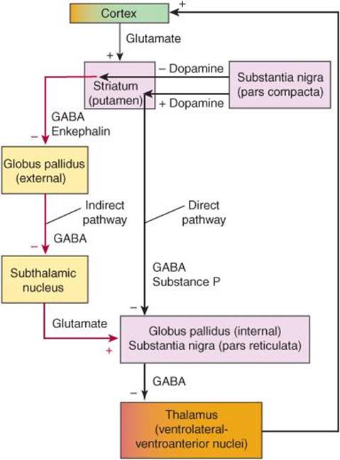
Figure 11-2. Functional circuitry between the cerebral cortex, basal ganglia, and thalamus. The major neurotransmitters and their excitatory (+) or inhibitory (–) effects are indicated. In Parkinson disease, there is degeneration of the pars compacta of the substantia nigra, leading to overactivity in the indirect pathway (red) and increased glutamatergic output from the subthalamic nucleus. (From Aminoff MJ. Pharmacologic management of parkinsonism and other movement disorders. In Katzung BG, Masters SB, Trevor AJ, eds. Basic and Clinical Pharmacology. 11th ed. New York, NY: McGraw-Hill; 2009.)

Figure 11-3. Schematic representation of the sequence of neurons involved in parkinsonism. Top: Dopaminergic neurons (red) originating in the substantia nigra normally inhibit the GABAergic output from the striatum (caudate and putamen), whereas cholinergic neurons (green) exert an excitatory effect. Bottom: In parkinsonism, there is a selective loss of dopaminergic neurons (dashed, red). This leads to increased inhibitory output from the striatum. (From Aminoff MJ. Pharmacologic management of parkinsonism and other movement disorders. In Katzung BG, Masters SB, Trevor AJ (eds). Basic and Clinical Pharmacology. 12th ed. New York, NY: McGraw-Hill; 2012.)
CLINICAL FINDINGS
Tremor
The 4- to 6-Hz tremor of parkinsonism is characteristically most conspicuous at rest; it increases at times of emotional stress and often improves during voluntary activity. It commonly begins in the hand or foot, where it takes the form of rhythmic flexion–extension of the fingers or of the hand or foot—or of rhythmic pronation–supination of the forearm. It frequently involves the face in the area of the mouth as well. Although it may ultimately be present in all of the limbs, it is not uncommon for the tremor to be confined to one limb—or to the two limbs on one side—for months or years before it becomes more generalized. In some patients, tremor never becomes prominent.
Rigidity
Rigidity or increased tone, that is, increased resistance to passive movement, is a characteristic clinical feature of parkinsonism. The disturbance in tone is responsible for the flexed posture of many patients with parkinsonism. The resistance is typically uniform throughout the range of movement at a particular joint and affects agonist and antagonist muscles alike—in contrast to the findings in spasticity, where the increase in tone is often greatest at the beginning of the passive movement (clasp-knife phenomenon) and more marked in some muscles than in others. In some instances, the rigidity in parkinsonism is described as cogwheel rigidity because of ratchet-like interruptions of passive movement that may be due, in part, to the presence of tremor.
Hypokinesia
The most disabling feature of this disorder is hypokinesia (sometimes called bradykinesia or akinesia)—a slowness of voluntary movement and a reduction in automatic movement, such as swinging the arms while walking. The patient’s face is relatively immobile (hypomimia or masklike facies), with widened palpebral fissures, infrequent blinking, a certain fixity of facial expression, and a smile that develops and fades slowly. The voice is of low volume (hypophonia) and tends to be poorly modulated. Fine or rapidly alternating movements are impaired, but power is not diminished if time is allowed for it to develop. The handwriting is small (micrographia), tremulous, and hard to read.
Abnormal Gait & Posture
The patient generally finds it difficult to get up from bed or an easy chair and tends to adopt a flexed posture on standing (Figure 11-4). It is often difficult to start walking, so the patient may lean farther and farther forward while walking in place before being able to advance. The gait itself is characterized by small, shuffling steps and absence of the arm swing that normally accompanies locomotion; there is generally some unsteadiness on turning, and there may be difficulty in stopping. Retained arm swing, wide-based gait, or marked imbalance at an early stage suggests a nonparkinsonian disorder. In advanced cases, the patient tends to walk with increasing speed to prevent a fall (festinating gait) because of the altered center of gravity that results from the abnormal posture.
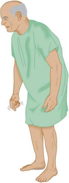
Figure 11-4. Typical flexed posture of a patient with parkinsonism.
Other Motor Abnormalities
There is often mild blepharoclonus (fluttering of the closed eyelids) and occasionally blepharospasm (involuntary closure of the eyelids). The patient may drool, perhaps because of impairment of swallowing. There is typically no alteration in the tendon reflexes (although a mild hyperreflexia may occur on the affected side in asymmetric parkinsonism), and the plantar responses are flexor. Repetitive tapping (approximately twice per second) over the bridge of the nose produces a sustained blink response (Myerson sign); the response is not sustained in normal subjects.
Nonmotor Manifestations
Anosmia is an early symptom (but may arise from many other causes, and is therefore not a specific indicator of Parkinson disease). Cognitive decline, executive dysfunction, and personality changes are common, as also are depression and anxiety. A sense of fatigue may be prominent, and some patients complain of pain or sensory disturbances. Dysautonomic symptoms are often troublesome, especially urinary urgency and urge incontinence, and constipation; postural hypotension relates most commonly to dopaminergic therapy or inactivity but may also reflect baroreflex failure or denervation of cardiac muscle. Sleep disorders including REM behavior disorder are common. Seborrheic dermatitis may occur.
DIFFERENTIAL DIAGNOSIS
The diagnosis may be difficult to make in mild cases. Some degree of slowing is normal in the elderly, and certain otherwise normal people have a deliberate slowness about them.
Depression
Depression may be accompanied by a somewhat expressionless face, poorly modulated voice, and reduction in voluntary activity; it can thus simulate parkinsonism. Moreover, the two diseases often coexist in the same patient. A trial of antidepressant drug treatment may be helpful if diagnostic uncertainty cannot be resolved by the presence of more widespread neurologic signs indicative of parkinsonism.
Essential (Benign Familial) Tremor
This has been considered separately (see earlier discussion). An early age at onset, a family history of tremor, the relationship of the tremor to activity, a beneficial effect of alcohol on the tremor, and a lack of other neurologic signs distinguish this disorder from parkinsonism. Furthermore, essential tremor commonly affects the head (causing a nod or head shake); parkinsonism typically affects the lower jaw rather than the head.
Dystonia
A dystonic tremor may also be mistaken for parkinsonism, particularly when the dystonia is mild or unrecognized.
Diffuse Lewy Body Disease
This disorder occurs especially in patients aged between 60 and 80 years and is marked clinically by the combination of a rapidly progressing neurobehavioral syndrome of dementia and hallucinations and extrapyramidal motor features characteristic of Parkinson disease. Myoclonus may also be seen. There is only an incomplete response to levodopa, but patients are extremely sensitive to parkinsonian complications of neuroleptics as well as to the side effects of antiparkinsonian drugs. It is discussed further in Chapter 5 and later in this chapter.
Wilson Disease
Wilson disease (discussed in more detail later) can also lead to a parkinsonian syndrome, but other varieties of abnormal movements are usually present as well. Moreover, the early age at onset and the presence of Kayser-Fleischer rings should distinguish Wilson disease from Parkinson disease, as should the abnormalities in serum and urinary copper and serum ceruloplasmin that occur in Wilson disease.
Huntington Disease
Huntington disease may occasionally be mistaken for parkinsonism when it presents with rigidity and akinesia, but a family history of Huntington disease or an accompanying dementia, if present, should suggest the correct diagnosis, which can be confirmed by genetic studies.
Striatonigral Degeneration
This rare disorder is one subtype of multisystem atrophy (MSA) and is now referred to as MSA-P. It is associated with neuronal loss in the putamen, globus pallidus, and caudate nucleus and presents with bradykinesia and rigidity. Antiparkinsonian drugs are typically ineffective. Striatonigral degeneration may be associated with cerebellar degeneration (Chapter 8), in which case the term MSA-C is applied. When autonomic insufficiency is a conspicuous accompaniment, the eponymous designation of Shy-Drager syndrome is sometimes used. This latter syndrome is characterized by parkinsonian features, autonomic insufficiency (leading to postural hypotension, anhidrosis, disturbance of sphincter control, and impotence), and signs of more widespread neurologic involvement (pyramidal or lower motor neuron signs and often a cerebellar deficit). MRI reveals a hypointense putamen with a hyperintense rim. There is no treatment for the motor deficit (although a modest response to antiparkinsonian agents may occur), but the postural hypotension may respond to a liberal salt diet, fludrocortisone 0.1 to 1 mg/d, midodrine (an α-adrenergic receptor agonist) 10 mg three times daily, wearing waist-high elastic hosiery, and sleeping with the head up at night.
Progressive Supranuclear Palsy
Bradykinesia and rigidity may be present in this disorder, but its most characteristic features are early postural instability and falls, loss of voluntary control of eye movements (especially vertical gaze), frontotemporal dementia, pseudobulbar palsy, dysarthria, and axial dystonia. The disorder responds poorly, if at all, to antiparkinsonian drugs. It is discussed further in Chapter 5 and later in this chapter.
Corticobasal Degeneration
Corticobasal degeneration is characterized clinically by both cortical and basal ganglionic dysfunction. Rigidity, bradykinesia, tremor, postural disturbances, and dystonia are accompanied by such additional deficits as cortical sensory loss, apraxia, focal reflex myoclonus, dementia, or aphasia. Symptoms are often strikingly asymmetric. Treatment with antiparkinsonian medication is usually unrewarding, although some patients do respond to Sinemet (see later).
Creutzfeldt-Jakob Disease
This prion disease may be accompanied by parkinsonian features, but dementia is usually present, myoclonic jerks are common, and ataxia is sometimes prominent; there may be pyramidal or cerebellar signs and visual disturbances, and the EEG findings of periodic discharges are usually characteristic.
Normal Pressure Hydrocephalus
This condition leads to a gait disturbance (often mistakenly attributed to parkinsonism), urinary incontinence, and dementia. CT scanning reveals dilation of the ventricular system of the brain without cortical atrophy. The disorder may follow head injury, intracranial hemorrhage, or meningoencephalitis, but the cause is often obscure. Surgical shunting procedures to bypass any obstruction to the flow of cerebrospinal fluid (CSF) are often beneficial. Normal pressure hydrocephalus is discussed in more detail in Chapter 5.
TREATMENT
Early parkinsonism requires no drug treatment, but it is important to discuss with the patient the nature of the disorder and the availability of medical treatment if symptoms become more severe, and to encourage activity. Treatment, when indicated, is directed toward restoring the dopaminergic–cholinergic balance in the striatum by blocking the effect of acetylcholine with anticholinergic drugs or by enhancing dopaminergic transmission (Figure 11-5).
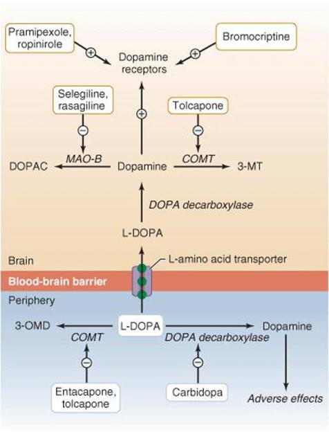
Figure 11-5. Pharmacologic basis of antiparkinsonian dopaminergic therapy. (From Aminoff MJ. Pharmacologic management of parkinsonism and other movement disorders. In Katzung BG, Masters SB, Trevor AJ (eds). Basic and Clinical Pharmacology. 12th ed. New York, NY: McGraw-Hill; 2012.)
Anticholinergic Drugs
Muscarinic anticholinergic drugs are more helpful in alleviating tremor and rigidity than hypokinesia but are generally less effective than dopaminergic drugs (see later). A number of preparations are available, and individual patients tend to favor different drugs. Among the most commonly prescribed drugs are trihexyphenidyl and benztropine (Table 11-7). Anticholinergic drugs are best avoided in the elderly because of their side effects, which include dry mouth, constipation, urinary retention, defective pupillary accommodation, and confusion. Treatment is started with a small dose of one of the anticholinergics; the dosage is then gradually increased until benefit occurs or side effects limit further increments. If treatment is not helpful, the drug is withdrawn and another anticholinergic preparation is tried.

Table 11-7. Drugs used in the treatment of Parkinson disease.
Amantadine
Amantadine can be given for mild parkinsonism either alone or in combination with an anticholinergic agent. Its precise mode of therapeutic action is unclear, but its pharmacologic effects include blockade of NMDA-preferring glutamate and muscarinic cholinergic receptors and stimulation of dopamine release. Amantadine improves all the motor features of parkinsonism, its side effects (restlessness, confusion, skin rashes, edema, disturbances of cardiac rhythm) are relatively uncommon, its effects are exerted rapidly, and it is given in a standard dose of 100 mg orally twice daily. Unfortunately, however, many patients fail to respond to this drug, or its benefit is short-lived. Amantadine may also be useful in reducing the sense of extreme fatigue experienced by some patients and for iatrogenic dyskinesias in patients with advanced disease (100 mg two or three times daily).
Levodopa
Levodopa, which is converted in the body to dopamine (Figure 11-5), ameliorates all the major clinical features of parkinsonism and, unlike the anticholinergic drugs, is often particularly helpful against hypokinesia. There remains disagreement about the best time to introduce levodopa therapy. Concerns that levodopa may lose its effectiveness with time (as opposed to with advance of the disease) are misplaced, but response fluctuations sometimes occur after it has been used for several years, and these may be particularly disabling and difficult to manage. It may thus be wise to defer the introduction of levodopa for as long as possible and then use dopamine agonists (discussed later) in conjunction with it to keep the levodopa dose as low as possible. The medication is best taken about 30 to 45 minutes before meals or 2 hours after meals to maximize absorption and uptake into the brain.
The most common side effects of levodopa are nausea, vomiting, hypotension, abnormal movements (dyskinesias), restlessness, and confusion. Cardiac arrhythmias occur occasionally. The late dyskinesias and behavioral side effects occur as dose-related phenomena, but reduction in dose may diminish any therapeutic benefit. Treatment with olanzapine, quetiapine, or risperidone may relieve confusion and psychotic mental disturbances without blocking the effects of levodopa or exacerbating parkinsonism. Clozapine, a dibenzodiazepine derivative that does not block the therapeutic effects of dopaminergic medication, may also relieve confusion and psychotic mental disturbances and, in some instances, the dyskinesias, but requires regular monitoring of the leukocyte count.
Another late complication of levodopa therapy is response fluctuation such as the wearing-off effect, in which deterioration occurs shortly before the next dose is to be taken, or the on–off phenomenon, in which abrupt but transient fluctuations in the severity of parkinsonism occur at frequent intervals during the day, apparently without any relationship to the last dose of levodopa. This sometimes disabling problem can be controlled only partly by varying the dosing intervals, restricting dietary protein intake, or providing treatment with dopamine agonists. It may relate to discontinuous (pulsatile) levels of cerebral dopamine.
Carbidopa is a drug that inhibits dopa decarboxylase, the enzyme responsible for the breakdown of levodopa to its active metabolite, dopamine (Figure 11-5), but does not cross the blood–brain barrier. Accordingly, if levodopa is given in combination with carbidopa, the breakdown of levodopa is limited outside the brain. The daily dose of levodopa required for benefit and the incidence of nausea, vomiting, hypotension, and cardiac irregularities can be reduced if levodopa is taken in combination with carbidopa. Carbidopa is generally combined with levodopa in a fixed proportion (1:10 or 1:4) as Sinemet.Treatment is started with a small dose, such as Sinemet 10/100 (mg) or Sinemet 25/100 (mg) orally three times daily, and the dose is gradually increased, depending on the response. Most patients ultimately require Sinemet 25/250 (mg) three or four times daily. Carbidopa should total at least 75 mg/d. A controlled-release (CR) formulation of Sinemet may reduce response fluctuations and the dosing frequency. A tablet of carbidopa-levodopa (25/100,10/100, 25/250) that disintegrates in the mouth and is then swallowed with the saliva (Parcopa) is also available. It is best taken about 1 hour before meals.
Levodopa therapy (either alone or in conjunction with carbidopa) is contraindicated in patients with narrow-angle glaucoma or psychotic illness and should be avoided in patients receiving monoamine oxidase type A (MAO-A) inhibitors. It should also be used with care in patients with active peptic ulcers or suspected malignant melanomas.
Dopamine Agonists
The older agonists are ergot derivatives such as bromocriptine, which stimulates dopamine D2 receptors. It is perhaps slightly less effective than levodopa in relieving the symptoms of parkinsonism but is less likely to cause dyskinesias or the on–off phenomenon. Bromocriptine is now used infrequently, as more effective dopamine agonists are available. The dose is built up gradually depending on response and tolerance; maintenance doses are usually between 2.5 and 10 mg orally three times daily. Side effects are similar to those of levodopa, but psychiatric effects such as delusions or hallucinations are especially common, and bromocriptine is therefore contraindicated in patients with a history of psychotic disorder. Relative contraindications to its use are recent myocardial infarction, severe peripheral vascular disease, and active peptic ulceration. Pericardial, pleural, and retroperitoneal fibroses are rare, ergot-related side effects.
Pergolide is another ergot derivative and dopamine receptor agonist; unlike bromocriptine, it activates both D1 and D2 receptors. Its indications, side effects, and contraindications are similar to those described above for bromocriptine, and it is unclear whether either compound is clinically superior to the other. Pergolide was used for many years but has been associated with the development of valvular heart disease in approximately one-third of patients and has therefore been withdrawn by the manufacturer.
The newer dopamine agonists are not ergot derivatives. They seem to be as effective as the older agonists but are without their potential ergot-related adverse effects and may be used in early or advanced Parkinson disease. Pramipexole is started at 0.125 mg three times daily; the daily dose is doubled after 1 week and again after another week; it is then increased by 0.75 mg each week according to response and tolerance. A common maintenance dose is between 0.5 and 1.5 mg three times daily. Ropinirole is started at 0.25 mg three times daily, and the total daily dose increased at weekly intervals by 0.75 mg until the fourth week and by 1.5 mg thereafter. Most patients need between 2 and 8 mg three times daily for benefit. Adverse effects of these medications include fatigue, somnolence, nausea, peripheral edema, dyskinesias, confusion, hallucinations, and orthostatic hypotension. An irresistible urge to sleep at inappropriate times sometimes occurs and may lead to injury. Disturbances of impulse control may lead to such behaviors as compulsive gambling or abnormal sexual activity. Extended-release preparations of both pramipexole and ropinirole are available.
Apomorphine hydrochloride, a nonselective dopamine receptor agonist administered by subcutaneous injection, may help rescue patients with advanced parkinsonism and severe “off” episodes of akinesia despite optimized oral therapy. Side effects include severe nausea and vomiting, somnolence, hallucinations, chest pain, and hyperhidrosis; dyskinesias may be enhanced. It should not be prescribed by physicians who are unfamiliar with its potential complications and interactions.
Catechol-O-Methyltransferase Inhibitors
Catechol-O-methyltransferase (COMT) is one of two principal enzymes involved in the metabolic breakdown of dopamine (Figure 11-5); the other is monoamine oxidase, discussed later. COMT inhibitors may be used to reduce the dose requirements of and any response fluctuations to Sinemet. Their use leads to more sustained plasma levels of levodopa, with improved transport into the blood and across the blood–brain barrier. Side effects include diarrhea, confusion, dyskinesias, and abnormalities of liver function tests. Two of these inhibitors are in widespread use. Tolcapone is taken in a daily dose of 100 or 200 mg three times daily. Acute hepatic necrosis has occurred in rare instances in patients receiving this medication and, accordingly, entacapone (200 mg) taken with Sinemet up to five times daily is generally preferred.
A commercial preparation named Stalevo is now available that combines levodopa with both carbidopa and entacapone. In addition to the convenience of simplifying the drug regime and requiring the consumption of fewer tablets, it is priced at or below the price of its individual components. It is available in three combinations: Stalevo 50 (50 mg levodopa plus 12.5 mg carbidopa and 200 mg entacapone), Stalevo 100 (100 mg, 25 mg, and 200 mg, respectively), and Stalevo 150 (150 mg, 37.5 mg, and 200 mg, respectively). More sustained plasma levels of levodopa may lead to more continuous delivery of levodopa to the brain, with a consequent reduction in the risk of response fluctuations and dyskinetic complications. However, initiating levodopa therapy with Stalevo rather than carbidopa-levodopa failed to delay the time of onset or reduce the frequency of dyskinesia; indeed, it seemed that dyskinesias occurred sooner and with increased frequency.
Monoamine Oxidase Inhibitors
Selegiline, an irreversible monoamine oxidase type B (MAO-B) inhibitor, inhibits the metabolic breakdown of dopamine (Figure 11-5). It thus enhances the antiparkinsonian effect of levodopa and may reduce mild on–off fluctuations in responsiveness. Some clinical studies suggest that selegiline may also delay the progression of Parkinson disease, although the evidence is incomplete in this regard; when used for neuroprotection, selegiline is best kept for patients with mild disease. The dose is 5 mg orally twice daily, usually given early in the day to avoid insomnia.
Rasagiline is a more potent and selective, well-tolerated, irreversible MAO-B inhibitor that is taken in a dose of 0.5 or 1 mg once daily. It is effective in the initial treatment of early parkinsonism and in addition is effective as adjunctive therapy in patients with more advanced disease and response fluctuations to levodopa. It may also slow disease progression, although the evidence for this is ambiguous.
Ablative Surgery
Surgical treatment of parkinsonism by thalamotomy or pallidotomy is often helpful when patients become unresponsive to pharmacologic measures or develop intolerable adverse reactions to antiparkinsonian medication. Lesions of the internal segment of the globus pallidus, for example, will attenuate its unbalanced inhibitory output (Figure 11-2). Surgery is sometimes helpful in relatively young patients with predominantly unilateral tremor and rigidity that have failed to respond to medication; thalamotomy is more helpful for tremor, and pallidotomy for hypokinesia. Diffuse vascular disease or dementia is a contraindication to this approach. The rate of significant complications is less than 5% after unilateral pallidotomy or thalamotomy, but approximately 20% or more after bilateral procedures, which are therefore best avoided. Candidates for surgery should have classic Parkinson disease, be cognitively intact, have previously responded well to pharmacologic treatment, and have developed response fluctuations with a significant amount of off-time. Patients with atypical parkinsonism, a poor previous response to antiparkinsonian drugs, cognitive impairment, psychiatric disorders, or unrealistic expectations of surgery should not be selected. Ablative surgery has now largely been replaced by high-frequency stimulation of target structures.
Deep Brain Stimulation
High-frequency stimulation of the globus pallidus or subthalamic nucleus may help all the cardinal motor features of parkinsonism to a similar degree as ablative surgery, and reduces the time spent in the off-state in patients with response fluctuations. Gait disturbances and akinesia may be helped by stimulation of the pedunculopontine nucleus. Deep brain stimulation has the advantage of being reversible, of having a much lower morbidity than ablative surgical procedures (especially when bilateral procedures are contemplated), and of causing minimal damage to the brain. It is thus preferred over ablative procedures. It is generally avoided in patients with atypical parkinsonism or dementia.
Cellular Therapies
Autologous or fetal adrenal medullary tissue or fetal substantia nigra has been transplanted to the putamen or caudate nucleus, in the belief that the transplanted tissue can continue to synthesize and release dopamine. In a controlled trial involving intracerebral transplantation of human embryonic mesencephalic tissue containing dopaminergic neurons, benefit occurred in patients younger than 60 years, but not in older subjects; among those initially responding favorably, severe uncontrolled dyskinesias and dystonias developed more than 1 year later in some patients despite reduction or discontinuation of antiparkinsonian medication and was attributed to a relative excess of dopamine from continued fiber outgrowth from the transplant. In a second such trial, benefit was inconsequential, but again, dyskinetic complications occurred and were sometimes incapacitating. An additional concern is that Lewy body pathology has been shown to spread to the transplanted tissue in some cases. Research is currently focused on potential cellular therapies involving neural stem cells.
Protective Therapy
Attempts have been made to slow the progression of Parkinson disease by influencing the mechanisms involved in cell death. In addition to treatment with monoamine oxidase inhibitors such as selegiline or rasagiline (which also have antiapoptotic properties), candidate therapies include those that enhance mitochondrial function or cell energetics (eg, creatine or coenzyme Q10), limit glutamate toxicity, inhibit inflammatory responses (eg, minocycline), or have antiapoptotic effects. A number of clinical trials are currently examining these therapeutic possibilities, as well as the potential regenerative effect of growth factor replacement.
General Measures, Physical Therapy, & Aids for Daily Living
Cognitive abnormalities and psychiatric symptoms may be helped by rivastigmine (3-12 mg daily), donepezil (5-10 mg daily), or memantine (5-10 mg daily); psychosis or hallucinations by adjustment of dopaminergic regimen or addition of atypical antipsychotics (eg, quetiapine); excessive daytime sleepiness by modafinil (100-400 mg daily); REM sleep behavior disorder by clonazepam (0.5-2 mg at night); and a hyperactive bladder by oxybutynin (5-15 mg daily) or tolterodine (2-4 mg daily). Constipation may respond to stool softeners or osmotic laxatives and fatigue to amanta-dine. Physical therapy and speech therapy (Lee Silverman technique) are beneficial to many patients with parkinsonism, and the quality of life can often be improved by providing simple aids to daily living. Such aids may include extra rails or banisters placed strategically about the home for additional support, table cutlery with large handles, nonslip rubber table mats, devices to amplify the voice, and chairs that will gently eject the occupant at the push of a button.
LEWY BODY DISEASE
CLINICAL FINDINGS
Up to 15% of all patients with dementia have diffuse Lewy body disease (also discussed in Chapter 5), which typically has its age of onset between 50 and 85 years. Cognitive changes leading to dementia are conspicuous and usually precede or occur shortly after the appearance of parkinsonian deficits. Cognitive function may fluctuate markedly over the 24-hour period. Visual hallucinations are common but may not be distressing to the patient. Many patients have unexplained periods of markedly increased confusion or delirium. Parkinsonian deficits become increasingly severe with time, but tremor is often relatively inconspicuous compared with bradykinesia and rigidity. Postural hypotension and syncope are common. The disorder is characterized pathologically by the occurrence of Lewy bodies diffusely in cortical and subcortical structures. In some instances, mutations in the α-synuclein or β-synuclein genes have been described.
DIFFERENTIAL DIAGNOSIS
Parkinson disease differs from Lewy body disease in that cognitive function is preserved, at least until a later stage, in Parkinson disease, and motor involvement is more likely to be asymmetric in onset, with more conspicuous tremor. The marked variability over short periods of time and the accompanying motor deficit differentiate Lewy body disease from Alzheimer disease. Imaging in Lewy body disease reveals generalized cortical atrophy.
TREATMENT
Management of Lewy body disease is difficult because levodopa induces hallucinations and exacerbates the cognitive and behavioral disturbances while providing only limited benefit to the motor disturbance. Anticholinergic drugs are best avoided because they also may exacerbate cognitive dysfunction. The dementia and behavioral abnormalities often respond favorably to cholinesterase inhibitors. Antipsychotic medication is usually poorly tolerated; if necessary, however, low doses of atypical antipsychotics, such as quetiapine (up to 50 mg daily) can be prescribed. Education and support of caregivers are important.
PROGRESSIVE SUPRANUCLEAR PALSY
PATHOGENESIS
Progressive supranuclear palsy is an idiopathic degenerative disorder, a tauopathy (Chapter 5), that primarily affects subcortical gray matter regions of the brain. There is much overlap clinically and pathologically with cortico-basal degeneration (see later discussion). The principal neuropathologic finding is neuronal degeneration with the presence of neurofibrillary tangles in the midbrain, pons, basal ganglia, and dentate nuclei of the cerebellum. Associated neurochemical abnormalities include decreased concentrations of dopamine and its metabolite homovanillic acid in the caudate nucleus and putamen. Pathogenesis seems to relate to both a genetic predisposition and mitochondrial dysfunction.
CLINICAL FINDINGS
The classic clinical features are gait disturbance with early falls, supranuclear ophthalmoplegia, pseudobulbar palsy, axial dystonia with or without extrapyramidal rigidity of the limbs, and dementia. Men are affected twice as often as women, and the disorder has its onset between ages 45 and 75 years.
Supranuclear ophthalmoplegia is characterized by prominent failure of voluntary vertical gaze, with later paralysis of horizontal gaze; oculocephalic and oculovestibular reflexes are preserved. Vertical saccades may initially be slowed. Postural instability, marked akinesia, and unexplained falls also occur early and may precede vertical gaze palsies. In addition, the neck often assumes an extended posture (axial dystonia in extension), with resistance to passive flexion. Rigidity of the limbs and bradykinesia may mimic Parkinson disease, but tremor is less common or conspicuous. A coexisting pseudobulbar palsy produces facial weakness, dysarthria, dysphagia, and often exaggerated jaw jerk and gag reflexes; there may also be exaggerated and inappropriate emotional responses (pseudobulbar affect). Hyperreflexia, extensor plantar responses, and cerebellar signs are sometimes seen. The dementiaof progressive supranuclear palsy is characterized by forgetfulness, slowed thought processes, alterations of mood and personality, and impaired calculation and abstraction.
Some patients with pathologically verified disease have pure akinesia or a clinical phenotype resembling cortico-basal degeneration, with dystonia, apraxia, and cortical sensory loss.
DIFFERENTIAL DIAGNOSIS
Parkinson disease differs in that voluntary downward and horizontal gaze are not usually lost, axial posture tends to be characterized by flexion rather than extension, tremor is common, the course is less fulminant, and antiparkinsonian medications are more often effective. MRI may show midbrain atrophy (hummingbird sign) in progressive supranuclear palsy.
TREATMENT
Dopaminergic preparations are occasionally of benefit for rigidity and bradykinesia, especially in the first 1 or 2 years. Anticholinergics such as amitriptyline 50 to 75 mg orally at bedtime or benztropine 6 to 10 mg/d orally have been reported to improve speech, gait, and pathologic laughing or crying. Treatment of pseudobulbar affect with a commercial preparation containing dextromethorphan and quinidine sulfate (20 mg/10 mg capsules) has recently been approved in the United States. Methysergide 8 to 12 mg/d orally may ameliorate dysphagia. There is no treatment for the dementia. Treatment is supportive.
PROGNOSIS
The disorder typically follows a progressive course, with death from aspiration or inanition within 2 to 12 (usually 6-10) years.
CORTICOBASAL DEGENERATION
CLINICAL FINDINGS
Corticobasal degeneration is a rare, nonfamilial, degenerative disorder, a tauopathy that occurs in middle-aged or elderly persons of either sex. It is characterized pathologically by the presence of abnormal intracellular filamentous deposits containing tau protein. It sometimes simulates Parkinson disease when bradykinesia and rigidity are conspicuous features. Postural-action tremor may also occur, but the usual cause of profound disability is limb apraxiaand clumsiness rather than extrapyramidal deficits. Other features of the established disorder include speech disturbances (aphasic, apraxic, or dysarthric), acalculia, cortical sensory deficits (eg, neglect syndromes), stimulus-sensitive myoclonus, alien limb phenomenon (the tendency for a limb to move semipurposefully, involuntarily, and without the knowledge of its owner), dysphagia, postural disturbances, dystonic features, and ultimately cognitive decline and behavioral changes. Frontal release signs, brisk tendon reflexes, and extensor plantar responses may also be encountered. There is increased saccadic latency, but saccades are of normal velocity.
DIFFERENTIAL DIAGNOSIS
The disorder is distinguished from Parkinson disease by the marked apraxia that often leads to a useless limb, difficulty in opening or closing the eyes, or speech disturbances. The presence of pyramidal and cortical deficits in addition to any extrapyramidal dysfunction also helps in this regard, but definitive diagnosis can be made only at autopsy. The disorder is sometimes impossible to distinguish clinically from progressive supranuclear palsy. MRI may show cortical, callosal, and midbrain atrophy and enlargement of the third ventricle. SPECT reveals hypoper-fusion in regions of the frontal and parietal lobes.
TREATMENT
No specific therapy exists, and treatment is generally supportive. Antiparkinsonian medication is sometimes helpful for treating bradykinesia and rigidity, but often the response is disappointing. There is no treatment for the limb apraxia. Botulinum toxin may help focal dystonic features. Physical therapy is sometimes worthwhile.
PROGNOSIS
The disorder follows a progressive course, leading to increasing disability and dependence. Death typically follows within 10 years, often sooner, from aspiration pneumonia.
HUNTINGTON DISEASE
EPIDEMIOLOGY
Huntington disease is a hereditary disorder of the nervous system characterized by the gradual onset and subsequent progression of chorea and dementia. It occurs throughout the world and in all ethnic groups. Its prevalence rate is approximately 5 per 100,000 population. Symptoms usually do not appear until adulthood (typically between 30 and 50 years of age), by which time these patients have often started families of their own; thus the disease continues from one generation to the next.
GENETICS
Huntington disease is an autosomal dominant disorder due to a mutation in the huntingtin gene (HTT). The disease shows complete penetrance, so that offspring of an affected individual have a 50% chance of developing it. Additional features of the inheritance of Huntington disease include anticipation, meaning that there is a trend toward earlier onset in successive generations, and paternal descent, which refers to the tendency for anticipation to be most pronounced in individuals who inherit the disease from their father. Both of these phenomena are related to the unstable nature of the mutation responsible for Huntington disease—expansion of a CAG trinucleotide repeat that codes for a polyglutamine tract. The repeat can expand during gametogenesis, especially in the male germ-line. This leads to an abnormal protein with longer and longer polyglutamine tracts. Normal subjects have between 9 and 37 CAG repeats, whereas nearly all patients with Huntington disease have more than 40. The age of disease onset depends on the length of the CAG repeat, but genetic polymorphisms are also associated with age of onset.
In cases in which a positive family history cannot be obtained, it must be remembered that the early death of a parent may make the history incomplete and that relatives often conceal the familial nature of the disorder. In addition, a certain degree of eccentric behavior, clumsiness, or restlessness may be regarded as normal by lay people and medical personnel unfamiliar with the disorder. The family history cannot therefore be regarded as negative until all close relatives of the patient have been examined by the physician personally. Nevertheless, apparently sporadic cases are occasionally encountered.
PATHOLOGY
Postmortem examination of patients with Huntington disease reveals cell loss, particularly in the cerebral cortex and corpus striatum (Figure 11-6). In the latter region, medium-sized spiny neurons that contain γ-aminobutyric acid (GABA) and enkephalin and project to the external segment of the globus pallidus are affected earliest, but other classes of neurons are eventually involved as well. Biochemical studies have shown that concentrations of the inhibitory neurotransmitter GABA, its biosynthetic enzyme glutamic acid decarboxylase (GAD), and acetylcholine and its biosynthetic enzyme choline acetyltransferase are all reduced in the basal ganglia of patients with Huntington disease. The concentration of dopamine is normal or slightly increased. Changes in the concentrations of certain neuropeptides in the basal ganglia have also been found. PET has shown reduced glucose utilization, even in an anatomically normal caudate nucleus.

Figure 11-6. Schematic representation of the sequence of neurons involved in Huntington disease. A: Dopaminergic neurons (red) originating in the substantia nigra normally inhibit the GABAergic output from the striatum (caudate and putamen), whereas cholinergic neurons (green) exert an excitatory effect. B: In Huntington disease, GABAergic neurons (black) are preferentially lost, resulting in reduced inhibitory output from the striatum. (From Aminoff MJ. Pharmacologic management of parkinsonism and other movement disorders. In Katzung BG, Masters SB, Trevor AJ (eds). Basic and Clinical Pharmacology. 12th ed. New York, NY: McGraw-Hill; 2012.).
CLINICAL FINDINGS
Symptoms usually begin in the fourth or fifth decade, and the disease is progressive, with an average life span after onset of approximately 15 years.
Initial Symptoms
Either abnormal movements or intellectual changes may be the initial symptom, but ultimately both are present. Neurodegeneration commences many years earlier and may be accompanied by subtle cognitive, psychiatric, or motor changes that are only apparent in retrospect.
1. Dementia—The earliest mental changes often consist of irritability, moodiness, and antisocial behavior, but a more obvious dementia subsequently develops. This is characterized at an early stage by selective and progressive impairment of attention and executive function, consistent with frontostriatal pathology.
2. Chorea—Movement disturbance may be characterized initially by no more than an apparent fidgetiness or restlessness, but grossly abnormal choreiform movements are eventually seen.
3. Atypical forms—Especially in cases developing during childhood—but occasionally in adult-onset cases as well—the clinical picture is dominated by progressive rigidity and akinesia, with little or no chorea. This is known as the Westphal variant, and the correct diagnosis is suggested by the accompanying dementia and positive family history. Epilepsy and cerebellar ataxia are frequent features of the juvenile form but not of adult cases.
Genetic Testing
Genetic testing now provides an accurate and definitive means of establishing the diagnosis and also permits the presymptomatic detection of the disease, if desired. It should be preceded and followed by genetic counseling.
Imaging
CT scanning or MRI often demonstrates atrophy of the cerebral cortex and caudate nucleus in established cases. Reduction in striatal metabolic rate may be demonstrated by PET.
DIFFERENTIAL DIAGNOSIS
Conditions that should be considered in the differential diagnosis of Huntington disease are listed in Table 11-2. Tardive dyskinesia (discussed later under drug-induced movement disorders), which is most common, can usually be identified from the history. Laboratory studies can exclude most medical disorders associated with chorea. Other hereditary disorders in which chorea is a conspicuous feature are described later. The gene defects, where known, are listed in Table 11-5.
Huntington disease-like (HDL) disorders resemble Huntington disease but are not associated with abnormal CAG trinucleotide repeat number of the huntingtin gene. Autosomal dominant (HDL1 and HDL2) and recessive forms (HDL3) have been described. HDL1 is associated with a 192-nucleotide insertion, resulting in an expanded octapeptide repeat, in the prion protein gene (PRNP). HDL2 is caused by an expanded CAG/CTG repeat in the junctophilin-3 gene (JPH3).
Benign hereditary chorea is inherited in an autosomal dominant manner or occurs de novo and is characterized by choreiform movements that develop in early childhood, do not progress during adult life, and are not associated with dementia. An autosomal recessive form may also exist. In patients with mutations in the gene coding for thyroid transcription factor-1, hypothyroidism and pulmonary abnormalities may also be present (brain–thyroid–lung syndrome).
Familial chorea sometimes occurs in association with circulating acanthocytes (spiny red blood cells), but examination of a wet blood film will clearly distinguish this disorder. Other clinical features of chorea-acanthocytosisinclude orolingual ticlike dyskinesias, vocalizations, mild intellectual decline, seizures, peripheral neuropathy, and muscle atrophy. Parkinsonian features are sometimes present. Unlike certain other disorders associated with circulating acanthocytes, there is no disturbance of β-lipoprotein concentration in the peripheral blood.
Paroxysmal choreoathetosis may occur on a familial basis, but the intermittent nature of the symptoms and their relationship to movement or emotional stress usually distinguish this disorder from Huntington disease.
Wilson disease can be distinguished from Huntington disease by the mode of inheritance, the presence of Kayser-Fleischer rings, and abnormal serum copper and ceruloplasmin levels.
Dentatorubral-pallidoluysian atrophy, another dominantly inherited CAG repeat disorder that is clinically similar to Huntington disease, is distinguished by genetic testing.
The age at onset of symptoms usually distinguishes Huntington disease from certain rare inherited childhood disorders characterized by choreoathetosis.
When the early symptoms constitute progressive intellectual failure, it may not be possible to distinguish Huntington disease from other varieties of dementia unless the family history is characteristic or the movement disorder becomes noticeable.
TREATMENT & PROGNOSIS
There is no cure for Huntington disease, which, as a rule, terminates fatally 10 to 20 years after clinical onset. There is no treatment for the dementia, but the movement disorder may respond to drugs that interfere with dopaminergic inhibition of striatal output neurons. These include drugs that deplete dopamine from nerve terminals, such as reserpine 0.5 to 5 mg/d orally or tetrabenazine 12.5 to 50 mg orally three times daily, and dopamine D2-receptor–blocking drugs such as haloperidol 0.5 to 4 mg orally four times daily or atypical antipsychotic agents such as quetiapine (increasing from 25 mg daily up to 100 mg twice daily as tolerated). Quetiapine is also used to treat psychosis or disruptive behavior, as are olanzapine or risperidone. In some patients, clozapine can be tried but necessitates weekly blood counts. Drugs that potentiate GABAergic or cholinergic neurotransmission are generally ineffective. The role of deep brain stimulation is uncertain. Selective serotonin reuptake inhibitors may help to reduce depression, aggressiveness, and agitation. Social services are helpful in management.
PREVENTION
Patients should be advised of the risk of transmitting the disease to offspring, and living offspring should receive genetic counseling. The use of genetic markers for detection of presymptomatic Huntington disease may present ethical concerns about adverse psychologic reactions or potential misuse of such information by others to the individual’s detriment.
DENTATORUBRAL-PALLIDOLUYSIAN ATROPHY
This disorder, which is inherited in an autosomal dominant manner, is rare except in Japan. It is characterized by dementia, choreoathetosis, ataxia, and myoclonic epilepsy. The mutant gene, atrophin 1 (ATN1), is distinct from that in Huntington disease, despite the similarity of clinical pheno-type. Mutant ATN1 contains an expanded CAG repeat, the size of which correlates with age at onset and disease severity. Treatment is symptomatic, as for Huntington disease.
SYDENHAM CHOREA & PANDAS
Sydenham chorea occurs principally in children and adolescents as a complication of a previous group A hemolytic streptococcal infection. It is the most common cause of chorea developing acutely in children. The underlying pathologic feature is probably an arteritis. In approximately 30% of cases, it appears 2 or 3 months after an episode of rheumatic fever or polyarthritis, but in other patients no such history can be obtained. There is usually no recent history of sore throat and no fever. The disorder may have an acute or insidious onset, usually subsiding within the following 4 to 6 months. It may recur during pregnancy (chorea gravidarum), however, or in patients taking oral contraceptive preparations.
Sydenham chorea is characterized by abnormal choreiform movements that are sometimes unilateral and, when mild, may be mistaken for restlessness or fidgetiness. There may be accompanying behavioral changes, with the child becoming irritable or disobedient. Obsessive-compulsive symptoms and emotional lability also occur. In 30% of cases there is evidence of cardiac involvement, but the sedimentation rate and antistreptolysin O titer are usually normal. The diagnosis is supported by the presence of other manifestations of rheumatic fever and by the absence of any other cause for the chorea. Cerebral MRI or CT findings are usually normal. PET and SPECT studies show reversible basal ganglia hypermetabolism.
The traditional treatment is bed rest, sedation, and prophylactic antibiotic therapy even if there are no other signs of acute rheumatism. A course of intramuscular penicillin is generally recommended, and continuous prophylactic oral penicillin daily until approximately age 20 years is also frequently advised to prevent streptococcal infections. If necessary, chorea can be treated with valproic acid, risperidone, or other dopamine receptor–blocking drugs, such as haloperidol, depending on its severity. In severe cases unresponsive to other measures, a course of corticosteroids may be effective. The prognosis is essentially that of the cardiac complications.
PANDAS (pediatric autoimmune neuropsychiatric disorders associated with streptococcal infections) is the acronym used to refer to the association of obsessive-compulsive or tic disorders of variable severity with streptococcal infections in children. In addition, dystonia, chorea and dystonic choreoathetosis may be sequelae of streptococcal infection. The etiology is unclear but may be similar to that of Sydenham chorea, relating to poststreptococcal autoimmunity.
PRIMARY GENERALIZED TORSION DYSTONIA
This disorder is characterized by dystonic movements and postures and an absence of other neurologic signs. The birth and developmental histories are normal. Before the diagnosis can be made, other possible causes of dystonia must be excluded on clinical grounds and by laboratory investigations.
PATHOGENESIS
Primary generalized torsion dystonia may be inherited as an autosomal dominant (with variable penetrance of 30%-40%), autosomal recessive, or X-linked recessive disorder (Table 11-5). Molecular genetic techniques permit identification of carriers of the responsible deletion in torsin 1A, an ATP-binding protein of uncertain function. Other cases seem to occur on a sporadic basis. Changes in the concentrations of norepinephrine, serotonin, and dopamine have been demonstrated in a variety of brain regions, but their role in the pathogenesis of dystonia is uncertain. Onset may be in childhood or later life, and this disorder remains as a lifelong affliction. The diagnosis is made on clinical grounds.
CLINICAL FINDINGS
History
When onset is in childhood, a family history is usually obtainable. Symptoms generally commence in the legs. Progression is likely, and the disorder leads to severe disability from generalized dystonia.
With onset in adult life, a positive family history is not likely to be obtained. The initial symptoms are usually in the arms or axial structures. Generalized dystonia may ultimately develop in approximately 20% of patients with adult-onset dystonia, but severe disability does not usually occur.
Examination
The disorder is characterized by abnormal movements and postures that are typically exacerbated by voluntary activity. For example, the neck may be twisted to one side (torticollis), the arm held in a hyperpronated position with the wrist flexed and fingers extended, the leg held extended with the foot plantar-flexed and inverted, or the trunk held in a flexed or extended position. There is often facial grimacing, and other characteristic facial abnormalities may also be encountered, including blepharospasm (characterized by spontaneous, involuntary forced closure of the eyelids for a variable period of time; difficulty in eye opening; repetitive blinking) and oromandibular dystonia(involuntary jaw closure, opening, or deviation). This consists of spasms of the muscles about the mouth, causing, for example, involuntary opening or closing of the mouth; pouting, pursing, or retraction of the lips; retraction of the platysma muscle; and roving or protruding movements of the tongue. The combination of blepharospasm and oromandibular dystonia is sometimes referred to as Meige syndrome.
DIFFERENTIAL DIAGNOSIS
It is important to exclude other causes of dystonia (Table 11-3) before a diagnosis of primary torsion dystonia is made. A normal developmental history before the onset of abnormal movements, together with the absence of other neurologic signs and normal results of laboratory investigations, is important in this regard. Drug-induced dystonia can generally be excluded by the history. Acquired brain lesions may be revealed by imaging studies. In patients with primary torsion dystonia that begins before the age of 30 years, genetic testing in conjunction with genetic counseling is helpful by obviating the need for other diagnostic studies and facilitating further advice and management. Testing patients who are older at onset may also be warranted in those with a family history of early-onset disease.
TREATMENT
The abnormal movements may be helped, at least in part, by drugs. A dramatic response to levodopa suggests a variant of classic torsion dystonia, discussed separately later. Anticholinergic drugs given in the highest doses that can be tolerated (typically, trihexyphenidyl 40-50 mg/d orally in divided doses) may be very effective. Diazepam is occasionally helpful. Phenothiazines, haloperidol, or tetrabenazine may be worthwhile; however, at effective doses, these drugs usually lead to a mild parkinsonian syndrome. Other drugs that are sometimes helpful are baclofen and carbamazepine. Stereotactic thalamotomy may help patients with predominantly unilateral dystonia that particularly involves the limbs. Deep brain stimulation of the globus pallidus has shown benefit in a number of patients and should be considered in medically refractory cases as a means of reducing the movement impairment and associated disability. Potential adverse events include infection at the stimulator site, broken leads, hemorrhage, affective changes, and dysarthria.
COURSE & PROGNOSIS
If all cases are considered together, approximately one-third of patients eventually become so severely disabled that they are confined to chair or bed, and another one-third are affected only mildly. In general, severe disability is more likely to occur when the disorder commences in childhood.
DOPA-RESPONSIVE DYSTONIA
This disorder is inherited in an autosomal dominant manner with incomplete penetrance or, rarely, as an autosomal recessive trait (Segawa syndrome) (Table 11-5). Symptom onset is typically in childhood but may occur later. Girls are affected more commonly than boys. Disabling dystonia may be accompanied by bradykinesia and rigidity that sometimes leads to a mistaken diagnosis of juvenile Parkinson disease; diurnal worsening of symptoms is common. Extensor plantar response or other evidence of upper motor neuron involvement may occur. Some patients have focal dystonias or minor functional deficits, whereas others become chair-bound if untreated. Remarkable recovery occurs with low doses of levodopa, to which patients are particularly sensitive. Because of the wide variation in age and manner of presentation, all children with an unexplained extrapyramidal motor disorder and all patients with symptoms that might relate to dopa-responsive dystonia probably merit a trial of levodopa therapy.
DYSTONIA-PARKINSONISM
An X-linked recessive form of dystonia-parkinsonism (sometimes called lubag) has been identified in men from the Philippines. Female heterozygotes are reported to have mild dystonia or chorea. The response to pharmacotherapy is often disappointing. Another variety with autosomal dominant inheritance has been described in the United States, with rapid evolution of symptoms and signs over hours, days, or weeks, but slow progression thereafter. It may first manifest during childhood or adulthood, often after a period of stress. Levodopa therapy is ineffective.
MYOCLONIC DYSTONIA
This is an autosomal dominant disorder with incomplete penetrance and variable expression in which patients exhibit rapid jerks in addition to more sustained abnormal postures. The legs are often spared. The jerks may respond to alcohol. The EEG is normal. The disorder appears to be distinct from classic idiopathic torsion dystonia. Its onset is usually before the age of 20 years, and it usually has a benign, slowly progressive course over many years. Genetic studies suggest that myoclonic dystonia and essential myoclonus are allelic disorders.
FOCAL TORSION DYSTONIA
A number of the dystonic features of idiopathic torsion dystonia may also occur as isolated phenomena. They are probably best regarded as focal dystonias that occur as formes frustes of idiopathic torsion dystonia in patients with a positive family history or that represent a focal manifestation of its adult-onset form when there is no family history. In addition, focal adult-onset dystonia may exhibit autosomal dominant inheritance.
Both blepharospasm and oromandibular dystonia can occur as isolated focal dystonias. Familial blepharospasm inherited as an autosomal dominant trait has been described, but the gene remains unmapped.
Spasmodic torticollis usually begins in the fourth or fifth decade and is characterized by a tendency for the neck to twist to one side. This often occurs episodically in early stages, but eventually the neck is held continuously to one side. Sensory tricks (eg, light touch of the face) may help to reduce the intensity of symptoms. Neck and shoulder pain are common, and a head tremor may be present. Although the disorder is usually lifelong once it develops, spontaneous remission does occur occasionally, especially in the first 18 months after onset. Medical treatment is generally unsatisfactory. A trial of the drugs used in treating idiopathic torsion dystonia is worthwhile, as some patients do obtain undoubted benefit. Local injection of botulinum toxin into the overactive muscles may produce benefit for up to several months; it can be repeated as needed. It is the most effective treatment available for this disorder. Selective section of the spinal accessory (XI) nerve and the upper cervical nerve roots is sometimes helpful for patients in whom the neck is markedly deviated to the side, but recurrence of the abnormal posture is frequent.
Writer’s cramp is characterized by dystonic posturing of the hand and forearm when the hand is used for writing and sometimes other tasks such as playing the piano or using a screwdriver or table cutlery. Drug treatment is usually unrewarding, but anticholinergic medications, baclofen, or benzodiazepines help occasionally. Injections of botulinum toxin into the involved muscles are sometimes helpful, but hand or arm weakness may be a troublesome consequence. It is often necessary for patients to learn to use the other hand for writing or other fine motor tasks, although in some cases this hand also becomes affected. The use of a pen with a large handle is sometimes worthwhile; in other instances, patients may have to use a keyboard.
HEREDODEGENERATIVE DYSTONIA
A large group of disorders are characterized by dystonia and other neurologic features, such as dementia, ataxia, dyskinesias, or parkinsonism. This includes Wilson disease, which is discussed separately.
Fahr disease consists of idiopathic basal ganglia calcification associated with dystonia, parkinsonism, and behavioral disturbances; autosomal dominant inheritance occurs in some families.
Hallervorden-Spatz disease, now designated neurode-generation with brain iron accumulation 1 (NBIA1) or pantothenate kinase-associated neurodegeneration (PKAN), is characterized by extrapyramidal and cognitive abnormalities, dysarthria, dysphagia, and ocular abnormalities (eg, gaze palsies, optic atrophy). Deposition of iron and other pigments in the globus pallidus leads to a characteristic MRI appearance on T2-weighted MRI called the “eye of the tiger” sign (Figure 11-7). The disorder has autosomal recessive inheritance and results from mutations in the gene for pantothenate kinase 2 (PANK2).

Figure 11-7. Hallervorden-Spatz disease showing the “eye of the tiger” sign. T2-weighted image shows bilateral symmetric hyperintense signal changes in the anterior medial globus pallidus representing gliosis, demyelination, neuronal loss, and axonal swelling. The surrounding hypointensity in the globus pallidus is secondary to iron deposition. (Courtesy of A. DiBernardo.)
Chorea-acanthocytosis is characterized by some combination of dystonia, chorea, orofacial dyskinesias, tics, hyporeflexia, amyotrophy, and cognitive abnormalities. Dysexecutive syndromes, obsessive-compulsive disorder, depression, and psychosis may all occur. The peripheral blood contains circulating acanthocytes (spiny red cells) but a normal lipid profile. The disorder has autosomal recessive inheritance.
Certain mitochondrionopathies may also be associated with dystonia, such as Leber hereditary optic atrophy.
PSYCHOGENIC DYSTONIA
Dystonia may occur as a somatoform or conversion disorder. Anxiety, depression, a personality disorder, or some combination of these and other psychiatric disturbances may be present. However, anxiety and depression are common consequences of dystonia that has an organic basis. Features that help to support a diagnosis of psychogenic dystonia include variable and inconsistent findings, findings that are incongruent with those of organic dystonia, a known psychologic precipitating factor, excessive pain, a prior history of somatoform disorder, other psychogenic signs (eg, nonanatomic sensory loss), multiple somatizations, abnormal posturing that disappears with distraction, lack of sensory tricks (to relieve the dystonia) and overflow dystonia, onset in the lower limbs in adults, and an impairment of function that is out of proportion to the dystonia and is selective in a manner that is difficult to explain (eg, limits the ability to work but not the ability to dress and attend to other activities of daily living). Symptoms may be relieved by psychotherapy, suggestion, or treatment with a placebo. Video surveillance may reveal a discrepancy between reported disability and the patient’s actual clinical state. Treatment is of the underlying psychiatric disorder.
PAROXYSMAL DYSKINESIAS
In this group of disorders, dystonia and dyskinesias occur episodically, often on a familial basis. The familial disorders are classified by whether they are induced by movement, as discussed later. Paroxysmal dyskinesias may also occur in patients with frontal lobe or basal ganglia pathology, multiple sclerosis, cerebral palsy, thyroid dysfunction, or idiopathic hypoparathyroidism; an MRI, EEG, and laboratory studies may then be necessary to clarify the nature of the underlying disorder. Treatment is of the underlying disorder.
PAROXYSMAL DYSTONIC CHOREOATHETOSIS (NONKINESIGENIC DYSKINESIA)
Dystonia, chorea, and athetosis lasting from a few minutes to several hours characterize this disorder, which is inherited as an autosomal dominant trait with incomplete penetrance. Affected patients are likely to harbor mutations in the myofibrillogenesis regulator 1 (MR1) gene. Attacks may occur several times daily and are precipitated by caffeine, alcohol, fatigue, hunger, and emotional stress but not by movement. Onset may be in childhood or early adulthood. Examination between episodes is normal.
PAROXYSMAL KINESIGENIC CHOREOATHETOSIS
This disorder occurs on a sporadic basis or as an autosomal dominant trait. The gene has been mapped to chromosome 16. There may be a history of convulsions in infancy. Attacks begin in the first or second decade, last for seconds to minutes, and are precipitated by sudden movement. They often respond to anticonvulsant medication.
PAROXYSMAL EXERCISE-INDUCED DYSKINESIAS
In this rare disorder, which may be sporadic or familial, dystonia is brought on by exercise (as opposed to the initiation of movement) and affects the exercised limb. Onset is usually before the age of 30 years; attacks last for several minutes to hours and are poorly responsive to medication. The disorder has been related to mutations in the solute carrier family 2 (facilitated glucose transporter), member 1 gene (SLC2A1).
WILSON DISEASE
PATHOGENESIS
Wilson disease is an autosomal recessive disorder of copper metabolism that produces neurologic and hepatic dysfunction. The affected gene (ATP7B) codes for the beta poly-peptide of copper-transporting ATPase. Although the precise nature of the biochemical abnormality in Wilson disease is unknown, its pathogenesis appears to involve decreased binding of copper to the transport protein ceruloplasmin. As a result, large amounts of unbound copper enter the circulation and are subsequently deposited in tissues, including the brain, liver, kidney, and cornea. Studies of mitochondrial function and aconitase activity suggest that free radical formation and oxidative damage, perhaps through mitochondrial copper accumulation, are important in the pathogenesis of the disease.
CLINICAL FINDINGS
Mode of Presentation
Wilson disease usually presents in childhood or young adult life. The average age at onset is approximately 11 years for patients presenting with hepatic dysfunction and 19 years for those with initial neurologic manifestations, but the disease may begin as late as the sixth decade. Hepatic and neurologic presentations are about equally common, and most patients, if untreated, eventually develop both types of involvement. Rare presentations include joint disease, fever, hemolytic anemia, and behavioral disturbances.
Non-Neurologic Findings
Ocular and hepatic abnormalities are the most prominent non-neurologic manifestations of Wilson disease. The most common ocular finding is Kayser-Fleischer rings (Figure 11-8): bilateral brown corneal rings that result from copper deposition in Descemet membrane. The rings are present in virtually all patients with neurologic involvement but may be detectable only by slit lamp examination. Hepatic involvement leads to chronic cirrhosis, which may be complicated by splenomegaly, esophageal varices with hematemesis, or fulminant hepatic failure. Splenomegaly may cause hemolytic anemia and thrombocytopenia.

Figure 11-8. Kayser-Fleischer ring in Wilson disease. This corneal ring is brown and located at the outer edge of the gray-blue iris. Its darkness increases as the outer border (limbus) of the cornea is approached. (Usatine R, Smith MA, Mayeux EJ Jr, Chumley H, Tysinger J, eds. The Color Atlas of Family Medicine. New York, NY: McGraw-Hill; 2008.)
Neurologic Findings
Neurologic findings in Wilson disease reflect the disproportionate involvement of the caudate nucleus, putamen, cerebral cortex, and cerebellum. Neurologic signs include resting or postural tremor, choreiform movements of the limbs, facial grimacing, rigidity, hypokinesia, dysarthria, dysphagia, abnormal (flexed) postures, and ataxia. Seizures may also occur. Psychologic disorders in Wilson disease include dementia, characterized by mental slowness, poor concentration, and memory impairment; disorders of affect, behavior, or personality; and (rarely) psychosis with hallucinations. There is a tendency for a dystonic or parkinsonian picture with hyperreflexia and extensor plantar responses to predominate when the disease begins before the age of 20 years—and for older patients to exhibit wild tremor, chorea, or ballismus. Symptoms may progress rapidly, especially in younger patients, but are more often gradual in development with periods of remission and exacerbation.
DIFFERENTIAL DIAGNOSIS
When Wilson disease presents as a neurologic disorder, other conditions that must be considered in the differential diagnosis include multiple sclerosis and juvenile-onset Huntington disease.
INVESTIGATIVE STUDIES
Investigation may reveal abnormal liver function blood tests and aminoaciduria as a result of renal tubular damage. The levels of serum copper and ceruloplasmin (an α2-globulin to which 90% of the circulating copper is bound) are low, and 24-hour urinary copper excretion is generally increased. Liver biopsy reveals a huge excess of copper; it also usually reveals cirrhosis. No single laboratory feature is reliable in isolation. Brain CT scanning or MRI (Figure 11-9) may show cerebrocortical atrophy and abnormalities in the basal ganglia. The MRI abnormalities include the “face of the giant panda” sign in the midbrain and sometimes a “face of the miniature panda” in the pontine tegmentum.

Figure 11-9. MRI of a 31-year-old woman with Wilson disease. T2 hyperintensity involving the basal ganglia and thalamus bilaterally is shown. Other images showed T2 hyperintensity of the dorsal mid-brain and central pons, T1 shortening involving the basal ganglia bilaterally, and diffuse cerebral atrophy. (Image courtesy of A. Gean.)
TREATMENT
The optimal means of removing copper from the brain and other organs is disputed. Most physicians use penicillamine, a copper-chelating agent that promotes extraction of copper from tissue deposition sites, even though instances of penicillamine-induced worsening have been described. Treatment should be started as early as possible and customarily employs approximately 1.5 g/d of orally administered penicillamine, taken in divided doses about 1 to 2 hours before meals to maximize absorption. The response to treatment may take several months and can be monitored by serial slit lamp examinations and blood chemistries. Side effects of penicillamine include nausea, nephrotic syndrome, myasthenia gravis, arthropathy, pemphigus, diverse blood dyscrasias, and a lupuslike syndrome.
Trientine hydrochloride, another chelating agent, can be given in a daily dose of 1 to 1.5 g (divided into two or three doses) and is less likely than penicillamine to cause drug reactions or neurologic deterioration. Treatment with tetrathiomolybdate may be even more effective in preserving neurologic function, but it is not yet available commercially.
Restriction of dietary copper (to less than 2 mg daily) is important but is insufficient as sole treatment. Administration of zinc acetate (150 mg/d orally in divided doses) can decrease copper absorption.
Treatment must be continued for the lifetime of the patient. Most patients treated early can expect a complete or nearly complete recovery. Liver transplantation may be required in cases with fulminant hepatic failure.
Siblings of affected patients should be screened for presymptomatic Wilson disease with neurologic and slit lamp examinations and determination of serum ceruloplasmin levels. If no abnormalities are found, serum copper and urinary copper excretion should be assayed and liver biopsy performed if necessary. Mutation analysis allows for screening family members when the index case has a known mutation. If these investigations reveal pre-clinical Wilson disease, therapy should be instituted as described previously for symptomatic disease.
DRUG-INDUCED MOVEMENT DISORDERS
PARKINSONISM
Parkinsonism frequently complicates treatment with dopamine-depleting agents such as reserpine or dopamine-receptor antagonists such as phenothiazines or butyrophenones. In the case of antipsychotic drugs, the risk of this complication is greatest when agents are used that are potent D2-receptor antagonists with little anticholinergic effect, such as piperazine phenothiazines, butyrophenones, and thioxanthenes (Table 11-8). In addition, women and elderly patients appear to be at somewhat increased risk. Tremor is relatively uncommon. Hypokinesia tends to be symmetric and the most conspicuous neurologic feature. These points, together with the history of drug ingestion, often point to the iatrogenic nature of the disorder. Signs usually develop within 3 months after starting the offending drug and disappear over weeks or months after discontinuance.

Table 11-8. Antipsychotic drug-induced extrapyramidal side effects.
Depending on the severity of symptoms and the necessity for continuing antipsychotic drug therapy, several strategies are available for treating drug-induced parkinsonism. These include slow tapering and eventual withdrawal of the antipsychotic drug, substituting an antipsychotic agent less likely to cause extrapyramidal reactions (Table 11-8), or adding an anticholinergic drug such as trihexyphenidyl or benztropine (Figure 11-10). Levodopa is of no help if the neuroleptic drugs are continued; it may be helpful if these drugs are discontinued but may aggravate the psychotic disorder for which they were originally prescribed.

Figure 11-10. Mechanisms and treatment of drug-induced parkinsonism. Symptoms result from pharmacologic blockade of dopamine receptors by antipsychotic drugs (1), which mimics the degeneration of nigrostriatal dopamine (DA) neurons seen in idiopathic parkinsonism (dashed line). Symptoms may be relieved by the administration of muscarinic anticholinergic drugs (2) or by substituting an antipsychotic drug with anticholinergic properties. These measures restore the normal balance between dopaminergic and cholinergic (ACh) transmission in the striatum.
ACUTE DYSTONIA OR DYSKINESIA
Acute dystonia or dyskinesia (such as blepharospasm, torticollis, or facial grimacing) is an occasional complication of dopamine receptor antagonist treatment, generally occurring within 1 week after introduction of such medication and often within 48 hours. Men and younger patients show increased susceptibility to this complication. The pathophysiologic basis of the disturbance is unclear, but intravenous treatment with an anticholinergic drug (eg, benztropine 2 mg or diphenhydramine 50 mg) usually alleviates it.
AKATHISIA
Akathisia is a state of motor restlessness characterized by an inability to sit or stand still, which is relieved by moving about. It is a very common movement disorder induced by chronic treatment with antipsychotic drugs and occurs more often in women than in men. It may be seen as a tardive phenomenon after the discontinuation of neuroleptics. Akathisia is treated in the same manner as drug-induced parkinsonism.
TARDIVE DYSKINESIA
Tardive dyskinesia may develop after long-term treatment with antipsychotic dopamine receptor antagonist drugs or with metoclopramide. It is commonly encountered in chronically institutionalized psychiatric patients, and the risk of developing tardive dyskinesia appears to increase with advancing age.
Pathogenesis
The manner in which chronic drug treatment promotes a movement disorder is unknown. Drug-induced supersensitivity of striatal dopamine receptors has been proposed but is unlikely to be responsible for several reasons. Supersensitivity always accompanies chronic antipsychotic drug treatment, whereas tardive dyskinesia does not. Supersensitivity may occur early in the course of treatment, whereas tardive dyskinesia does not develop for at least 3 months. In addition, supersensitivity is invariably reversible when drugs are discontinued; tardive dyskinesia is not.
The clinical features of tardive dyskinesia, particularly its persistent nature, are more suggestive of an underlying degenerative abnormality. Such an abnormality may involve GABA neurons, because GABA and its synthesizing enzyme, glutamic acid decarboxylase, are depleted in the basal ganglia after chronic treatment of animals with antipsychotic drugs, and GABA levels in cerebrospinal fluid (CSF) are decreased in patients with tardive dyskinesia.
Pathology
No consistent pathologic features have been found in the brains of patients with tardive dyskinesia, although inferior olive atrophy, degeneration of the substantia nigra, and swelling of large neurons in the caudate nucleus have been described in some cases.
Clinical Findings
The clinical disorder is characterized by abnormal chore-oathetoid movements that are often especially conspicuous about the face and mouth in adults and tend to be more obvious in the limbs in children. The onset of dyskinesia is generally not until months, or years, after the start of treatment with the responsible agent. Tardive dyskinesia may be impossible to distinguish from such disorders as Huntington disease or idiopathic torsion dystonia unless a history of drug exposure is obtained.
Prevention
Tardive dyskinesia is easier to prevent than to cure. Antipsychotic drugs should be prescribed only on clear indication, and their long-term use should be monitored, with periodic drug holidays to determine whether the need for treatment continues. Drug holidays may also help to unmask incipient dyskinesias—which, curiously, tend to worsen when the drug is withdrawn. Antipsychotic medication should be gradually withdrawn if possible when dyskinesia appears during a drug holiday, as this may allow remission to occur.
Treatment
Treating the established disorder is generally unsatisfactory, although the disorder may resolve spontaneously, especially in children or young adults. Antidopaminergic agents such as haloperidol or phenothiazines suppress the abnormal movements, but their use for this purpose is not recommended because they may aggravate the underlying disorder. Treatment with reserpine 0.25 mg, gradually increased to 2 to 4 mg/d orally, or tetrabenazine 12.5 mg, gradually increased to as much as 200 mg/d orally, taken in divided doses, may be helpful. Both these drugs deplete monoamine neurotransmitters, including dopamine.
A number of other pharmacologic approaches have been suggested and may help in individual cases; these include treatment with carbamazepine, baclofen, valproate, lithium, clonazepam, and alprazolam. Calcium-channel blockers have also been advocated. However, evidence of benefit with these various drugs is inconclusive. Anticholinergic drugs should be avoided, as they may exacerbate the dyskinesia.
Occasional patients with severe dyskinesia have been treated with deep brain stimulation of the globus pallidus or subthalamic nucleus. In patients requiring continued treatment for psychosis, clozapine, risperidone, olanzapine, or quetiapine should be used in place of the typical antipsychotics.
OTHER TARDIVE SYNDROMES
A variety of other late and often persistent movement disorders may appear during the course of antipsychotic drug treatment.
Tardive dystonia is usually segmental in distribution, affecting two or more contiguous body parts, such as the face and neck or arm and trunk. It is less often focal; when this is the case, the head and neck are particularly apt to be affected, producing blepharospasm, torticollis, or oromandibular dystonia. Generalized dystonia is least common and tends to occur in younger patients. Treatment is as for tardive dyskinesia, except that anticholinergic drugs may also be helpful; focal dystonias may also respond to local injection of botulinum A toxin.
Tardive akathisia (characterized by a feeling of restlessness and a need to move about, with an inability to sit or stand still) can also occur; it is treated in the same manner as drug-induced parkinsonism.
Tardive tic, a drug-induced disorder resembling Gilles de la Tourette syndrome (see later), is characterized by multifocal motor and vocal tics. It can be treated in the same manner as Gilles de la Tourette syndrome if symptoms do not remit spontaneously.
Tardive tremor and tardive myoclonus may also occur.
Rabbit syndrome is a neuroleptic-induced disorder characterized by rhythmic vertical movements about the mouth, resembling the chewing movements of a rabbit; the tongue is spared. Anticholinergic drugs may be helpful in its treatment.
NEUROLEPTIC MALIGNANT SYNDROME
This rare complication of treatment with antipsychotic drugs (neuroleptics) is manifested by rigidity, fever, altered mental status, and autonomic dysfunction. Haloperidol is implicated most often, but the syndrome can complicate treatment with any antipsychotic drug; whether concomitant treatment with lithium or anticholinergic drugs increases the risk is uncertain. Symptoms typically develop over 1 to 3 days and can occur at any time during the course of treatment.
The differential diagnosis includes infection, which must be excluded in any febrile patient. Neuroleptic malignant syndrome resembles malignant hyperthermia (Chapter 9), but the latter disorder develops over minutes to hours rather than days and is associated with the administration of inhalational anesthetics or neuromuscular blocking agents rather than antipsychotics.
Treatment of neuroleptic malignant syndrome includes withdrawal of antipsychotic drugs, lithium, and anticholinergics; reduction of body temperature with antipyretics and artificial cooling; and rehydration. Dantrolene (Chapter 9) may be beneficial, as may dopamine agonists, levodopa preparations, or amantadine. The mortality rate is as high as 20%.
OTHER DRUG-INDUCED MOVEMENT DISORDERS
Levodopa produces a wide variety of abnormal movements as a dose-related phenomenon in patients with parkinsonism. They can be reversed by withdrawing the medication or reducing the dose. Choreamay also develop in patients receiving a variety of other medications, including dopamine agonists, anticholinergic drugs, buspirone, phenytoin, carbamazepine, amphetamines, methylphenidate, lithium, and oral contraceptives; it resolves with discontinuance of the responsible drug. Dystonia has resulted from administration of dopamine agonists, lithium, serotonin reuptake inhibitors, carbamazepine, and metoclopramide; and postural tremor from administration of theophylline, caffeine, lithium, thyroid hormone, tricyclic antidepressants, valproic acid, and isoproterenol.
GILLES DE LA TOURETTE SYNDROME
Gilles de la Tourette syndrome, characterized by chronic—typically lifelong—multiple motor and verbal tics, is of unknown cause and does not relate to social class, ethnic group, perinatal abnormalities, birth trauma, or birth order. Symptoms begin before 21 years of age, most often by the age of 11, and the course is one of remission and relapse. Most cases are sporadic, although there is occasionally a family history, and partial expression of the trait may occur in siblings or offspring of patients. Inheritance has been attributed to an autosomal dominant gene with variable penetrance, but inheritance is complex, and risk alleles have been difficult to identify. The prevalence in the United States has been estimated to be 0.05%. The disorder occurs in all racial groups and is more common in males than females.
PATHOGENESIS
The pathogenesis is obscure, but the corticostriato-thalamo-cortical pathways seem to be involved. Dopaminergic excess in the brains of patients with Gilles de la Tourette syndrome has been postulated, mainly because of the beneficial effects that dopamine-blocking drugs can have on the tics. The administration of dopamine receptor agonists often fails to produce the exacerbation of symptoms that might be anticipated from this hypothesis, however.
Analysis of linkage in a two-generation pedigree has led to the identification of a rare mutation in the HDC gene encoding histidine decarboxylase, the rate-limiting enzyme in histamine biosynthesis. Such findings suggest a role for histaminergic neurotransmission in this pathogenesis and modulation of Gilles de la Tourette syndrome and tics.
No structural basis for the clinical disorder has been recognized. Only a few cases have come to autopsy, and the findings are conflicting.
CLINICAL FINDINGS
Symptoms usually commence between ages 2 and 21 years. The first signs consist of motor tics in 80% of cases and vocal tics in 20%; there may be either a single tic or multiple tics. When the initial sign is a motor tic, it most commonly involves the face, as in sniffing, blinking, or forced eye closure. It is generally not possible to make the diagnosis at this stage.
All patients ultimately develop a number of different motor tics and involuntary vocal tics, the latter commonly consisting of grunts, barks, hisses, throat-clearing or coughing, and the like. Vocal tics sometimes take the form of verbal utterances including coprolalia (vulgar or obscene speech), which occurs eventually in approximately half of all patients. There may also be echolalia (parroting the speech of others), echopraxia (imitation of others’ movements), and palilalia (repetition of words or phrases). The tics vary over time in severity, character, and the muscle groups involved. In 40% to 50% of cases, some of the tics involve self-mutilation with such activities as severe nail-biting or hair-pulling, picking at the nose, or biting the lips or tongue. Sensory tics, consisting of pressure, tickling, and warm or cold sensations, also occur. Behavioral disorders, including obsessive-compulsive disorder, attention deficit disorder, learning difficulties, and impulse control disorders, are common in patients with Gilles de la Tourette syndrome, but their precise relationship to the tic disorder is uncertain.
Examination usually reveals no other abnormalities, and the history is therefore of paramount importance. Videotaping of the patient in the home environment may be helpful. There is a higher than expected incidence of left-handedness or ambidexterity. In approximately 50% of cases, the EEG shows minor nonspecific abnormalities of no diagnostic relevance.
DIFFERENTIAL DIAGNOSIS
The differential diagnosis includes the various movement disorders that can present in childhood. Other disorders characterized by tics (see earlier discussion) are distinguished by resolution of the tics by early adulthood, by the restricted number of tics, or by the context in which the tics occur. Laboratory tests are typically normal, but examination of a wet blood film for acanthocytes, thyroid function tests, and serum copper and ceruloplasmin determination should be considered to exclude other causes. Imaging studies usually are unnecessary unless there are abnormalities other than tics on neurologic examination.
Wilson disease can simulate Gilles de la Tourette syndrome; it must be excluded because it responds well to medical treatment. In addition to a movement disorder, Wilson disease produces hepatic involvement, Kayser-Fleischer corneal rings, and abnormalities of serum copper and ceruloplasmin, which are absent in Gilles de la Tourette syndrome.
Sydenham chorea can be difficult to recognize if there is no recent history of rheumatic fever or polyarthritis and no clinical evidence of cardiac involvement, but this disorder is a self-limiting one, usually clearing in 3 to 6 months.
Bobble-head syndrome, which can be difficult to distinguish from Gilles de la Tourette syndrome, is characterized by rapid, rhythmic bobbing of the head in children with progressive hydrocephalus.
COMPLICATIONS
Gilles de la Tourette syndrome is often unrecognized for years, the tics being attributed to psychiatric illness or attention-seeking behavior, or mistaken for some other form of abnormal movement. They may also be mistaken for a general medical disorder, as when sniffing and throat clearing are attributed to allergies. Indeed, in many cases the correct diagnosis is finally made by the family rather than the physician. In consequence, patients are often subjected to unnecessary and expensive treatment before the true nature of the disorder is recognized. Psychiatric disturbances, sometimes culminating in suicide, may occur because of the cosmetic and social embarrassment produced by the tics. Drug therapy can lead to a number of side effects, as discussed next.
TREATMENT
Treatment is symptomatic and, if effective, must be continued indefinitely. Education of the patient, family members, and teachers is important. Extra break periods at school and additional time for test taking are often helpful. Cognitive behavioral therapy or other forms of behavioral intervention may be helpful.
Clonidine, an alpha2-adrenergic receptor agonist, has been reported to ameliorate motor or vocal tics in roughly 50% of children so treated. It may act by reducing activity in noradrenergic neurons arising in the locus ceruleus. It is started in a dose of 2 to 3 μg/kg/d, increasing after 2 weeks to 4 μg/kg/d and then, if necessary, to 5 μg/kg/d taken to a daily maximum of 0.3 or 0.4 mg in divided doses. It may cause an initial transient fall in blood pressure. The most frequent side effect is sedation. Other adverse reactions include reduced or excessive salivation and diarrhea. Guanfacine can also be used, starting with 0.5 mg at bedtime and increasing to a maximum of 2 mg twice daily. These alpha-adrenergic agonists have less troublesome side effects than the typical antipsychotics, which are the only therapies for this disorder approved by the US Food and Drug Administration. There are reports that topiramate or tetrabenazine may also be helpful.
Atypical antipsychotics, including risperidone and aripiprazole, are sometimes beneficial, and may be preferred over the typical agents. When a typical antipsychotic is required, haloperidol is generally regarded as the drug of choice. It is started at a low daily dose (0.25 mg), which is gradually increased by 0.25 mg every 4 or 5 days until there is maximum benefit with a minimum of side effects or until side effects limit further increments. A total daily dose of 2 to 8 mg is usually optimal, but higher doses are sometimes necessary. Side effects include extrapyramidal movement disorders, sedation, dryness of the mouth, blurred vision, and gastrointestinal disturbances. Pimozide, another dopaminergic-receptor antagonist, may be helpful in patients who are either unresponsive to or cannot tolerate haloperidol. Treatment is started with 1 mg/d and the dose is increased by 1 mg every 5 days; most patients require 7 to 16 mg/d.
Phenothiazines such as fluphenazine are sometimes helpful for the management of tics, as also occasionally are dopamine agonists.
Injection of botulinum toxin A at the site of the most problematic tics may be worthwhile.
Treatment of any associated attention deficit disorder may include the use of a clonidine patch, guanfacine, methylphenidate, dextroamphetamine, desipramine, or atomoxetine, whereas obsessive-compulsive disorder may require selective serotonin reuptake inhibitors or clomipramine.
Patients occasionally respond favorably to clonazepam or carbamazepine, but diazepam, barbiturates, phenytoin, and cholinergic agonists (eg, deanol) are usually not helpful.
Neurosurgical treatment, for example, by prefrontal leucotomy, anterior cingulotomy, or thalamotomy, has not been helpful, but bilateral deep brain stimulation of various target sites has reportedly been worthwhile in otherwise intractable cases.
ACQUIRED HEPATOCEREBRAL DEGENERATION
Acquired hepatocerebral degeneration produces a neurologic disorder associated with extrapyramidal, cerebellar, and pyramidal signs as well as dementia. Extrapyramidal signs include rigidity, rest tremor, chorea, athetosis, and dystonia. This condition is discussed in Chapter 5.
RESTLESS LEGS SYNDROME
Restless legs syndrome is characterized by an unpleasant creeping discomfort that is perceived as arising deep within the legs and occasionally in the arms as well. Such symptoms tend to occur when patients are relaxed, especially while lying down or sitting, and lead to a need to move about. They are often particularly troublesome at night and may delay the onset of sleep. A sleep disorder associated with periodic movements during sleep may also occur and can be documented by polysomnographic recording. The cause is unknown, although the disorder may have a genetic predisposition; several genetic loci have been associated with the syndrome. The disorder seems especially common among pregnant women and is not uncommon among uremic or diabetic patients with neuropathy. Most patients, however, have no obvious predisposing cause.
Symptoms sometimes resolve after correction of coexisting iron-deficiency anemia, and they may respond to treatment with drugs such as dopamine agonists, levodopa, gabapentin, pregabalin, or opiates. Benzodiazepines are also sometimes helpful.
Dopaminergic therapy is the treatment of choice. Levodopa-carbidopa (100/25 or 200/50) taken approximately 1 hour before bedtime is helpful; a dopamine agonist is preferred by some and can be used in place of levodopa-carbidopa if augmentation occurs. The term augmentation refers to the earlier onset or greater intensity of symptoms, a reduced latency to symptom onset when at rest, and a briefer response to medication. Augmentation seems to occur especially in relation to levodopa therapy, prompting initial use of a dopamine agonist in place of levodopa when dopaminergic therapy is required. When augmentation occurs, the daily dose of levodopa should be reduced or divided; alternatively, especially when higher doses are being taken, a dopamine agonist should be substituted (pramipexole 0.125 to 0.75 mg or ropinirole 0.25 to 4.0 mg once daily). When augmentation occurs in patients taking an agonist, the daily dose should be reduced or divided, or the patient switched to other medications.
If opiates are required, those with long half-lives or low addictive potential are preferred. Oxycodone is often effective; the dose varies with the patient. Gabapentin can be used instead of opioids and is taken once or twice daily, typically in the evening and before sleep. It is started at 300 mg daily, and the dose is then built up depending on response and tolerance (to approximately 1,800 mg daily). Clinical studies suggest that pregabalin, 150 to 300 mg daily taken in divided doses, is also effective.
REFERENCES
General
Abdo WF, van de Warrenburg BP, Burn DJ, Quinn NP, Bloem BR. The clinical approach to movement disorders. Nat Rev Neurol. 2010;6:29-37.
Angot E, Steiner JA, Hansen C, Li JY, Brundin P. Are synucleinopathies prion-like disorders? Lancet Neurol. 2010;9:1128-1138.
Barton B, Zauber SE, Goetz CG. Movement disorders caused by medical disease. Semin Neurol. 2009;29:97-110.
Benabid AL. What the future holds for deep brain stimulation. Expert Rev Med Devices. 2007;4:895-903.
Berti V, Pupi A, Mosconi L. PET/CT in diagnosis of movement disorders. Ann N Y Acad Sci. 2011;1228:93-108.
Bordelon YM, Smith M. Movement disorders in pregnancy. Semin Neurol. 2007;27:467-475.
Christine CW, Aminoff MJ. Movement disorders associated with general medical diseases. In: Aminoff MJ, ed. Neurology and General Medicine. 4th ed. New York, NY: Churchill Livingstone Elsevier; 2008:1095-1121.
Dijk JM, Tijssen MA. Management of patients with myoclonus: available therapies and the need for an evidence-based approach. Lancet Neurol. 2010;9:1028-1036.
Feigin A, Eidelberg D. Gene transfer therapy for neurodegenerative disorders. Mov Disord. 2007;22:1223-1228.
Kenney C, Hunter C, Jankovic J. Long-term tolerability of tetrabenazine in the treatment of hyperkinetic movement disorders. Mov Disord. 2007;22:193-197.
Litvan I, Bhatia KP, Burn DJ, et al. Movement Disorders Society Scientific Issues Committee report: SIC Task Force appraisal of clinical diagnostic criteria for parkinsonian disorders. Mov Disord. 2003;18:467-486.
Mochizuki H. Gene therapy for Parkinson’s disease. Expert Rev Neurother. 2007;7:957-960.
Nath A, Jankovic J, Pettigrew LC. Movement disorders and AIDS. Neurology. 1987;37:37-41.
Panzer J, Dalmau J. Movement disorders in paraneoplastic and autoimmune disease. Curr Opin Neurol. 2011;24:346-353.
Perlmutter JS, Mink JW. Deep brain stimulation. Annu Rev Neurosci. 2006;29:229-257.
Pierre JM. Extrapyramidal symptoms with atypical antipsychotics: incidence, prevention and management. Drug Saf. 2005;28:191-208.
Schrag A, Lang AE. Psychogenic movement disorders. Curr Opin Neurol. 2005;18:399-404.
van Rooijen DE, Geraedts EJ, Marinus J, Jankovic J, van Hilten JJ. Peripheral trauma and movement disorders: a systematic review of reported cases. J Neurol Neurosurg Psychiatry. 2011;82:892-898.
Walker RH, Jung HH, Dobson-Stone C, et al. Neurologic pheno-types associated with acanthocytosis. Neurology 2007;68:92-98.
Walterfang M, Evans A, Looi JC, et al. The neuropsychiatry of neuroacanthocytosis syndromes. Neurosci Biobehav Rev. 2001;35:1275-1283.
Watts RL, Koller WC, eds. Movement Disorders. New York, NY: McGraw-Hill; 2005.
Familial, or Benign, Essential Tremor
Benito-León J, Louis ED. Essential tremor: emerging views of a common disorder. Nat Clin Pract Neurol. 2006;2:666-678.
Deng H, Le W, Jankovic J. Genetics of essential tremor. Brain. 2007;130:1456-1464.
Deuschl G, Raethjen J, Hellriegel H, Elble R. Treatment of patients with essential tremor. Lancet Neurol. 2011;10:148-161.
Elble RJ. Characteristics of physiologic tremor in young and elderly adults. Clin Neurophysiol. 2003;114:624-635.
Flora ED, Perera CL, Cameron AL, Maddern GJ. Deep brain stimulation for essential tremor: a systematic review. Mov Disord. 2010;25:1550-1559.
Lorenz D, Deuschl G. Update on pathogenesis and treatment of essential tremor. Curr Opin Neurol. 2007;20:447-452.
Louis ED. Essential tremor: evolving clinicopathological concepts in an era of intensive post-mortem enquiry. Lancet Neurol. 2010; 9:613-622.
Pahwa R, Lyons KE. Essential tremor: differential diagnosis and current therapy. Am J Med. 2003;115:134-142.
Sadeghi R, Ondo WG. Pharmacological management of essential tremor. Drugs. 2010;70:2215-2228.
Sethi KD. Tremor. Curr Opin Neurol. 2003;16:481-485.
Zesiewicz TA, Elble RJ, Louis ED, et al. Evidence-based guideline update: treatment of essential tremor: report of the Quality Standards subcommittee of the American Academy of Neurology. Neurology. 2011;77:1752-1755.
Zesiewicz TA, Chari A, Jahan I, Miller AM, Sullivan KL. Overview of essential tremor. Neuropsychiatr Dis Treat. 2010;6:401-418.
Parkinsonism
Ahlskog JE. Slowing Parkinson’s disease progression: recent dopamine agonist trials. Neurology. 2003;60:381-389.
Ahlskog JE. Parkinson’s disease: is the initial treatment established? Curr Neurol Neurosci Rep. 2003;3:289-295.
Allain H, Bentué-Ferrer D, Akwa Y. Disease-modifying drugs and Parkinson’s disease. Prog Neurobiol. 2008;84:25-39.
Aminoff MJ. Treatment should not be initiated too soon in Parkinson’s disease. Ann Neurol. 2006;59:562-564.
Antonini A, Barone P, Ceravolo R, Fabbrini G, Tinazzi M, Abbruzzese G. Role of pramipexole in the management of Parkinson’s disease. CNS Drugs. 2010;24:829-841.
Baldwin CM, Keating GM. Rotigotine transdermal patch: a review of its use in the management of Parkinson’s disease. CNS Drugs. 2007;21:1039-1055.
Boeve BF. Parkinson-related dementias. Neurol Clin. 2007;25: 761-781.
Cummings J, Winblad B. A rivastigmine patch for the treatment of Alzheimer’s disease and Parkinson’s disease dementia. Expert Rev Neurother. 2007;7:1457-1463.
Follett KA, Weaver FM, Stern M, et al. Pallidal versus subthalamic deep-brain stimulation for Parkinson’s disease. N Engl J Med. 2010;362:2077-2091.
Foroud T, Uniacke SK, Liu L, et al. Heterozygosity for a mutation in the parkin gene leads to later onset Parkinson disease. Neurology. 2003;60:796-801.
Forsayeth J, Bankiewicz KS, Aminoff MJ. Gene therapy for Parkinson’s disease: where are we now and where are we going? Expert Rev Neurother. 2010;10:1839-1845.
Freed CR, Greene PE, Breeze RE, et al. Transplantation of embryonic dopamine neurons for severe Parkinson’s disease. N Engl J Med. 2001;344:710-719.
Gasser T. Update on the genetics of Parkinson’s disease. Mov Disord. 2007;22(suppl 17):S343-S350.
Goldstein DS. Dysautonomia in Parkinson’s disease: neurocardiological abnormalities. Lancet Neurol. 2003;2:669-676.
Gottwald MD, Aminoff MJ. Therapies for dopaminergic-induced dyskinesias in Parkinson disease. Ann Neurol. 2011;69:919-927.
Hallett M, Litvan I. Evaluation of surgery for Parkinson’s disease: a report of the Therapeutics and Technology Assessment Subcommittee of the American Academy of Neurology. The task force on surgery for Parkinson’s disease. Neurology. 1999; 53:1910-1921.
Haq IU, Lewitt PA, Fernandez HH. Apomorphine therapy in Parkinson’s disease: a review. Expert Opin Pharmacother. 2007;8:2799-2809.
Jankovic J, Stacy M. Medical management of levodopa-associated motor complications in patients with Parkinson’s disease. CNS Drugs. 2007;21:677-692.
Kenney C, Fernandez HH, Okun MS. Role of deep brain stimulation targeted to the pedunculopontine nucleus in Parkinson’s disease. Expert Rev Neurother. 2007;7:585-589.
Lang AE. A critical appraisal of the premotor symptoms of Parkinson’s disease: potential usefulness in early diagnosis and design of neuroprotective trials. Mov Disord. 2011,26:775-783.
Lees AJ, Hardy J, Revesz T. Parkinson’s disease. Lancet. 2009;373: 2055-2066.
Lester DB, Rogers TD, Blaha CD. Acetylcholine–dopamine interactions in the pathophysiology and treatment of CNS disorders. CNS Neurosci Ther. 2010;16:137-162.
LeWitt PA. Levodopa for the treatment of Parkinson’s disease. N Engl J Med. 2008;359:2468-2476.
Lopez G, Sidransky E. Autosomal recessive mutations in the development of Parkinson’s disease. Biomark Med. 2010;4:713-721.
Olanow CW, Rascol O, Hauser R, et al. A double-blind, delayed-start trial of rasagiline in Parkinson’s disease. N Engl J Med. 2009;361:1268-1278.
Pahwa R, Factor SA, Lyons KE, et al. Practice Parameter: treatment of Parkinson disease with motor fluctuations and dyskinesia (an evidence-based review): report of the Quality Standards Subcommittee of the American Academy of Neurology. Neurology. 2006;66:983-995.
Potenza MN, Voon V, Weintraub D. Drug Insight: impulse control disorders and dopamine therapies in Parkinson’s disease. Nat Clin Pract Neurol. 2007;3:664-672.
Savica R, Rocca WA, Ahlskog JE. When does Parkinson disease start? Arch Neurol. 2010;67:798-801.
Schapira AH. Treatment options in the modern management of Parkinson disease. Arch Neurol. 2007;64:1083-1088.
Sidransky E, Nalls MA, Aasly JO, et al. Multicenter analysis of glucocerebrosidase mutations in Parkinson’s disease. N Engl J Med. 2009;361: 1651-1661.
Spillantini MG, Goedert M. The alpha-synucleinopathies: Parkinson’s disease, dementia with Lewy bodies, and multiple system atrophy. Ann NY Acad Sci. 2000;920:16-27.
Stepens A, Loqina I, Liquts V, et al. A parkinsonian syndrome in methcathinone users and the role of manganese. N Engl J Med. 2008;358:1009-1017.
Stocchi F, Rascol O, Kieburtz K, et al. Initiating levodopa/carbidopa therapy with and without entacapone in early Parkinson disease: the STRIDE-PD study. Ann Neurol. 2010;68:18-27.
Stoessl AJ. Neuroimaging in Parkinson’s disease. Neurotherapeutics. 2011;8:72-81.
Suchowersky O, Gronseth G, Perlmutter J, et al. Practice parameter: neuroprotective strategies and alternative therapies for Parkinson disease (an evidence-based review). Report of the Quality Standards Subcommittee of the American Academy of Neurology. Neurology. 2006;66:976-982.
Tolosa E, Wenning G, Poewe W. The diagnosis of Parkinson’s disease. Lancet Neurol. 2006;5:75-86.
Volkmann J. Update on surgery for Parkinson’s disease. Curr Opin Neurol. 2007;20:465-469.
Voon V, Fox SH. Medication-related impulse control and repetitive behaviors in Parkinson disease. Arch Neurol. 2007; 64:1089-1096.
Wenning GK, Colosimo C, Geser F, Poewe W. Multiple system atrophy. Lancet Neurol. 2004;3:93-103.
Yekhlef F, Ballan G, Macia F, Delmer O, Sourgen C, Tison F. Routine MRI for the differential diagnosis of Parkinson’s disease, MSA, PSP, and CBD. J Neural Transm. 2003;110:151-169.
Zesiewicz TA, Sullivan KL, Arnulf I, et al. Practice parameter: treatment of nonmotor symptoms of Parkinson disease: report of the Quality Standards Subcommittee of the American Academy of Neurology. Neurology. 2010;74:924-931.
Progressive Supranuclear Palsy
Esper CD, Weiner WJ, Factor SA. Progressive supranuclear palsy. Rev Neurol Dis. 2007;4:209-216.
Golbe LI, Ohman-Strickland PA. A clinical rating scale for progressive supranuclear palsy. Brain. 2007;130:1552-1565.
Josephs KA, Dickson DW. Diagnostic accuracy of progressive supranuclear palsy in the Society for Progressive Supranuclear Palsy brain bank. Mov Disord. 2003;18:1018-1026.
Lubarsky M, Juncos JL. Progressive supranuclear palsy: a current review. Neurologist. 2008;14:79-88.
Osaki Y, Ben-Shlomo Y, Lees AJ, et al. Accuracy of clinical diagnosis of progressive supranuclear palsy. Mov Disord. 2004;19:181-189.
Pearce JM. Progressive supranuclear palsy (Steele-Richardson-Olszewski syndrome): a short historical review. Neurologist. 2007;13:302-304.
Stamelou M, de Silva R, Arias-Carrión O, et al. Rational therapeutic approaches to progressive supranuclear palsy. Brain. 2010;133:1578-1590.
Williams DR, Lees AJ. Progressive supranuclear palsy: clinico-pathological concepts and diagnostic challenges. Lancet Neurol. 2009;8:270-279.
Williams DR, Holton JL, Strand K, Revesz T, Lees AJ. Pure akinesia with gait freezing: a third clinical phenotype of progressive supranuclear palsy. Mov Disord. 2007;22:2235-2241.
Corticobasal Degeneration
Boeve BF, Lang AE, Litvan I. Corticobasal degeneration and its relationship to progressive supranuclear palsy and frontotemporal dementia. Ann Neurol. 2003;54(suppl 5):S15-S19.
Graham NL, Bak TH, Hodges JR. Corticobasal degeneration as a cognitive disorder. Mov Disord. 2003;18:1224-1232.
Koyama M, Yaqishita A, Nakata Y, Hayashi M, Bandoh M, Mizutani T. Imaging of corticobasal degeneration syndrome. Neuroradiology. 2007;49:905-912.
Lang AE. Corticobasal degeneration: selected developments. Mov Disord. 2003;18(suppl 6):S51-S56.
Ling H, O’Sullivan SS, Holton JL, et al. Does corticobasal degeneration exist? A clinicopathological re-evaluation. Brain. 2010;133:2045-2057.
Murray R, Neumann M, Forman MS, et al. Cognitive and motor assessment in autopsy-proven corticobasal degeneration. Neurology. 2007;68:1274-1283.
Piboolnurak P, Waaters CH. Corticobasal degeneration. Curr Treat Options Neurol. 2003;5:161-168.
Sha S, Hou C, Viskontas IV, Miller BL. Are frontotemporal lobar degeneration, progressive supranuclear palsy and corticobasal degeneration distinct diseases? Nat Clin Pract Neurol. 2006;2:658-665.
Wadia PM, Lang AE. The many faces of corticobasal degeneration. Parkinsonism Relat Disord. 2007;13(suppl 3): S336-S340.
Lewy Body Disease
Burn DJ. Cortical Lewy body disease and Parkinson’s disease dementia. Curr Opin Neurol. 2006;19:572-579.
Ferman TJ, Boeve BF. Dementia with Lewy bodies. Neurol Clin. 2007;25:741-760.
Johansen KK, White LR, Sando SB, Aasly JO. Biomarkers: Parkinson disease with dementia and dementia with Lewy bodies. Parkinsonism Relat Disord. 2010;16:307-315.
Kovari E, Horvath J, Bouras C. Neuropathology of Lewy body disorders. Brain Res Bull. 2009;80:203-210.
Louis ED, Klatka LA, Liu Y, Fahn S. Comparison of extrapyramidal features in 31 pathologically confirmed cases of diffuse Lewy body disease and 34 pathologically confirmed cases of Parkinson’s disease. Neurology. 1997;48:376-380.
McKeith IG. Clinical Lewy body syndromes. Ann NY Acad Sci. 2000;920:1-8.
Sonnen JA, Postupna N, Larson EB, et al. Pathologic correlates of dementia in individuals with Lewy body disease. Brain Pathol. 2010;20:654-659.
Tarawneh R, Galvin JE. Distinguishing Lewy body dementias from Alzheimer’s disease. Expert Rev Neurother. 2007;7: 1499-1516.
Weisman D, McKeith I. Dementia with Lewy bodies. Semin Neurol. 2007;27:42-47.
Zaccai J, McCracken C, Brayne C. A systematic review of prevalence and incidence studies of dementia with Lewy bodies. Age Ageing. 2005;34:561-566.
Huntington Disease & Hereditary Choreas
Alberch J, Pérez-Navarro E, Canals JM. Neurotrophic factors in Huntington’s disease. Prog Brain Res. 2004;146:195-229.
Bachoud-Levi AC, Rémy P, Nguyen JP, et al. Motor and cognitive improvements in patients with Huntington’s disease after neural transplantation. Lancet. 2000;356:1975-1979.
Bartenstein P, Weindl A, Spiegel S, et al. Central motor processing in Huntington’s disease. A PET study. Brain. 1997;120:1553-1567.
Beal MF, Hantraye P. Novel therapies in the search for a cure for Huntington’s disease. Proc Natl Acad Sci U S A. 2001;98:3-4.
Frank S, Jankovic J. Advances in the pharmacological management of Huntington’s disease. Drugs. 2010;70:561-571.
Ho AK, Sahakian BJ, Brown RG, et al. Profile of cognitive progression in early Huntington’s disease. Neurology. 2003;61: 1702-1706.
Hogarth P. Huntington’s disease: a decade beyond gene discovery. Curr Neurol Neurosci Rep. 2003;3:279-284.
Kleiner-Fisman G, Lang AE. Benign hereditary chorea revisited: a journey to understanding. Mov Disord. 2007;22:2297-2305.
MacDonald ME, Gines S, Gusella JF, Wheeler VC. Huntington’s disease. Neuromolecular Med. 2003;4:7-20.
Nakamura K, Aminoff MJ. Huntington’s disease: clinical characteristics, pathogenesis and therapies. Drugs Today (Barc). 2007;43:97-116.
Roos RA. Huntington’s disease: a clinical review. Orphanet J Rare Dis. 2010;5:40.
Rosenblatt A, Abbott MH, Gourley LM, et al. Predictors of neuropathological severity in 100 patients with Huntington’s disease. Ann Neurol. 2003;54:488-493.
Ross CA, Tabrizi SJ. Huntington’s disease: from molecular pathogenesis to clinical treatment. Lancet Neurol. 2011;10:83-98.
Sturrock A, Leavitt BR. The clinical and genetic features of Huntington disease. J Geriatr Psychiatry Neurol. 2010;23:243-259.
Walker FO. Huntington’s disease. Lancet. 2007;369:218-228.
Weir DW, Sturrock A, Leavitt BR. Development of biomarkers for Huntington’s disease. Lancet Neurol. 2011;10:573-590.
Zuccato C, Valenza M, Cattaneo E. Molecular mechanisms and potential therapeutical targets in Huntington’s disease. Physiol Rev. 2010;90:905-981.
Sydenham Chorea
Bonthius DJ, Karacay B. Sydenham’s chorea: not gone and not forgotten. Semin Pediatr Neurol. 2003;10:11-19.
Cardoso F. Sydenham’s chorea. Handb Clin Neurol. 2011;100: 221-229.
Idiopathic & Focal Torsion Dystonia
Asmus F, Gasser T. Inherited myoclonus-dystonia. Adv Neurol. 2004;94:113-119.
Bhidayasiri R. Dystonia: genetics and treatment update. Neurologist. 2006;12:74-85.
Bhidayasiri R, Tarsy D. Treatment of dystonia. Expert Rev Neurother. 2006;6:863-886.
Bressman S. Genetics of dystonia. J Neural Transm Suppl. 2006;70:489-495.
Coubes P, Roubertie A, Vayssiere N, Hemm S, Echenne B. Treatment of DYT1-generalised dystonia by stimulation of the internal globus pallidus. Lancet. 2000;355:2220-2221.
Defazio G, Brancati F, Valente EM, et al. Familial blepharospasm is inherited as an autosomal dominant trait and relates to a novel unassigned gene. Mov Disord. 2003;18:207-212.
Geyer HL, Bressman SB. The diagnosis of dystonia. Lancet Neurol. 2006;5:780-790.
Hallett M. Pathophysiology of dystonia. J Neural Transm Suppl. 2006;70:485-488.
Jankovic J. Treatment of dystonia. Lancet Neurol. 2006;5:864-872.
Jarman PR, Wood NW. Genetics of movement disorders and ataxia. J Neurol Neurosurg Psychiatry. 2002;73:22-26.
Kramer PL, Mineta M, Klein C, et al. Rapid-onset dystoniaparkinsonism: linkage to chromosome 19q13. Ann Neurol. 1999;46:176-182.
Kupsch, A Benecke R, Müller J, et al. Pallidal deep-brain stimulation in primary generalized or segmental dystonia. N Engl J Med. 2006;355:1978-1990.
Leube B, Hendgen T, Kessler KR, Knapp M, Benecke R, Auburger G. Evidence for DYT7 being a common cause of cervical dystonia (torticollis) in central Europe. Am J Med Genet. 1997;74:529-532.
Müller U. The monogenic primary dystonias. Brain. 2009;132:2005-2025.
Nemeth AH. The genetics of primary dystonias and related disorders. Brain. 2002;125:695-721.
Ozelius LJ, Bressman SB. Genetic and clinical features of primary torsion dystonia. Neurobiol Dis. 2011;42:127-135.
Pittock SJ, Joyce C, O’Keane V, et al. Rapid-onset dystonia-parkinsonism: a clinical and genetic analysis of a new kindred. Neurology. 2000;55:991-995.
Schmidt A, Klein C. The role of genes in causing dystonia. Eur J Neurol. 2010;17(suppl 1):65-70.
Sibbing D, Asmus F, König IR, et al. Candidate gene studies in focal dystonia. Neurology. 2003;61:1097-1101.
Skidmore F, Reich SG. Tardive Dystonia. Curr Treat Options Neurol. 2005;7:231-236.
Standaert DG. Update on the pathology of dystonia. Neurobiol Dis. 2011;42:148-151.
Tarsy D, Simon DK. Dystonia. N Engl J Med. 2006;355:818-829.
Paroxysmal Dyskinesias
Bruno MK, Hallett M, Gwinn-Hardy K, et al. Clinical evaluation of idiopathic paroxysmal kinesigenic dyskinesia: new diagnostic criteria. Neurology. 2004; 63:2280.
Bruno MK, Lee HY, Auburger GW, et al. Genotype-phenotype correlation of paroxysmal nonkinesigenic dyskinesia. Neurology. 2007;68:1782-1789.
Ghezzi D. Paroxysmal non-kinesigenic dyskinesia is caused by mutation of the MR-1 mitochondrial targeting sequence. Hum Molec Genet. 2009;18:1058-1064.
Strzelczyk A, Bürk K, Oertel WH. Treatment of paroxysmal dyskinesias. Expert Opin Pharmacother. 2011;12:63-72.
Wilson Disease
Brewer GJ, Johnson V, Dick RD, Kluin KJ, Fink JK, Brunberg JA. Treatment of Wilson disease with ammonium tetrathiomolybdate. Arch Neurol. 1996;53:1017-1025.
El-Youssef M. Wilson’s disease. Mayo Clin Proc. 2003;78:1126-1136.
Gow PJ, Smallwood RA, Angus PW, Smith AL, Wall AJ, Sewell RB. Diagnosis of Wilson’s disease: an experience over three decades. Gut. 2000;46:415-419.
Huster D. Wilson disease. Best Pract Res Clin Gastroenterol. 2010;24:531-539.
Jacobs DA, Markowitz CE, Liebeskind DS, Galetta SL. The “double panda sign” in Wilson’s disease. Neurology. 2003;61:969.
Lorincz MT. Neurologic Wilson’s disease. Ann N Y Acad Sci. 2010;1184:173-187.
Mak CM, Lam CW. Diagnosis of Wilson’s disease: a comprehensive review. Crit Rev Clin Lab Sci. 2008;45:263-290.
Murray KF, Lam D, Kowdley KV. Current and future therapy in haemochromatosis and Wilson’s disease. Expert Opin Pharmacother. 2003;4:2239-2251.
Roberts EA, Schilsky ML, American Association for Study of Liver Diseases (AASLD). Diagnosis and treatment of Wilson disease: an update. Hepatology. 2008;47:2089-2111.
Schilsky ML. Wilson disease: current status and the future. Biochimie. 2009;91:1278-1281.
Drug-Induced Movement Disorders
Fernandez HH, Friedman JH. Classification and treatment of tardive syndromes. Neurologist. 2003;9:16-27.
Gerber PE, Lynd LD. Selective serotonin-reuptake inhibitor–induced movement disorders. Ann Pharmacother. 1998;32:692-698.
Gratz SS, Simpson GM. Neuroleptic malignant syndrome. CNS Drugs. 1994;2:429-439.
Jankovic J. Tardive syndromes and other drug-induced movement disorders. Clin Neuropharmacol. 1995;18:197-214.
Jimenez-Jimenez FJ, Puertas I, de Toledo-Heras M. Drug-induced myoclonus: frequency, mechanisms and management. CNS Drugs. 2004;18:93-104.
Kanovsky P, Streitová H, Bares M, Hortová H. Treatment of facial and orolinguomandibular tardive dystonia by botulinum toxin A: evidence of a long-lasting effect. Mov Disord. 1999;14:886-888.
Llorca PM, Chereau I, Bayle FJ, Lancon C. Tardive dyskinesias and antipsychotics: a review. Eur Psychiatry. 2002;17: 129-138.
Schwartz M, Hocherman S. Antipsychotic-induced rabbit syndrome: epidemiology, management and pathophysiology. CNS Drugs. 2004;18:213-220.
Soares-Weiser KV, Joy C. Miscellaneous treatments for neuroleptic-induced tardive dyskinesia. Cochrane Database Syst Rev. 2003;2:CD000208.
Soares-Weiser K, Rathbone J. Calcium-channel blockers for neuroleptic-induced tardive dyskinesia. Cochrane Database Syst Rev. 2004;1:CD000206.
Soares KVS, McGrath JJ. The treatment of tardive dyskinesia—a systemic review and meta-analysis. Schizophr Res. 1999; 39:1-16.
Welter ML, Grabli D, Vidailhet M. Deep brain stimulation for hyperkinetic disorders: dystonia, tardive dyskinesia, and tics. Curr Opin Neurol. 2010;23:420-425.
Zesiewicz TA. Drug-induced hyperkinetic movement disorders by nonneuroleptic agents. Handb Clin Neurol. 2011;100: 347-363.
Tics & Gilles de la Tourette Syndrome
Berardelli A, Currà A, Fabbrini G, Gilio F, Manfredi M. Pathophysiology of tics and Tourette syndrome. J Neurol. 2003;250:781-787.
Bestha DP, Jeevarakshagan S, Madaan V. Management of tics and Tourette’s disorder: an update. Expert Opin Pharmacother. 2010;11:1813-1822.
Bloch M, State M, Pittenger C. Recent advances in Tourette syndrome. Curr Opin Neurol. 2011;24:119-125.
Chouinard S, Ford B. Adult onset tic disorders. J Neurol Neurosurg Psychiatry. 2000;68:738-743.
Ercan-Sencicek AG, Stillman AA, Ghosh AK, et al. L-histidine decarboxylase and Tourette’s syndrome. N Engl J Med. 2010;362:1901-1908.
Hariz MI, Robertson MM. Gille de la Tourette syndrome and deep brain stimulation. Eur J Neurosci. 2010;32: 1128-1134.
Jankovic J, Kurlan R. Tourette syndrome: evolving concepts. Mov Disord. 2011;26:1149-1156
Kimber TE. An update on Tourette syndrome. Curr Neurol Neurosci Rep. 2010;10:286-291
Kuperman S. Tics and Tourette’s syndrome in childhood. Semin Pediatr Neurol. 2003;10:35-40.
Kurlan R. Clinical practice. Tourette’s syndrome. N Engl J Med. 2010;363:2332-2338.
Lavenstein BL. Treatment approaches for children with Tourette’s syndrome. Curr Neurol Neurosci Rep. 2003;3:143-148.
Pringsheim T, Steeves T. Pharmacological treatment for attention deficit hyperactivity disorder (ADHD) in children with comorbid tic disorders. Cochrane Database Syst Rev. 2011; 4:CD007990.
Servello D, Porta M, Sassi M, Brambilla A, Robertson MM. Deep brain stimulation in 18 patients with severe Gilles de la Tourette syndrome refractory to treatment: the surgery and stimulation. J Neurol Neurosurg Psychiatry. 2008;79:136-142.
Shprecher D, Kurlan R. The management of tics. Mov Disord. 2009;24:15-24.
Singer HS, Minzer K. Neurobiology of Tourette’s syndrome: concepts of neuroanatomic localization and neurochemical abnormalities. Brain Dev. 2003;25(suppl 1):S70-S84.
Singer HS. Treatment of tics and Tourette syndrome. Curr Treat Options Neurol. 2010;12:539-561.
Restless Legs Syndrome
Earley CJ, Allen RP, Hening W. Restless legs syndrome and periodic leg movements in sleep. Handb Clin Neurol. 2011;99:913-948.
Garcia-Borreguero D, Larrosa O, Williams AM, et al. Treatment of restless legs syndrome with pregabalin: a double-blind, placebo-controlled study. Neurology. 2010;74:1897-1904.
Garcia-Borreguero D, Allen RP, Benes H, et al. Augmentation as a treatment complication of restless legs syndrome: concept and management. Mov Disord. 2007;22(suppl 18): S476-S484.
Kemlink D, Polo O, Montagna P, et al. Family-based association study of the restless legs syndrome loci 2 and 3 in a European population. Mov Disord. 2007;22:207-212.
Kushida CA. Clinical presentation, diagnosis, and quality of life issues in restless legs syndrome. Am J Med. 2007;120(suppl 1): S4-S12.
McCormack PL, Siddiqui MA. Pramipexole: in restless legs syndrome. CNS Drugs. 2007;21:429-437.
Oertel WH, Trenkwalder C, Zucconi M, et al. State of the art in restless legs syndrome therapy: practice recommendations for treating restless legs syndrome. Mov Disord. 2007;22 (suppl 18):S466-S475.
Splinter MY. Rotigotine: transdermal dopamine agonist treatment of Parkinson’s disease and restless legs syndrome. Ann Pharmacother. 2007;41:285-295.
Winkelman JW, Allen RP, Tenzer P, Hening W. Restless legs syndrome: nonpharmacologic and pharmacologic treatments. Geriatrics. 2007;62:13-16.