
Episodic Loss of Consciousness
Seizures
Syncope
Approach to Diagnosis
Events at Onset of Spell
Events During the Spell
Events after the Spell
Seizures
Etiology
Primary Neurologic Disorders
Systemic Disorders
Pseudoseizures
Classification & Clinical Findings
Classification
Generalized Seizures
Partial Seizures
Diagnosis
Treatment
Principles of Treatment
Anticonvulsant Drugs
Treatment Strategies
Discontinuing Anticonvulsants
Complications of Epilepsy & Anticonvulsant Therapy
Prognosis
Syncope
Vasovagal Syncope (Simple Faints)
Cardiovascular Syncope
Cardiac Arrest
Tachyarrhythmias
Bradyarrhythmias
Cardiac Inflow Obstruction
Cardiac Outflow Obstruction
Hypertrophic Cardiomyopathy
Dissecting Aortic Aneurysm
Pulmonary Hypertension & Pulmonary Embolus
Cerebrovascular Syncope
Basilar Artery Insufficiency
Subclavian Steal Syndrome
Migraine
Takayasu Disease
Carotid Sinus Syncope
Orthostatic Hypotension
Miscellaneous Causes of Syncope
Hyperventilation Syncope
Cough Syncope
Micturition Syncope
Glossopharyngeal Neuralgia
Psychogenic Syncope
References
EPISODIC LOSS OF CONSCIOUSNESS
Consciousness is lost when the function of both cerebral hemispheres or the brainstem reticular activating system is compromised. Episodic dysfunction of these anatomic regions produces transient, and often recurrent, loss of consciousness. There are two major causes of episodic loss of consciousness.
SEIZURES
Seizures are disorders characterized by temporary neurologic signs or symptoms resulting from abnormal, paroxysmal, hypersynchronous electrical neuronal activity in the cerebral cortex.
SYNCOPE
Syncope is loss of consciousness due to a reduced supply of blood to the cerebral hemispheres or brainstem. It can result from pancerebral hypoperfusion caused by vasovagal reflexes, orthostatic hypotension, or decreased cardiac output or from selective hypoperfusion of the brainstem resulting from vertebrobasilar ischemia.
It is important to distinguish seizures from syncope because they have different causes, diagnostic approaches, and treatment.
APPROACH TO DIAGNOSIS
The initial step in evaluating a patient who has suffered a lapse of consciousness is to determine whether the setting in which the event occurred, or associated symptoms or signs, suggests that it was a direct result of a disease requiring prompt attention, such as hypoglycemia, meningitis, head trauma, cardiac arrhythmia, or acute pulmonary embolism. The number of spells and their similarity or dissimilarity should be established. If all spells are identical, then a single pathophysiologic process can be assumed. The following major differential features should be ascertained.
EVENTS AT ONSET OF SPELL
Prodromal Symptoms (Aura)
A detailed inquiry should always be made about prodromal and initial symptoms. A witness is critical. The often brief, stereotyped premonitory symptoms (aura) at the onset of some seizures may localize the central nervous system (CNS) abnormality responsible for seizures. Note that more than one type of aura may occur in a given patient.
Posture When Loss of Consciousness Occurs
Orthostatic hypotension and simple faints occur in the upright or sitting position. Episodes that also or only occur in the recumbent position suggest seizure or cardiac arrhythmia as a likely cause, although syncope induced by strong emotional stimuli (eg, phlebotomy) can also occur in recumbency.
Relationship to Physical Exertion
Syncope induced by exertion is usually due to cardiac outflow obstruction (eg, aortic stenosis, obstructive hypertrophic cardiomyopathy, atrial myxoma) or arrhythmias.
Focal Motor or Sensory Symptoms
Focal motor or sensory phenomena (eg, involuntary jerking of one hand, hemifacial paresthesias, or forced head turning) suggest a seizure originating in the contralateral frontoparietal cortex.
Affective or Cognitive Symptoms
A sensation of fear, olfactory or gustatory hallucinations, or visceral or déjà vu sensations are commonly associated with seizures originating in the temporal lobe.
Presyncopal Symptoms
Progressive light-headedness, dimming of vision, and faintness, which indicate diffuse CNS dysfunction, are associated with decreased cerebral blood flow (eg, simple faints, cardiac arrhythmias, orthostatic hypotension).
EVENTS DURING THE SPELL
Tonic Stiffening and Clonic Movement
Generalized tonic–clonic (grand mal, or major motor) seizures are characterized by loss of consciousness, accompanied initially by tonic stiffening and subsequently by clonic (jerking) movements of the extremities.
Flaccidity
Cerebral hypoperfusion usually produces flaccid unresponsiveness.
Brief Stiffening or Jerking
Cerebral hypoperfusion can also result in stiffening or jerking movements, especially if hypoperfusion is prolonged because the patient is prevented from falling or otherwise assuming a recumbent posture. This phenomenon, sometimes referred to as convulsive syncope, is self-limited and does not require anticonvulsant treatment. Loss of consciousness from hypoperfusion rarely lasts more than 10 to 20 seconds and is not followed by postictal confusion unless severe and protracted brain ischemia has occurred.
EVENTS AFTER THE SPELL
Prompt Recovery of Consciousness
Recovery from a simple faint is characterized by a prompt return to consciousness, with full lucidity, within 20-30 seconds.
Brief Confusion
A period of confusion, disorientation, or agitation (postictal state) follows a generalized tonic–clonic seizure. The period of confusion usually lasts only minutes. Although such behavior is often strikingly evident to witnesses, it may not be recalled by the patient.
Prolonged Confusion
Prolonged alteration of consciousness (prolonged postictal state) may follow status epilepticus. It may also occur after a single seizure in patients with diffuse structural cerebral disease (eg, dementia, other cognitive impairment, or encephalitis) or metabolic encephalopathy.
Tongue Biting
Biting of the lateral aspect of the tongue is highly specific for generalized tonic–clonic seizure and may be noted by the patient after such a spell.
Urinary Incontinence
On regaining consciousness, patients may discover that they have been incontinent of urine, but this can occur during either seizure or syncope. Fecal incontinence is uncommon.
SEIZURES
A seizure is a transient disturbance of cerebral function caused by an abnormal neuronal discharge. Epilepsy, a group of disorders characterized by recurrent seizures, is a common cause of episodic loss of consciousness; the prevalence of epilepsy in the general population is about 1%, and the lifetime probability of a seizure is approximately 10%.
An actively convulsing patient or a reported seizure in a known epileptic usually poses no diagnostic difficulty. However, because most seizures occur outside the hospital unobserved by medical personnel, the diagnosis often must be established retrospectively. The two historic features most suggestive of a seizure are the aura associated with seizures of focal onset and the postictal confusional state that follows generalized tonic–clonic seizures (see later).
ETIOLOGY
Seizures can result from either primary CNS dysfunction or an underlying metabolic derangement or systemic disease. This distinction is critical, because therapy must be directed at the underlying disorder as well as at seizure control. A list of common neurologic and systemic disorders that produce seizures is presented in Table 12-1. The age of the patient may help in establishing the cause of seizures (Figure 12-1).

Table 12-1. Common causes of seizures of new onset.
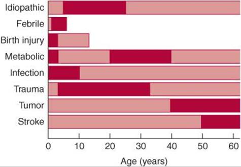
Figure 12-1. Causes of seizures as a function of age at onset. Bars show the range of ages at which seizures from a given cause typically begin; darker shading indicates peak incidence.
The genetic contribution to epilepsy and its response to treatment is complex. A single epileptic syndrome (eg, juvenal myoclonic epilepsy) can result from mutations in several different genes and, conversely, mutations in a single gene (eg, SCN1A sodium channel subunit) can cause several epilepsy phenotypes. Genes implicated in susceptibility to epilepsy include those coding for sodium, calcium, potassium, and chloride channels; nicotinic cholinergic, GABA, and G protein-coupled receptors; and enzymes.
PRIMARY NEUROLOGIC DISORDERS
Benign Febrile Convulsions
Benign febrile convulsions occur in 2% to 5% of children aged 6 months to 5 years, usually during the first day of a febrile illness (temperature > 100.4°F or 38°C), and in the absence of CNS infection (meningitis or encephalitis). There may be a family history of benign febrile convulsions or other types of seizures. Mutations in several genes have been linked to febrile convulsions, including the G protein-coupled receptor MASS1; the inositol monophosphatase IMPA2; SCN1A, SCN1B, and SCN2A sodium channel subunits; KCNQ2, KCNQ3, and KCNA1 potassium channel subunits; and GABRG2 and GABRDGABA receptor subunits.
Benign febrile convulsions usually last for less than 10 to 15 minutes and lack focal features. Approximately two-thirds of patients experience a single seizure, and fewer than one-tenth have more than three. Seizures occurring during the first hour of fever in children younger than 18 months or in children with a family history of febrile seizures are associated with an increased risk for recurrence; 90% of recurrences occur within 2 years of the initial episode. The differential diagnosis includes meningitis and encephalitis (Chapter 4); if present, these should be treated as described elsewhere in this volume.
Because benign febrile convulsions are usually self-limited, treatment is often unnecessary; prolonged convulsions (≥15 minutes) can be treated with diazepam 0.3 mg/kg orally, intramuscularly, or intravenously or 0.6 mg/kg rectally. Such treatment may decrease the risk of recurrence. The probability of developing a chronic seizure disorder is 2% to 6% and is highest in patients with persistent neurologic abnormalities; prolonged, focal, or multiple seizures; or a family history of nonfebrile seizures. Long-term administration of phenobarbital to reduce the risk of subsequent afebrile seizures is rarely indicated, as the risk of nonfebrile seizures is not altered.
Idiopathic (Cryptogenic) Seizures
These account for two-thirds of new-onset seizures in the general population. The age range is broad, from the second to the seventh decade (Figure 12-1). The risk of recurrence in the next 5 years is approximately 35% after a first unprovoked seizure. A second seizure increases the risk of recurrence to approximately 75%. Most recurrences occur in the first year. Genes implicated in idiopathic generalized epilepsy include the mitochondrial NAD-dependent malic enzyme ME2 and the CACNA1A and CACNB4 calcium channel subunits.
Head Trauma
Head trauma is a common cause of epilepsy, particularly when it occurs perinatally or is associated with a depressed skull fracture or intracerebral or subdural hematoma. Seizures that occur within the first week after nonpenetrating head injuries are not predictive of a chronic seizure disorder, however. Although patients with serious head injuries are often treated prophylactically with anticonvulsant drugs, this practice has been questioned, because a reduction in the incidence of posttraumatic seizures has not been consistently observed.
Stroke
Stroke affecting the cerebral cortex produces seizures in 5% to 15% of patients and can occur after thrombotic or embolic infarction or intracerebral hemorrhage (Chapter 13). As in head trauma, early seizures are not necessarily indicative of chronic epilepsy, and long-term anticonvulsant therapy may not be required. Even without rupturing, vascular malformations may be associated with seizures, presumably as a result of their irritative effects on adjacent brain tissue.
Mass Lesions
Mass lesions, such as brain tumors (Chapter 6) or abscesses (Chapter 3), can present with seizures. Glioblastomas, astrocytomas, and meningiomas are the most common tumors associated with seizures, reflecting their high prevalence among tumors that affect the cerebral hemispheres.
Meningitis or Encephalitis
Bacterial (eg, Haemophilus influenzae or tuberculous), viral (eg, herpes simplex), fungal, or parasitic (eg, cysticercosis) infections (Chapter 4) can also cause seizures. Seizures in patients with AIDS are most often related to HIV-associated dementia, but also occur with toxoplasmosis or cryptococcal meningitis.
Developmental Anomalies
Cortical dysgenesis and neuronal migration disorders can predispose to epilepsy.
SYSTEMIC DISORDERS
Metabolic and other systemic disorders, including drug-overdose and drug-withdrawal syndromes, may be associated with seizures that abate with correction of the underlying abnormality. In these cases, the patient is not considered to have epilepsy.
1. Hypoglycemia (Chapter 4) can produce seizures, especially with serum glucose levels of 20 to 30 mg/dL, but neurologic manifestations of hypoglycemia are also related to the rate at which serum glucose levels fall.
2. Hyponatremia (Chapter 4) may be associated with seizures at serum sodium levels less than 120 mEq/L or at higher levels after a rapid decline.
3. Hyperosmolar states, including both hyperosmolar nonketotic hyperglycemia (Chapter 4) and hypernatremia, may lead to seizures when serum osmolality rises above approximately 330 mOsm/L.
4. Hypocalcemia (Chapter 4) with serum calcium levels in the range of 4.3 to 9.2 mg/dL can produce seizures with or without tetany.
5. Uremia (Chapter 4) can cause seizures, especially when it develops rapidly, but this tendency correlates poorly with absolute serum urea nitrogen levels.
6. Hepatic encephalopathy (Chapter 4) is sometimes accompanied by generalized or multifocal seizures.
7. Porphyria is a disorder of heme biosynthesis that produces both neuropathy (Chapter 9) and seizures. The latter may be difficult to treat because most anticonvulsants can exacerbate the metabolic abnormalities. Case reports attest to the safety and efficacy of gabapentin, oxcarbazepine, and levetiracetam in porphyria.
8. Drug overdose can exacerbate epilepsy or cause seizures in nonepileptic patients. Generalized tonic–clonic seizures are most common, but focal or multifocal partial seizures can also occur. The drugs most frequently associated with seizures are antidepressants, antipsychotics, cocaine, insulin, isoniazid, lidocaine, and methylxanthines (Table 12-2).
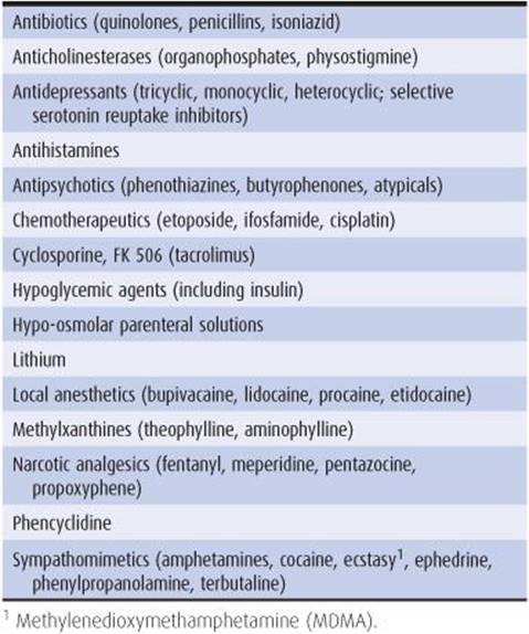
Table 12-2. Major categories of drugs reported to cause seizures.
9. Drug withdrawal, especially withdrawal from ethanol or sedative drugs (Chapter 4), may be accompanied by one or more generalized tonic–clonic seizures that usually resolve spontaneously. Alcohol withdrawal seizures occur within 48 hours after cessation or reduction of ethanol intake in 90% of cases and are characterized by brief flurries of one to six attacks that resolve within 12 hours. Acute abstinence from sedative drugs can also produce seizures in patients habituated to more than 600 to 800 mg/d of secobarbital or equivalent doses of other short-acting sedatives. Seizures from sedative drug withdrawal typically occur 2 to 4 days after abstinence but may be delayed for up to 1 week. Focal seizures are rarely due to alcohol or sedative drug withdrawal alone; they suggest an additional focal cerebral lesion that requires evaluation.
10. Global cerebral ischemia (Chapter 13) from cardiac arrest, cardiac arrhythmias, or hypotension may produce, at onset, a few tonic or tonic–clonic movements that resemble seizures, but they probably reflect abnormal brainstem activity instead. Global ischemia may also be associated with spontaneous myoclonus (Chapter 11) or, after consciousness returns, with myoclonus precipitated by movement (action myoclonus). Partial or generalized tonic–clonic seizures also occur; these may be manifested only by subtle movements of the face or eyes and must be recognized and treated. Nonetheless, isolated seizures after global cerebral ischemia do not necessarily indicate a poor outcome.
11. Hypertensive encephalopathy (Chapter 4) may be accompanied by generalized tonic–clonic or partial seizures.
12. Eclampsia refers to the occurrence of seizures or coma in a pregnant woman with hypertension, proteinuria, and edema (preeclampsia). As in hypertensive encephalopathy in nonpregnant patients, cerebral edema, ischemia, and hemorrhage may contribute to neurologic complications. Magnesium sulfate has been widely used to treat eclamptic seizures and may be superior for this purpose to anticonvulsants such as phenytoin.
13. Hyperthermia can result from infection, exposure (heat stroke), hypothalamic lesions, or drugs such as phencyclidine, as well as anticholinergics or neuroleptics (neuroleptic malignant syndrome; Chapter 11) and inhalational anesthetics or neuromuscular blocking agents (malignant hyperthermia; Chapter 9). Clinical features of severe hyperthermia (42°C, or 107°F) include seizures, confusional states or coma, shock, and renal failure. Treatment is with antipyretics and artificial cooling to reduce body temperature immediately to 39°C (102°F) and anticonvulsants and more specific therapy (eg, antibiotics for infection, dantrolene for malignant hyperthermia) where indicated. Patients who survive may be left with ataxia as a result of the special vulnerability of cerebellar neurons to hyperthermia.
PSEUDOSEIZURES
Attacks that resemble seizures (psychogenic seizures or pseudoseizures) may be manifestations of a psychiatric disturbance such as conversion disorder, somatization disorder, factitious disorder with physical symptoms, or malingering.
Pseudoseizures usually can be distinguished both clinically and by the electroencephalogram (EEG) findings. In patients with pseudoseizures resembling tonic–clonic attacks, there may be warning and preparation before the attack; there is usually no tonic phase, and the clonic phase consists of wild thrashing movements during which the patient rarely comes to harm or is incontinent. Ictal eye closure is common. In some instances, there are abnormal movements of all extremities without loss of consciousness; in others, there is shouting, uttering of obscenities, or goal-directed behavior during apparent loss of consciousness. There is no postictal confusion or abnormal clinical signs after the attack. The EEG, if recorded during an episode, does not show organized seizure activity, and postictal slowing does not occur. The differential diagnosis should include frontal lobe seizures, which may be marked by unusual midline movements (eg, pelvic thrusting or bicycling) and by very brief postictal states. Ictal EEG abnormalities may escape detection as well.
It is important to appreciate that some patients with pseudoseizures also have genuine epileptic attacks that require anticonvulsant medications, but these should be prescribed at an empirically appropriate dose. Psychiatric referral may be helpful.
CLASSIFICATION & CLINICAL FINDINGS
CLASSIFICATION
Seizures are classified as follows:
Generalized Seizures
1. Tonic–clonic (grand mal)
2. Absence (petit mal)
3. Other types (tonic, clonic, myoclonic, atonic)
Partial Seizures
1. Simple partial
2. Complex partial (temporal lobe, psychomotor)
3. Partial seizures with secondary generalization
GENERALIZED SEIZURES
Generalized Tonic–Clonic Seizures
Generalized tonic–clonic seizures are attacks in which consciousness is lost, usually without aura or other warning. When a warning does occur, it usually consists of nonspecific symptoms.
1. Tonic phase—The initial manifestations are unconsciousness and tonic contraction of limb muscles for 10 to 30 seconds, producing first flexion and then extension, particularly of the back and neck (Figure 12-2). Tonic contraction of the muscles of respiration may produce an expiration-induced vocalization (cry or moan) and cyanosis, and contraction of masticatory muscles may cause tongue trauma. The patient falls to the ground and may be injured.

Figure 12-2. Generalized tonic–clonic seizure, illustrating the appearance of the patient in the tonic (stiffening) and clonic (shaking) phases.
2. Clonic phase—The tonic phase is followed by a clonic (alternating muscle contraction and relaxation) phase of symmetric limb jerking that persists for an additional 30 to 60 seconds or longer. Ventilatory efforts return immediately after cessation of the tonic phase, and cyanosis clears. The mouth may froth with saliva. With time, the jerking becomes less frequent, until finally all movements cease and the muscles are flaccid. Sphincteric relaxation or detrusor muscle contraction may produce urinary incontinence.
3. Recovery—As the patient regains consciousness, there is postictal confusion and often headache. Full orientation commonly takes 10 to 30 minutes or even longer in patients with status epilepticus (see next section) or preexisting structural or metabolic brain disorders. Physical examination during the postictal state is usually otherwise normal in idiopathic epilepsy or seizures of metabolic origin, except that plantar responses may be transiently extensor (Babinski sign). The pupils always react to light, even when the patient is unconscious.
4. Status epilepticus—Status epilepticus is defined arbitrarily as seizures that continue for more than 30 minutes without ceasing spontaneously or that recur so frequently that full consciousness is not restored between successive episodes. Status epilepticus is a medical emergency because it can lead to permanent brain damage from hyperpyrexia, circulatory collapse, or excitotoxic neuronal damage if untreated.
Absence (Petit Mal) Seizures
These are genetically transmitted seizures that always begin in childhood and rarely persist into adolescence. Genes linked to childhood absence epilepsy include the CACNA1H calcium channel, CLCN2chloride channel, and GAGRB2 GABA receptor subunits. The spells are characterized by brief loss of consciousness (for 5-10 seconds) without loss of postural tone. Subtle motor manifestations, such as eye blinking or a slight head turning, are common. More complex automatic movements (automatisms) are uncommon. Full orientation immediately follows cessation of the seizure.
There may be as many as several hundred spells daily, leading to impaired school performance and social interactions, so that children may be mistakenly thought to be mentally retarded before the diagnosis of petit mal epilepsy is made. The spells are characteristically inducible by hyperventilation. The EEG shows a characteristic 3-per-second spike-and-wave pattern during the seizures (Figure 12-3). In most patients with normal intelligence and normal background activity on EEG, absence spells occur only during childhood; in other cases, however, the attacks continue into adult life, either alone or in association with other types of seizures.
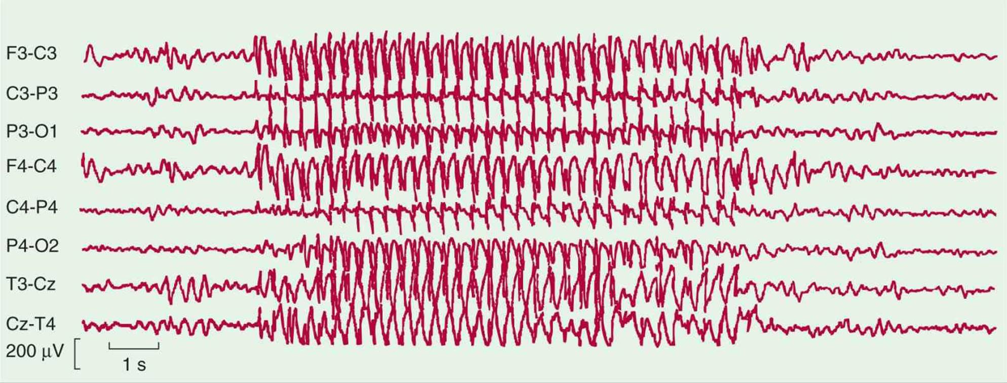
Figure 12-3. EEG of a patient with typical absence (petit mal) seizures, showing a burst of generalized 3-Hz spike-and-wave activity (center of record) that is bilaterally symmetric and bisynchronous. Odd-numbered leads indicate electrode placements over the left side of the head; even numbers, those over the right side.
Other Types of Generalized Seizures
These include tonic seizures (not followed by a clonic phase), clonic seizures (not preceded by a tonic phase), and myoclonic seizures.
1. Tonic seizures are characterized by continuous muscle contraction that can lead to fixation of the limbs and axial musculature in flexion or extension and are a cause of drop attacks; the accompanying arrest of ventilatory movements leads to cyanosis. Consciousness is lost, and there is no clonic phase to these seizures.
2. Clonic seizures are characterized by repetitive clonic jerking accompanied by loss of consciousness. There is no initial tonic component.
3. Myoclonic seizures are characterized by sudden, brief, shocklike contractions that may be localized to a few muscles or one or more extremities or may have a more generalized distribution. Juvenile myoclonic epilepsy is the most common cause, with onset usually in adolescence. Not all myoclonic jerks have an epileptic basis, however, as discussed in Chapter 11.
There is a family history of seizures in one-third of patients with myoclonic seizures. The disorder is genetically heterogeneous, with linkage in different families to genes for an EF-hand–containing calcium-binding protein (EFHC1), bromo domain–containing protein (BRD2), or calcium channel (CACNB4), chloride channel (CLCN2), or GABA receptor (GABRA1) subunit. Myoclonic seizures may also be associated with a variety of rare hereditary neurodegenerative disorders, including Unverricht-Lundborg disease (cystatin B [CSTB] mutations), Lafora body disease (laforin [EPM2A] or NHL repeat-containing gene 1 [NHLRC1] mutations), neuronal ceroid lipofuscinosis (late infantile, juvenile, or adult onset; palmityl-protein thioesterase-1 [PPT1], tripeptidyl peptidase I [TPP1], or battenin [CLN3] mutations), sialidosis (sialidase [NEU1] mutations), and mitochondrial encephalomyopathy (myoclonus epilepsy with ragged red fibers on skeletal muscle biopsy; mutations in any of several mitochondrial genes).
4. Atonic seizures result from loss of postural tone, sometimes after a myoclonic jerk, leading to a fall or drop attack. They are most common in developmental disorders such as the Lennox-Gastaut syndrome, which in some cases results from mutations affecting the mitogen-activated protein kinase 10 (MAPK10) gene.
PARTIAL SEIZURES
Simple Partial Seizures
Simple partial seizures begin with motor, sensory, or autonomic phenomena, depending on the cortical region affected. For example, clonic movements of a single muscle group in the face, a limb, or the pharynx may occur and may be self-limited; they may be recurrent or continuous or may spread to involve contiguous regions of the motor cortex (jacksonian march).
Autonomic symptoms may consist of pallor, flushing, sweating, piloerection, pupillary dilatation, vomiting, borborygmi, or hypersalivation. Psychic symptoms include distortions of memory (eg, déjà vu, the sensation that a new experience is familiar), forced thinking or labored thought processes, cognitive deficits, affective disturbances (eg, fear, depression, an inappropriate sense of pleasure), hallucinations, or illusions. During simple partial seizures, consciousness is preserved unless and until the seizure discharge spreads to other areas of the brain, producing tonic–clonic seizures (secondary generalization). The aura is the portion of the seizure that precedes loss of consciousness and of which the patient retains some memory. The aura is sometimes the sole manifestation of the epileptic discharge.
In the postictal state, a focal neurologic deficit such as hemiparesis (Todd paralysis) may persist for 30 minutes to 36 hours and indicates an underlying focal brain lesion.
Complex Partial Seizures
Complex partial seizures, formerly called temporal lobe or psychomotor seizures, are partial seizures in which consciousness, responsiveness, or memory is impaired. The seizure discharge usually arises from the temporal lobe or medial frontal lobe but can originate elsewhere. The symptoms take many forms but are usually stereotyped for the individual patient. Episodes may begin with an aura. Epigastric sensations are most common, but affective (fear), psychic (déjà vu), and sensory (olfactory hallucinations) symptoms also occur. Consciousness is then impaired. Seizures generally persist for 1 to 3 minutes. The motor manifestations of complex partial seizures are characterized by coordinated involuntary motor activity, termed automatism, which takes the form of orobuccolingual movements in approximately 75% of patients and other facial or neck or hand movements in approximately 50%. Sitting up or standing, fumbling with objects, and bilateral limb movements are less common. Secondary generalization may occur.
DIAGNOSIS
The diagnosis of seizures is based on clinical recognition of one of the seizure types described previously. The EEG can be a helpful confirmatory test in distinguishing seizures from other causes of loss of consciousness (Figure 12-4). However, a normal or nonspecifically abnormal EEG never excludes the diagnosis of epilepsy. Specific EEG features that suggest epilepsy include abnormal spikes, polyspike discharges, and spike-wave complexes.
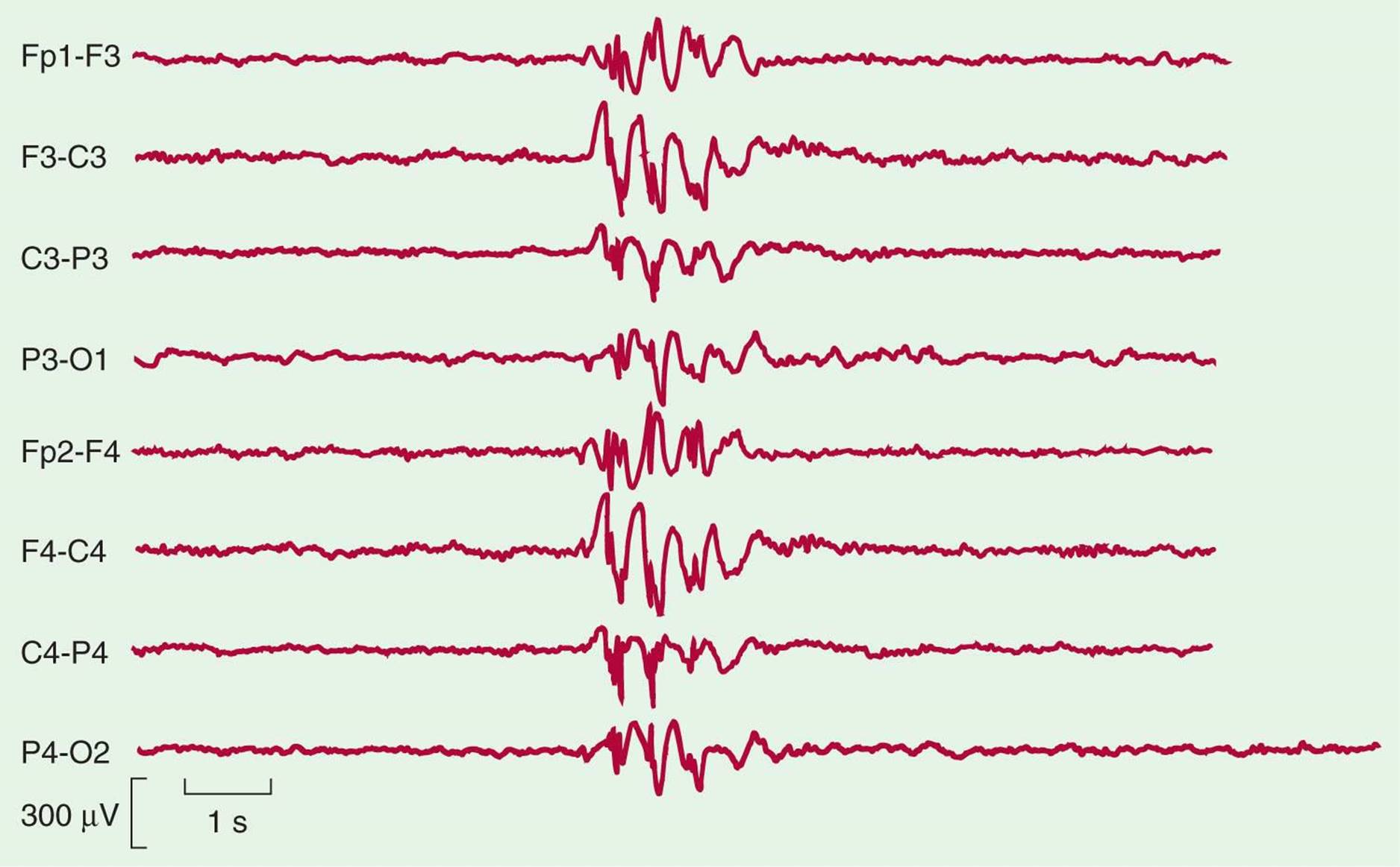
Figure 12-4. EEG of a patient with idiopathic (primary generalized) epilepsy. A burst of generalized epileptiform activity (center) is seen on a relatively normal background. These findings, obtained at a time when the patient was not experiencing seizures, support the clinical diagnosis of epilepsy. Odd-numbered leads indicate electrode placements over the left side of the head; even numbers, those over the right side.
A standard diagnostic evaluation of patients with recent onset of seizures is presented in Table 12-3. Metabolic and toxic disorders that can cause seizures (Table 12-1) should be excluded, because they do not require anticonvulsants.
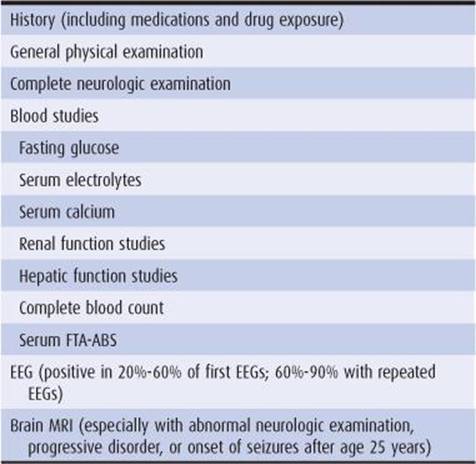
Table 12-3. Evaluation of a new seizure disorder in a stable patient.
Seizures with a clearly focal onset or those that begin after the age of 25 years require prompt evaluation to exclude the presence of a structural brain lesion. Magnetic resonance imaging (MRI) is essential for this purpose (computed tomography [CT] scan is not adequate). If no cause is found, the decision to begin chronic anticonvulsant therapy should be based on the probability of recurrence. After a single generalized tonic–clonic seizure, recurrence can be expected within 3 to 4 years in 30% to 70% of untreated adult patients.
TREATMENT
PRINCIPLES OF TREATMENT
Therapy should be directed toward the cause of the seizures, if known. Seizures associated with metabolic and systemic disorders usually respond poorly to anticonvulsants but cease with correction of the underlying abnormality. Acute withdrawal from alcohol and other sedative drugs produces self-limited seizures that, in general, require no anticonvulsant drug therapy. Acute head trauma and other structural brain lesions that result in seizures must be rapidly diagnosed and treated, and the associated seizures controlled by anticonvulsant drug therapy. Idiopathic epilepsy is treated with anticonvulsant medications.
There are four key principles of management:
1. Establish the diagnosis of epilepsy before starting drug therapy—Therapeutic trials of anticonvulsant drugs intended to establish or reject a diagnosis of epilepsy may yield incorrect diagnoses.
2. Choose the right drug for the seizure type—Absence seizures, for example, do not respond to most drugs used for complex partial or generalized tonic–clonic seizures.
3. Treat the seizures, rather than the serum drug levels—Control of seizures is achieved at different drug levels in different patients.
4. Evaluate one drug at a time—In most cases, seizures can be controlled with a single drug. Therefore, beginning therapy with multiple drugs may expose patients to increased drug toxicity without added therapeutic benefit.
ANTICONVULSANT DRUGS
Most anticonvulsant drugs act either by potentiating inhibitory (GABAergic) or by inhibiting excitatory (glutamatergic) synaptic transmission in the brain. Drugs acting at GABAergic synapses and their molecular targets are illustrated in Figure 12-5; drugs acting at glutamatergic synapses and their molecular targets are illustrated in Figure 12-6.

Figure 12-5. Known and potential sites of action of anticonvulsant drugs at inhibitory GABAergic synapses: (1) GABA transporters (tiagabine); (2) GABA transaminase (vigabatrin); (3) GABAA receptors (benzodiazepines, barbiturates); and (4) GABAB receptors. Abbreviations: GABA-T, GABA transaminase; GAT, GABA transporter; VG, voltage-gated ion channel; (N) and (T), calcium channel subtypes. Small circles in the intercellular space represent GABA molecules. (From Katzung BG, Masters SB, Trevor AJ, eds. Basic and Clinical Pharmacology. 11th ed. New York, NY: McGraw-Hill; 2009.)
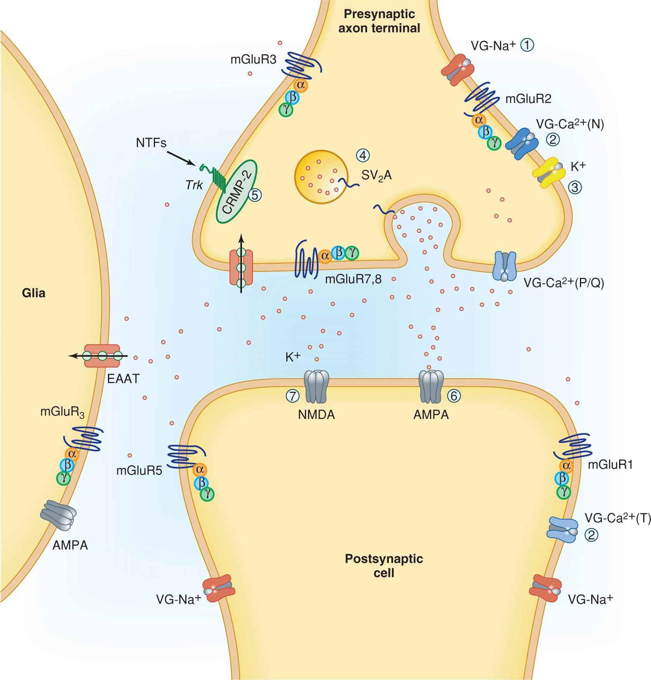
Figure 12-6. Known and potential sites of action of anticonvulsant drugs at excitatory glutamatergic synapses: (1) voltage-gated sodium channels (phenytoin, carbamazepine, lamotrigine); (2) voltage-gated calcium channels (ethosuximide, lamotrigine, gabapentin, pregabalin); (3) voltage-gated potassium channels; (4) synaptic vesicle glycoprotein 2A (levetiracetam); (5) collapsin-related mediator protein-2; (6) AMPA-preferring glutamate receptors (phenobarbital, topiramate, lamotrigine); (7) NMDA-preferring glutamate receptors (felbamate). Abbreviations: EAAT, excitatory amino acid transporter; mGluR, metabotropic glutamate receptor; (N), (P/Q) and (T), calcium channel subtypes; NTFs, neurotrophic factors. Small circles in the intercellular space represent glutamate molecules. (From Katzung BG, Masters SB, Trevor AJ, eds. Basic and Clinical Pharmacology. 11th ed. New York, NY: McGraw-Hill; 2009.)
Commonly used anticonvulsant drugs and their dosages and methods of administration are listed in Table 12-4.


Table 12-4. Summary of anticonvulsant drug therapy.
TREATMENT STRATEGIES
Most patients with epilepsy fall into one of the following treatment categories.
New Seizures
Most epileptologists do not recommend chronic anticonvulsant drug treatment after a single seizure unless an underlying cause is found that is not correctable and is likely to produce recurrent seizures (eg, brain tumor). However, recurrent seizures do require anticonvulsant treatment, and if such therapy is to be administered, the oral loading schedules presented in Table 12-4 can be used. Note that starting a drug at its daily maintenance dose produces stable serum drug levels only after approximately five half-lives have elapsed. Therefore, loading doses should be given to achieve therapeutic drug levels promptly in patients with frequent seizures.
1. Partial (including complex partial) or secondarily generalized tonic–clonic seizures—Phenytoin, carbamazepine, and lamotrigine are appropriate drugs of first choice for treating partial or secondarily generalized tonic–clonic seizures.
2. Generalized seizures—Valproic acid is preferred for all types of primary generalized seizures. Phenobarbital is also very effective in treating generalized tonic–clonic seizures in adults, but it is less helpful for treatment of complex partial seizures.
3. Absence seizures—Absence attacks of the petit mal variety are treated with valproic acid or ethosuximide. The former has the advantage of also providing protection against tonic–clonic seizures but has caused fatalities from hepatic damage in children younger than 10 (usually < 2) years of age.
3. Myoclonic seizures—These are treated with valproic acid, levetiracetam, zonisamide, or clonazepam.
As experience is gained with newer anticonvulsants (gabapentin, levetiracetam, pregabalin, vigabatrin, topira-mate, tiagabine, zonisamide), some will find indications as effective monotherapy for epilepsy. Levetiracetam, topira-mate, and zonisamide have broad spectra against both partial and generalized epilepsies. Oxcarbazepine can be used in monotherapy for partial and secondarily generalized tonic–clonic seizures. Gabapentin, tiagabine, and pregabalin should be avoided in patients with generalized seizures.
Recurrent Seizures on Drug Therapy
1. Determining serum levels of drugs—Blood levels of anticonvulsant drugs the patient has been taking should be measured in samples taken just before a scheduled dose. For a single breakthrough seizure, no acute change in medication is mandated, even if there has been no interruption of drug therapy and anticonvulsant drug levels are in the therapeutic range, but a slight increase in prescribed dose may be considered. If the history or serum drug levels suggest that treatment has been interrupted, the prescribed drug should be started again as for new seizures.
2. Changing to a different drug—A different anticonvulsant should be introduced only if seizures continue to occur after maximum therapeutic benefit has been achieved with the initial drug. This means that blood levels of the drug are in the therapeutic range and that drug toxicity precludes further dosage increments. An anticonvulsant that has failed to alter seizure frequency should be discontinued gradually once therapeutic levels of the new drug have been achieved. Transition to monotherapy with a different drug is recommended before trials of two-drug combination therapy.
3. Treating refractory seizures—In some patients, disabling seizures persist despite trials of all major anticonvulsants, alone and in combination, and at the highest doses the patient can tolerate. When no treatable cause can be found, seizures are not due to a progressive neurodegenerative disease, and medical treatment has been unsuccessful for at least 2 years, evaluation for possible surgical therapy should be considered.
Presurgical evaluation begins with a detailed history and neurologic examination to explore the cause of seizures and their site of origin within the brain and to document the adequacy of prior attempts at medical treatment. MRI and electrophysiologic studies are performed to identify the epileptogenic zone within the brain. Several electrophysiologic techniques can be used: EEG, in which cerebral electrical activity is recorded noninvasively from the scalp; intracranial or invasive EEG, in which activity is recorded from electrodes inserted (depth electrodes) into the brain or placed over the brain surface (subdural electrodes); and electrocorticography, which involves intraoperative recording from the surface of the brain. When an epileptogenic zone can be identified in this manner and its removal is not expected to produce undue neurologic impairment, surgical excision may be indicated.
Patients with complex partial seizures arising from a single temporal lobe are the most frequent surgical candidates; unilateral anterior temporal lobectomy abolishes seizures and auras in approximately 50% of these patients and significantly reduces their frequency in another 25%. Hemispherectomy and corpus callosum section are also sometimes used to treat intractable epilepsy. Left vagal (X) nerve stimulation has been shown to reduce seizure frequency by as much as 50% in adults and children with refractory epilepsy. The mechanism of action is unknown, but afferent responses from the vagus are received in the nucleus tractus solitarius in the medulla and project widely.
Transcranial magnetic stimulation and deep brain stimulation are evolving experimental treatments.
Multiple Seizures or Status Epilepticus
1. Early management—Status epilepticus is a medical emergency because of its potential for causing irreversible brain injury and death.
a. Immediate attention should be given to ensure that the airway is patent and the patient is positioned to prevent aspiration of stomach contents.
b. The laboratory studies listed in Table 12-5 should be ordered without delay.

Table 12-5. Emergency evaluation of serial seizures or status epilepticus.
c. Dextrose (50 mL of 50% solution) should be given intravenously.
d. Meningitis and encephalitis should be considered, especially if fever and meningeal signs are present, and a lumbar puncture performed if indicated. Patients should also undergo lumbar puncture if the cause of the seizures has not been determined, unless signs of increased intracranial pressure or of focal brain dysfunction are present. Postictal pleocytosis is detectable in cerebrospinal fluid (CSF) in approximately 2% of patients with single generalized tonic–clonic seizures (and approximately 15% of those with status epilepticus) in the absence of infection. The white blood cell count may be as high as 80/μL, with either polymorphonuclear or mononuclear predominance. Serum protein content may be slightly elevated, but glucose concentration is normal, and Gram stain is negative. The postictal pleocytosis resolves in 2 to 5 days.
2. Drug therapy to control seizures—Every effort must be made to establish a precise etiologic diagnosis so that treatment of the underlying disorder can be started. Because generalized seizure activity per se damages the brain if it persists for more than 30 minutes, drug therapy to terminate seizures should be instituted immediately. An outline for rapid pharmacologic control of multiple seizures is presented in Table 12-6.

Table 12-6. Drug treatment of status epilepticus in adults.
3. Management of hyperthermia—The systemic physiologic consequences of status epilepticus are related to increased motor activity and high levels of circulating catecholamines; they include hyperthermia (temperature elevation to 42-43°C [108-109°F] in the absence of infection), lactic acidosis (pH <7.00), and peripheral blood leukocytosis (elevation to 30,000 cells/μL). These derangements resolve after cessation of the seizures. Only hyperthermia, which is known to increase the risk of brain damage from status epilepticus, requires specific attention.
Severe hyperthermia must be treated with a cooling blanket and, if necessary, the induction of motor paralysis with a neuromuscular blocking agent. Mild or moderate hyperthermia (101-102°F), not requiring specific intervention, may persist for 24 to 48 hours after seizure cessation. Lactic acidosis resolves spontaneously over 1 hour and should not be treated. Infection should, of course, be excluded.
DISCONTINUING ANTICONVULSANTS
Patients (usually children) with epilepsy who are seizure-free on medication for 2 to 5 years may wish to discontinue anticonvulsant drugs. In patients with normal intelligence and a normal neurologic examination, the risk of seizure recurrence may be as low as 25%. Risk factors for recurrence include slowing or spikes (maximum risk with both present) on EEG. When anticonvulsants are to be withdrawn, one drug is eliminated at a time by tapering the dose slowly over approximately 6 weeks. Recurrence of seizures has been reported in approximately 20% of children and 40% of adults after medication withdrawal, in which case prior medication should be reinstituted at the previously effective levels.
COMPLICATIONS OF EPILEPSY & ANTICONVULSANT THERAPY
Complications of Epilepsy
When the diagnosis of epilepsy is made, the patient should be warned against working around moving machinery or at heights and reminded of the risks of swimming alone. The issue of driving must also be addressed. Many state governments have notification requirements when a diagnosis of epilepsy is made.
Side Effects of Anticonvulsant Drugs
The side effects of anticonvulsant drug therapy are summarized in Table 12-7. All anticonvulsant drugs may lead to blood dyscrasias, but carbamazepine and valproic acid has been associated with the highest incidence of hematologic and hepatic toxicity. For this reason, a complete blood count and liver function tests should be obtained before initiating administration of these drugs and at intervals during the course of treatment. The authors recommend performing these tests twice in the first weeks to months and every 6 to 12 months thereafter. Carbamazepine should be discontinued if the total neutrophil count falls below 1,500/mL or if aplastic anemia is suspected. Valproic acid should be terminated if symptoms of hepatotoxicity, such as nausea, vomiting, anorexia, or jaundice occur. Lamotrigine has a 1:1,000 incidence of Stevens-Johnson syndrome in the first 8 weeks. Most anticonvulsant drugs (especially barbiturates) affect cognitive function to some degree, even in therapeutic doses.

Table 12-7. Side effects of anticonvulsant drugs.
Drug Interactions
A variety of drugs alter the absorption or metabolism of anticonvulsants when given concomitantly. The changes in anticonvulsant levels are summarized in Table 12-8. Some anticonvulsants (carbamazepine, primidone, phenytoin, phenobarbital, topiramate, felbamate, and oxcarbazepine) induce the cytochrome P450 system, which may lead to reduced effectiveness of oral contraceptives.

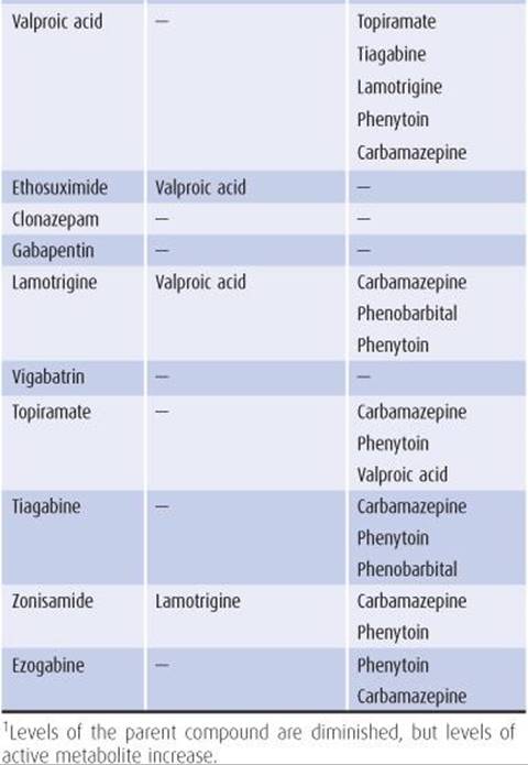
Table 12-8. Some major anticonvulsant drug interactions.
Epilepsy & Anticonvulsant Therapy in Pregnancy
1. Teratogenic effects of epilepsy—The incidence of stillbirth, microcephaly, mental retardation, and seizure disorders is increased in children born to epileptic mothers.
2. Teratogenic effects of anticonvulsant treatment—Anticonvulsant therapy during pregnancy is also associated with a greater than normal frequency of congenital malformations—especially cleft palate, cleft lip, and cardiac anomalies. Such malformations are about twice as common in the offspring of medicated than of unmedicated epileptic mothers, but because patients with more severe epilepsy are more likely to be treated, it is difficult to know whether epilepsy or its treatment is the more important risk factor.
3. Differences in teratogenesis among anticonvulsants—Among commonly used anticonvulsants, valproic acid and to some degree carbamazepine are associated with an increased incidence of neural tube defects (2% and 0.5%, respectively). Phenobarbital and phenytoin pose some teratogenic risk, but the extent of the risk is controversial. The fetal risks of the newer anticonvulsants are not known.
4. Folate deficiency—Several anticonvulsants can lower serum folate levels. As dietary deficiency of folic acid is associated with neural tube defects, folate supplementation (1 mg/d) should be provided for all women of child-bearing age who take antiepileptic drugs.
5. Withdrawing anticonvulsants before pregnancy—When an epileptic patient who has been seizure free for several years is contemplating pregnancy, an attempt should be made to assess whether anticonvulsant drugs can be safely withdrawn before conception. In contrast to generalized tonic–clonic seizures, partial and absence seizures present little risk to the fetus, and it may be possible to tolerate imperfect control of these seizures during pregnancy to avoid fetal drug exposure. If anticonvulsant therapy is continued during pregnancy, it is best to maintain treatment with a single drug that has been shown effective for the patient’s seizures, using a dose that avoids clinical toxicity. Status epilepticus is treated as described previously for nonpregnant patients.
6. Anticonvulsant levels in pregnancy—Plasma levels of anticonvulsant drugs may decrease during pregnancy because of the patient’s enhanced drug metabolism, and higher doses may be required to maintain control of seizures. It is therefore important to monitor drug levels closely in this setting.
PROGNOSIS
After a single unprovoked seizure, only one-third to one-half of patients will have a recurrence (ie, develop epilepsy). If a second seizure occurs, however, the recurrence rate approaches 75%, and anticonvulsants therefore should be started. With appropriate anticonvulsant drug treatment, seizures can be well controlled, although not always eliminated, in most epileptic patients. At the onset of treatment, patients should be seen every few months to monitor seizure frequency and make dose adjustments.
SYNCOPE
Syncope is episodic loss of consciousness associated with loss of postural tone. The pathophysiology is distinct from that of seizures, involving global hypoperfusion of the brain or brainstem. The most common causes of syncope are given in Table 12-9.

Table 12-9. Common causes of syncope and their prevalence.
VASOVAGAL SYNCOPE (SIMPLE FAINTS)
Vasovagal syncope occurs in all age groups. Precipitating factors include emotional stimulation, pain, the sight of blood, fatigue, medical instrumentation, blood loss, or prolonged motionless standing. Vagally mediated decreases in arterial blood pressure and heart rate combine to produce CNS hypoperfusion and subsequent syncope. Cerebral ischemia resulting in brief tonic–clonic movements can occur.
Vasovagal episodes generally begin while the patient is in a standing or sitting position and only rarely in a horizontal position (eg, with phlebotomy or intrauterine device insertion). A prodrome lasting 30 to 60 seconds usually precedes syncope and can include lassitude, yawning, light-headedness, nausea, pallor, diaphoresis, salivation, blurred vision, and tachycardia.
The patient, who then loses consciousness and falls to the ground, is pale and diaphoretic and has dilated pupils. Breathing continues. The eyes remain open and there is an upward turning of the globes. Bradycardia replaces tachycardia as consciousness is lost. During unconsciousness, abnormal movements may occur, particularly if the patient remains relatively vertical; these are mainly tonic or opisthotonic, but seizure-like tonic–clonic activity is occasionally seen, which can lead to a misdiagnosis of epilepsy. Urinary incontinence may also occur.
The patient recovers consciousness very rapidly (20-30 seconds) after assuming the horizontal position, but residual nervousness, dizziness, headache, nausea, pallor, diaphoresis, and an urge to defecate may be noted. A postictal confusional state with disorientation and agitation either does not occur or is very brief (<30 seconds). Syncope may recur, especially if the patient stands within the next 30 minutes.
Reassurance and a recommendation to avoid precipitating factors are usually the only treatment necessary.
Recurrent vasovagal syncope (currently termed neurally mediated or neurocardiogenic syncope) can be diagnosed by inducing syncope during head-up tilt-testing. The bradycardia and hypotension can be ameliorated by volume expansion (fludrocortisone 0.1-1 mg/d), by β-adrenergic blockade, or with the selective serotonin reuptake inhibitor paroxetine (10-40 mg/d); artificial pacing is ineffective.
CARDIOVASCULAR SYNCOPE
A cardiovascular cause is suggested when syncope occurs in a recumbent position, during or after physical exertion, or in a patient with known heart disease. Loss of consciousness related to cardiac disease is most often due to an abrupt decrease in cardiac output with resultant cerebral hypoperfusion. Such cardiac dysfunction can result from cardiac arrest, rhythm disturbances (either brady- or tachyarrhythmias), cardiac inflow or outflow obstruction, intracardiac right-to-left shunts, leaking or dissecting aortic aneurysms, or acute pulmonary embolism (Table 12-10).


Table 12-10. Cardiovascular causes of syncope.
CARDIAC ARREST
Cardiac arrest (ventricular fibrillation or asystole) from any cause will result in loss of consciousness in 3 to 5 seconds if the patient is standing or within 15 seconds if the patient is recumbent. Seizurelike activity and urinary and fecal incontinence may be seen as the duration of cerebral hypoperfusion increases.
TACHYARRHYTHMIAS
Supraventricular Tachyarrhythmias
Supraventricular tachyarrhythmias (atrial or junctional tachycardia, atrial flutter, or atrial fibrillation) may be paroxysmal or chronic.
Heart rates faster than 160 to 200/min reduce cardiac output by decreasing the ventricular filling period or inducing myocardial ischemia. Prolonged tachycardia of 180 to 200 beats or more per minute will produce syncope in 50% of normal persons in the upright posture; in patients with underlying heart disease, a heart rate of 135/min may impair cardiac output enough to induce loss of consciousness. Patients with sinus node dysfunction may develop profound bradycardia or even asystole on termination of their tachyarrhythmias. The diagnosis is established when arrhythmias are demonstrated during a symptomatic episode. Continuous electrocardiogram (ECG) or outpatient portable Holter monitoring may be required; event monitors triggered by the patient at the onset of symptoms may be particularly helpful.
Ventricular Tachyarrhythmias
Ventricular tachyarrhythmias (ventricular tachycardia or multiform, frequent, or paired premature ventricular contractions) are found on prolonged ECG monitoring in some patients with syncope. The syncope associated with ventricular tachycardia is characterized by a very brief prodrome (< 5 seconds). The duration of syncope and of the arrhythmia are closely linked. Frequent or repetitive premature ventricular contractions alone do not often coincide with syncopal symptoms but are predictive of sudden death.
Mitral Valve Prolapse
Mitral valve prolapse (click-murmur syndrome) is a common disorder associated with supraventricular and ventricular arrhythmias and with syncope in a small percentage of patients. Other symptoms include nonexertional chest pain, dyspnea, and fatigue. Serious ventricular arrhythmias and profound bradycardia may occur. The ECG may be normal or show nonspecific ST-T wave changes or frequent premature ventricular contractions. Diagnosis is by echocardiography.
Prolonged QT Syndrome
The congenital prolonged QT-interval syndrome consists of paroxysmal ventricular arrhythmias (often torsades de pointes), syncope, and sudden death and is inherited as an autosomal recessive condition associated with deafness or in an autosomal dominant form without deafness. Genes implicated in prolonged QT syndrome include potassium channels (KCNE1, KCNE2, KCNH2, KCNJ2, KCNJ5, KCNQ1), sodium channels (SCN4B, SCN5A), calcium channels (CACNA13), A kinase anchor protein 9 (AKAP9), ankyrin 2 (ANK2), caveolin 3 (CAV3), and α-1 syntrophin (SNTA1). Sporadic cases also occur. Quinidine, hypocalcemia, and hypokalemia can also produce QT prolongation. Hereditary cases may respond to β-blockers.
BRADYARRHYTHMIAS
Sinoatrial Node Disease
Sinoatrial node disease (eg, sick sinus syndrome) may cause syncope with profound sinus bradycardia, prolonged sinus pauses, or sinus arrest with a slow atrial, junctional, or idioventricular escape rhythm. It may be inherited as an autosomal recessive or dominant disorder, caused by mutations in the type V voltage-gated sodium channel alpha subunit (SCN5A) and hyperpolarization-activated cyclic nucleotide-gated potassium channel 4 (HCN4) genes, respectively. Patients should be promptly evaluated by a cardiologist, because a permanent pacemaker is necessary in many cases. In tachycardia-bradycardia syndrome, a form of sick sinus syndrome, both types of arrhythmias occur.
Complete Heart Block
Complete heart block (third-degree atrioventricular block) is a common cause of bradyarrhythmia producing syncope. Permanent atrioventricular conduction abnormalities are easily noted on a routine ECG, but intermittent conduction abnormalities may not be present on a random tracing. A normal PR interval on an ECG obtained after the episode does not exclude the diagnosis of transient complete heart block.
Patients with syncope and documented or suspected complete heart block should be promptly hospitalized. Patients with acute inferior myocardial infarctions are at high risk for atrioventricular block.
CARDIAC INFLOW OBSTRUCTION
Atrial or ventricular myxomas and atrial thrombi usually present with embolic events, but they may also produce a left ventricular inflow or outflow obstruction that results in a sudden decrease in cardiac output, followed by syncope. A history of syncope occurring with change in position is classic but uncommon. Echocardiography can confirm the diagnosis. Surgical removal of the myxoma is indicated.
With constrictive pericarditis or pericardial tamponade, any maneuver or drug that decreases heart rate or venous return can result in suddenly inadequate cardiac output and syncope.
CARDIAC OUTFLOW OBSTRUCTION
Aortic Stenosis
Loss of consciousness from congenital or acquired severe aortic stenosis usually occurs after exercise and is often associated with dyspnea, angina, and diaphoresis. The pathophysiology may involve acute left ventricular failure resulting in coronary hypoperfusion and subsequent ventricular fibrillation, or abrupt increases in left ventricular pressure that stimulate baroreceptors, leading to peripheral vasodilation. Echocardiography can help confirm the diagnosis.
Symptomatic aortic stenosis requires valve replacement; without treatment, survival after syncope from aortic stenosis is 18 months to 3 years.
Pulmonary Stenosis
Severe pulmonary stenosis can produce syncope, especially after exertion. A hemodynamic process similar to that occurring in aortic stenosis is responsible.
HYPERTROPHIC CARDIOMYOPATHY
Hypertrophic cardiomyopathy comprises a group of congenital cardiomyopathies inherited as autosomal dominant disorders of variable severity. Many different genes have been implicated. Symptoms usually begin between the second and fourth decades. Dyspnea is the most common presenting complaint, but syncope occurs in 30% of patients and is the presenting complaint in 10% of patients. Syncope characteristically develops during or after exercise, but orthostatic and posttussive episodes also occur. Syncope may be due to left ventricular outflow obstruction, inflow obstruction, or transient arrhythmias. The diagnosis can be confirmed by echocardiography. Propranolol may control symptoms.
DISSECTING AORTIC ANEURYSM
Approximately 5% to 10% of patients with acute aortic dissections present with isolated syncope; other neurologic abnormalities may or may not be present. In 15% of patients, the dissection is painless.
PULMONARY HYPERTENSION & PULMONARY EMBOLUS
Syncope, often exertional, may be the presenting symptom of pulmonary hypertension. A history of exertional dyspnea is usual, and blood gas analysis shows hypoxemia, even at rest. Syncope is the presenting symptom in approximately 20% of patients experiencing massive pulmonary embolism. Upon recovery, such patients often complain of pleuritic chest pain, dyspnea, and apprehension. Hypotension, tachycardia, tachypnea, and arterial hypoxemia frequently accompany these large emboli.
CEREBROVASCULAR SYNCOPE
Cerebrovascular disease (Chapter 13) is an often suspected but actually uncommon cause of episodic unconsciousness.
BASILAR ARTERY INSUFFICIENCY
Basilar artery transient ischemic attacks usually occur after the sixth decade. The symptom complex of diplopia, vertigo, dysphagia, dysarthria, various sensory or motor symptoms, drop attacks, and occipital headaches suggests diffuse brainstem ischemia. Attacks are typically sudden in onset and brief in duration (seconds to minutes), but when consciousness is lost, recovery is frequently prolonged (30-60 minutes or longer). Isolated unconsciousness without other symptoms of brainstem ischemia is rarely due to basilar artery insufficiency. Two-thirds of patients have recurrent attacks, and strokes eventually occur in approximately one-fifth of all cases. Treatment is discussed in Chapter 13.
SUBCLAVIAN STEAL SYNDROME
The subclavian steal syndrome results from subclavian or innominate artery stenosis that causes retrograde blood flow in the vertebral artery, diverting flow from the brainstem and producing hypoperfusion. The degree of subclavian artery stenosis that will produce symptoms is variable, but even minor (∼40%) stenosis may cause the syndrome in some patients. A difference between blood pressures measured in the two arms is nearly always found, the average difference being a 45 mm Hg decrease in systolic pressure in the arm supplied by the stenotic vessel. Stroke is rare. If this diagnosis is suspected, arteriography and surgical correction may be indicated.
MIGRAINE
Syncope occurs in ∼10% of patients with migraine during the headache, often on rapid rising to a standing position, suggesting that loss of consciousness is due to orthostatic hypotension. In some patients, basilar migraineproduces symptoms similar to those of basilar artery transient ischemic attacks. Antimigraine drug therapy (Chapter 6) is often effective in preventing attacks.
TAKAYASU DISEASE
Takayasu disease, sometimes referred to as pulseless arteritis, is a panarteritis of the great vessels that is most common in Asian women. Symptoms of cerebral hypoperfusion such as impaired vision, confusion, and syncope are often prominent. Precipitating factors include exercise, standing, or head movement. Examination reveals decreased or absent brachial pulses with low blood pressures in both arms. The erythrocyte sedimentation rate is moderately elevated in the acute stage. Corticosteroid treatment is indicated.
CAROTID SINUS SYNCOPE
Carotid sinus syncope is uncommon. Men are affected twice as often as women, and most affected individuals are more than 60 years old. Drugs known to predispose to carotid sinus syncope include propranolol, digitalis, and methyldopa. Pressure on the carotid sinus by a tight collar, a neck mass, enlarged cervical lymph nodes, or a tumor causes vagal stimulation, which inhibits the cardiac sinoa-trial and atrioventricular nodes and reduces sympathetic vascular tone. The resultant bradycardia or systemic hypotension may then produce syncope; pure cardioinhibitory or vasodepressor syncope also occurs.
Carotid sinus syncope may be mistakenly diagnosed when symptoms result from compression of a normal carotid artery contralateral to an occluded internal carotid artery. Under these circumstances, unilateral compression transiently interrupts the entire anterior cerebral circulation. Performing carotid sinus massage in an attempt to diagnose carotid sinus syncope in patients with carotid atherosclerotic disease entails a risk of distal embolization of atheromatous material and is therefore not recommended.
ORTHOSTATIC HYPOTENSION
Orthostatic hypotension occurs more often in men than in women and is most common in the sixth and seventh decades. It may, however, appear even in teenagers. Loss of consciousness usually occurs upon rapidly rising to a standing position, standing motionless for a prolonged period (especially after exercise), or standing after prolonged recumbency (especially in the elderly).
Numerous conditions can produce orthostatic hypotension (Table 12-11), which generally results from either hypovolemia or autonomic dysfunction. The latter may be due to drugs, autonomic neuropathy, or CNS disorders affecting sympathetic pathways in the hypothalamus, brainstem, or spinal cord.
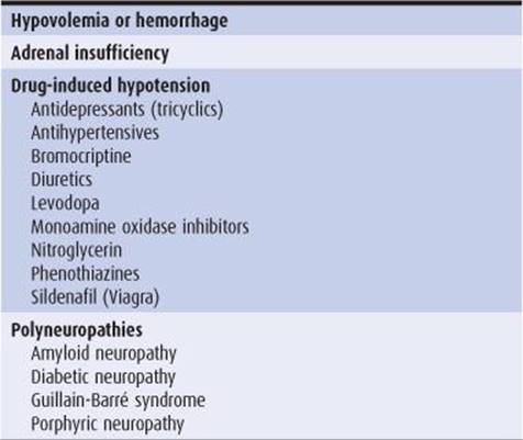

Table 12-11. Causes of orthostatic hypotension.
Orthostatic hypotension may also be a feature of neuro-degenerative disorders. Idiopathic orthostatic hypotension is associated with isolated degeneration of postganglionic sympathetic neurons. In Shy-Drager syndrome, degeneration of preganglionic sympathetic neurons occurs in combination with parkinsonian, pyramidal, cerebellar, or lower motor neuron signs. These disorders are discussed further in Chapter 11.
The diagnosis of orthostatic hypotension is established by demonstrating a drop in blood pressure of at least 20 mm Hg systolic or 10 mm Hg diastolic when the patient changes from the lying to the standing position for 3 minutes. In equivocal cases, tilt-table testing may be necessary. A detailed general physical and neurologic examination and laboratory studies (hematocrit, stool occult blood, serum glucose and electrolytes, FTA-ABS, nerve conduction studies) should be directed toward establishing the cause of the disorder.
Any medication that might be responsible should be discontinued if possible, and the patient should be instructed to stand up gradually, to elevate the head of the bed on blocks, and to use waist-high elasticized support hosiery. Other therapy is dictated by the specific cause of hypotension.
The potent mineralocorticoid fludrocortisone has been effective in idiopathic cases and in diabetic patients in doses beginning at 0.1 mg/d orally and increased gradually, as necessary, up to 1 mg/d orally. Its mode of action is unclear, but its benefit may relate to increased responsiveness to circulating norepinephrine, as well as an increased plasma volume. Side effects include recumbent hypertension. Alternatively the α-adrenergic receptor agonist midodrine (starting at 2.5 mg two or three times daily) can be used.
MISCELLANEOUS CAUSES OF SYNCOPE
HYPERVENTILATION SYNCOPE
Hyperventilation is a frequent cause of faintness or dizziness but rarely culminates in syncope. Common symptoms include light-headedness, shortness of breath, circumoral numbness and tingling, and muscular twitching. Symptoms result from hypocapnia, which produces cerebral vasoconstriction and CNS hypoperfusion. Patients are usually between 20 and 40 years of age, and women are affected far more frequently than men. The disorder is usually benign, with anxiety a prominent precipitant, but serious cardiopulmonary causes of hyperventilation or subjective dyspnea must be excluded. Symptoms commonly occur in the lying position, which can be diagnostically helpful. Patients often report prolonged unconsciousness, but on close questioning this rarely proves to be true. Hyperventilation at the examiner’s request often reproduces the symptoms.
COUGH SYNCOPE
Cough (tussive) syncope occurs chiefly in middle-aged men with chronic obstructive pulmonary disease but has also been reported in children. Coughing, which need not be prolonged, immediately precedes unconsciousness. Cough syncope may occur while the patient is supine. Prodromal symptoms are absent, and the duration of unconsciousness is brief—often only a few seconds. Full recovery of consciousness occurs promptly. A history of similar episodes is common, and symptoms may be reproduced by having the patient cough on request. The cause may be a decrease in cerebral blood flow from increased intracranial pressure, which results from transmission of increased intrathoracic pressure to the intracranial compartment via the spinal fluid or venous connections.
The condition is usually benign, and there is no specific treatment except for antitussive drugs such as dextromethorphan.
MICTURITION SYNCOPE
Micturition syncope occurs almost exclusively in men, probably because of the standing position for urination. Episodes can occur immediately before, during, or after micturition. They are more likely to occur at night after the prolonged recumbency of sleep and are due to peripheral pooling of blood plus a vagally induced bradycardia. Urination in a sitting position usually eliminates the symptoms.
GLOSSOPHARYNGEAL NEURALGIA
Glossopharyngeal neuralgia (Chapter 6) is a rare syndrome characterized by intermittent, agonizing, paroxysmal pain localized to the tonsillar pillar or occasionally to the external auditory meatus. The pain is triggered by contact with or movement of the tonsillar pillars, especially during swallowing or talking. Syncope results from activation of a glossopharyngeal-vagal reflex arc, producing transient bradyarrhythmia leading to cerebral hypoperfusion. Carbamazepine 400 to 1,000 mg/d orally will prevent pain and bradycardia in most patients.
PSYCHOGENIC SYNCOPE
Psychogenic syncope is a diagnosis of exclusion and is often made erroneously. Suggestive features are lack of any prodrome, possible secondary gain, bizarre postures and movements, lack of pallor, and a prolonged period of apparent unresponsiveness. Psychogenic spells rarely occur when the patient is alone and are rarely associated with incontinence or injury. Most patients are young or have a well-documented history of conversion disorder. Without such a history, diagnosis after the third decade is suspect.
The EEG during psychogenic unconsciousness is normal, without the slowing that typically occurs with cerebral hypoperfusion and follows unconsciousness from a seizure. Caloric testing (Chapter 3), which produces nystagmus in conscious patients, and tonic eye deviation in unconscious patients, can distinguish psychogenic unresponsiveness from coma caused by a metabolic or structural lesion.
REFERENCES
Seizures—General
Beyenburg S, et al. Acute confusion or altered mental status: consider nonconvulsive status epilepticus. Gerontology. 2007;53:388-396.
Browne TR, Holmes GL. Epilepsy. N Engl J Med. 2001; 344:1145.
Chang BS, Lowenstein DH. Epilepsy. N Engl J Med. 2003; 349:1257-1266.
Dulac O, et al. Early seizures: causal events or predisposition to adult epilepsy? Lancet Neurol. 2007;6:643-651.
Duncan JS, et al. Adult epilepsy. Lancet. 2006;367:1087-1100.
Glauser TA, et al. Ethosuximide, valproic acid, and lamotrigine in childhood absence epilepsy. N Engl J Med. 2010; 362:790-799.
Jetter GM, Cavazos JE. Epilepsy in the elderly. Semin Neurol. 2008;28:336-341.
Scharfman HE. The neurobiology of epilepsy. Curr Neurol Neurosci Rep. 2007;7:348-354.
Sillanpää M, et al. Long-term prognosis of seizures with onset in childhood. N Engl J Med. 1998;338:1715-1722.
Sisodiya S. Etiology and management of refractory epilepsies. Nat Clin Pract Neurol. 2007;3:320-330.
Theodore WH. The postictal state: effects of age and underlying brain dysfunction. Epilepsy Behav. 2010;19:118-120.
Seizures—Etiology
American Academy of Pediatrics. Neurodiagnostic evaluation of the child with a simple febrile seizure. Subcommittee on Febrile Seizures. Pediatrics. 2011;127:389-394.
Avbersek A, Sisodiya S. Does the primary literature provide support for clinical signs used to distinguish psychogenic non-epileptic seizures from epileptic seizures? J Neurol Neurosurg Psychiatry. 2010;81:719-725.
Brodie MJ, et al. Epilepsy in later life. Lancet Neurol. 2009;8: 1019-1030.
Brust JC. Seizures and substance abuse: treatment considerations. Neurology. 2006;67(suppl 4):S45-S48.
D’Ambrosio R, Perucca E. Epilepsy after head injury. Curr Opin Neurol. 2004;17:731-735.
Delanty N, et al. Medical causes of seizures. Lancet. 1998; 352:383-390.
Hughes JR. Alcohol withdrawal seizures. Epilepsy Behav. 2009;15:92-97.
Kurtz Z, et al. Epilepsy in young people: 23 year follow up of the British national child development study. BMJ. 1998;316: 339-342.
Ryvlin P, et al. Optimizing therapy of seizures in stroke patients. Neurology. 2006;67(suppl 4):S3-S9.
Shorvon S. The treatment of status epilepticus. Curr Opin Neurol. 2011;24:165-170.
Tetrault JM, O’Connor PG. Substance abuse and withdrawal in the critical care setting. Crit Care Clin. 2008;24:767-788.
Verity CM, et al. Long-term intellectual and behavioral outcomes of children with febrile convulsions. N Engl J Med. 1998;338:1723-1728.
Zenenberg RD, et al. Hyponatremia: evaluation and management. Hosp Pract. 2010;38:89-96.
Seizures—Treatment
Anonymous: the Brain Trauma Foundation. The American Association of Neurological Surgeons. The Joint Section on Neuro-trauma and Critical Care. Role of antiseizure prophylaxis following head injury. J Neurotrauma. 2000;17: 549-553.
Choi H, Sell RL, Lenert L, et al. Epilepsy surgery for pharmacoresistent temporal lobe epilepsy: a decision analysis. JAMA. 2008;300:2497-2505.
Christensen J, Pedersen MG, Pedersen CB, Sidenius P, Olsen J, Vestergaard M. Long-term risk of epilepsy after traumatic brain injury in children and young adults: a population-based cohort study. Lancet. 2009;373:1105-1110.
Collins NS, Shapiro RA, Ramsay RE. Elders with epilepsy. Med Clin North Am. 2006;90:945-966.
French JA, Pedley TA. Initial management of epilepsy. N Engl J Med. 2008;359:166-176.
Glauser TA, Cnaan A, Shinnar S, et al. Ethosuximide, valproic acid, and lamotrigine in childhood absence epilepsy. N Engl J Med. 2010;362:790-799.
Harden CL. Pregnancy and epilepsy. Semin Neurol. 2007;27: 453-459.
Jentink J, Loane MA, Dolk H, et al. Valproic acid monotherapy in pregnancy and major congenital malformations. N Engl J Med. 2010;362: 2185-2193.
Krauss G, Theodore WH. Treatment strategies in the postictal state. Epilepsy Behav. 2010;19:188-190.
Krumholz A, Wiebe S, Gronseth G, et al. Quality Standards Subcommittee of the American Academy of Neurology; American Epilepsy Society. Practice Parameter: evaluating an apparent unprovoked first seizure in adults (an evidence-based review): report of the Quality Standards Subcommittee of the American Academy of Neurology and the American Epilepsy Society. Neurology. 2007;69: 1996-2007.
Kuzniecky R, Devinsky O. Surgery insight: surgical management of epilepsy. Nat Clin Pract Neurol. 2007;3:673-681.
Kwan P, Schachter SC, Brodie MJ. Drug-resistant epilepsy. N Engl J Med. 2011; 365:919-926.
Leeman BA, Cole AJ. Advancements in the treatment of epilepsy. Annu Rev Med. 2008;59:503-523.
Loring DW; NEAD Study Group. Cognitive function at 3 years of age after fetal exposure to antiepileptic drugs. N Engl J Med. 2009;360:1597-1605.
Luciano AL, Shorvon SD. Results of treatment changes in patients with apparently drug-resistant chronic epilepsy. Ann Neurol. 2007;62:375-381.
Marson AG, Al-Kharusi AM, Alwaidh M. SANAD Study Group. The SANAD study of effectiveness of valproate, lamotrigine, or topiramate for generalised and unclassifiable epilepsy: an unblinded randomised controlled trial. Lancet. 2007;369: 1016-1026.
Marson AG, Al-Kharusi AM, Alwaidh M. SANAD Study Group. The SANAD study of effectiveness of carbamazepine, gabapentin, lamotrigine, oxcarbazepine, or topiramate for treatment of partial epilepsy: an unblinded randomised controlled trial. Lancet. 2007;369:1000-1015.
Meador KJ, et al. Function at 3 years of age after fetal exposure to antiepileptic drugs. N Engl J Med. 2009;360: 1597-1605.
Mølgaard-Nielsen D, Hyiid A. Newer-generation antiepileptic drugs and the risk of major birth defects. JAMA. 2011;305:1996-2002.
Pohlmann-Eden B, Beghi E, Camfield C, Camfield P. The first seizure and its management in adults and children. BMJ. 2006;332:339-342.
Prunetti P, Perucca E. New and forthcoming anti-epileptic drugs. Curr Opin Neurol. 2011;24:159-164.
Raspall-Chaure M, Neville BG, Scott RC. The medical management of the epilepsies in children: conceptual and practical considerations. Lancet Neurol. 2008;7:57-69.
Specchio LM, Tramacere L, La Neve A, Beghi E. Discontinuing antiepileptic drugs in patients who are seizure free on mono-therapy. J Neurol Neurosurg Psychiatry. 2002;72:22-25.
Spencer SS, Berg AT, Vickrey BG, et al. Multicenter Study of Epilepsy Surgery. Health-related quality of life over time since resective epilepsy surgery. Ann Neurol. 2007;62:327-334.
Syncope—General
Brenner RP. Electroencephalography in syncope. J Clin Neurophysiol. 1997;14:197-209.
Hood R. Syncope in the elderly. Clin Geriatr Med. 2007;23:351-361.
Lempert T, Bauer M, Schmidt D. Syncope: a videometric analysis of 56 episodes of transient cerebral hypoxia. Ann Neurol. 1994; 36:233-237.
Linzer M, Yang EH, Estes NA 3rd, Wang P, Vorperian VR, Kapoor WN. Diagnosing syncope. Part 1: value of history, physical examination, and electrocardiography. Clinical Efficacy Assessment Project of the American College of Physicians. Ann Intern Med. 1997;126:989-996.
Linzer M, Yang EH, Estes NA 3rd, Wang P, Vorperian VR, Kapoor WN. Diagnosing syncope. Part 2: unexplained syncope. Clinical Efficacy Assessment Project of the American College of Physicians. Ann Intern Med. 1997;127:76-86.
Mathias CJ, Deguchi K, Schatz I. Observations on recurrent syncope and presyncope in 641 patients. Lancet. 2001;357: 348-353.
Saqr L, Kumar MM. Neurocardiogenic syncope in the obstetric patient. Anaesthesia. 2007;62:79-84.
Schnipper JL, Kapoor WN. Diagnostic evaluation and management of patients with syncope. Med Clin North Am. 2001;85:423-456.
Smars PA, Decker WW, Shen WK. Syncope evaluation in the emergency department. Curr Opin Cardiol. 2007;22:44-48.
Wieling W, Thijs RD, van Dijk N, Wilde AA, Benditt DG, van Dijk JG. Symptoms and signs of syncope: a review of the link between physiology and clinical clues. Brain. 2009;132: 2630-2642.
Syncope—Vasovagal
Connolly SJ, Sheldon R, Thorpe KE. Pacemaker therapy for prevention of syncope in patients with recurrent severe vasovagal syncope: second Vasovagal Pacemaker Study (VPS II): a randomized trial. JAMA. 2003;289:2224-2229.
Di Girolamo E, Di Iorio C, Sabatini P, Leonzio L, Barbone C, Barsotti A. Effects of paroxetine hydrochloride, a selective serotonin reuptake inhibitor, on refractory vasovagal syncope: a randomized, double-blind, placebo-controlled study. J Am Coll Cardiol. 1999;33:1227-1230.
Tan MP, Parry SW. Vasovagal syncope in the older patient. J Am Coll Cardiol. 2008;51:599-606.
Syncope—Cardiovascular
Calkins H, Shyr Y, Frumin H, Schork A, Morady F. The value of the clinical history in the differentiation of syncope due to ventricular tachycardia, atrioventricular block, and neurocardiogenic syncope. Am J Med. 1995;98:365-373.
Heist EK, Ruskin JN. Drug-induced arrhythmia. Circulation. 2010;122:1426-1435.
Kühne M, Schaer B, Moulay N, Sticherling C, Osswald S. Holter monitoring for syncope: diagnostic yield in different patient groups and impact on device implantation. QJM. 2007;100:771-777.
Syncope—Orthostatic Hypotension
Freeman R. Neurogenic orthostatic hypotension. N Engl J Med. 2008;358:615-624.
Williams L, Frenneaux M. Syncope in hypertrophic cardiomyopathy: mechanisms and consequences for treatment. Europace. 2007;9:817-822.
Syncope—Carotid Sinus Syncope
Dey AB, Kenny RA. Drop attacks in the elderly revisited. QJM. 1997;90:605.
Tea SH, Mansourati J, L’Heveder G, Mabin D, Blanc JJ. New insights into the pathophysiology of carotid sinus syndrome. Circulation. 1996;93:1411-1416.
Syncope—Cerebrovascular
Delaney CP, Couse NF, Mehigan D, Keaveny TV. Investigation and management of subclavian steal syndrome. Br J Surg. 1994;81:1093-1095.
Franco Folino A. Cerebral autoregulation and syncope. Prog Cardiovasc Dis. 2007;50:49-80.
Koudstaal PJ, Algra A, Pop GA, Kappelle LJ, van Latum JC, van Gijn J. Risk of cardiac events in atypical transient ischaemic attack or minor stroke. The Dutch TIA Study Group. Lancet. 1992;340:630-633.
Syncope—Miscellaneous Causes
Evans RW. Neurologic aspects of hyperventilation syndrome. Semin Neurol. 1995;15:115-125.
Ferrante L, Artico M, Nardacci B, Fraioli B, Cosentino F, Fortuna A. Glossopharyngeal neuralgia with cardiac syncope. Neurosurgery. 1995;36:58-63.
Mattle HP, Nirkko AC, Baumgartner RW, Sturzenegger M. Transient cerebral circulatory arrest coincides with fainting in cough syncope. Neurology. 1995;45:498-501.
West J, Goodacre S, Sampson F. The value of clinical features in the diagnosis of acute pulmonary embolism: systematic review and meta-analysis. QJM. 2007;100:763-769.