
Approach to Diagnosis
History
General Physical Examination
Mental Status Examination
Neurologic Examination
Laboratory Investigations
Dementia
Differential Diagnosis
Neurodegenerative Proteinopathies
Alzheimer Disease
Frontotemporal Dementia
Corticobasal Degeneration
Progressive Supranuclear Palsy
Lewy Body Disease
Huntington Disease
Creutzfeldt-Jakob (Prion) Disease
Cerebrovascular Disease
Vascular Dementia
Chronic Subdural Hematoma
Other Cerebral Disorders
Normal-Pressure Hydrocephalus
Brain Tumor
Chronic Traumatic Encephalopathy
Systemic Disorders
Infection
Metabolic Disorders
Organ Failure
Pseudodementia
Amnestic Syndromes
Acute Amnesia
Head Trauma
Hypoxia or Ischemia
Bilateral Posterior Cerebral Artery Occlusion
Transient Global Amnesia
Alcoholic Blackouts
Wernicke Encephalopathy
Dissociative (Psychogenic) Amnesia
Chronic Amnesia
Alcoholic Korsakoff Amnestic Syndrome
Postencephalitic Amnesia
Brain Tumor
Paraneoplastic Limbic Encephalitis
References
Dementia is an acquired, generalized, and usually progressive impairment of cognitive function. Dementia differsfrom other disorders of cognitive function, such as coma (Chapter 3) or confusional states (Chapter 4), in that the level of consciousness (wakefulness or arousal) is preserved in dementia. Although the prevalence of dementia increases with advancing age (Figure 5-1), dementia is not an invariable consequence of aging and results instead from diseases involving the cerebral cortex, its subcortical connections, or both. Normal aging may be associated with minor alterations in neurologic function (Table 5-1) and with neuroanatomic changes, such as enlargement of cerebral ventricles and cortical sulci seen on computed tomography (CT) or magnetic resonance imaging (MRI) scans (Figure 5-2). However, these are not indicative of dementia. The term mild cognitive impairment (MCI) is sometimes used to describe deficits that are more severe than are customarily seen with normal aging but are insufficiently pronounced to warrant a diagnosis of dementia. Nevertheless, patients with MCI have an increased risk (approximately 10% per year) of developing dementia.

Figure 5-1. Relationship between advancing age and incidence and prevalence of dementia. (Reproduced from Halter JB, Ouslander JG, Tinetti ME. Hazzard’s Geriatric Medicine and Gerontology. 6th ed. New York, NY: McGraw-Hill; 2009.)

Table 5-1. Neurologic changes in normal aging.

Figure 5-2. CT scan in cerebrocortical atrophy, showing ventricular dilation (A) and prominent cortical sulci (B).
In contrast to dementia, which affects multiple spheres of cognitive function, more limited disorders of cognition may also occur. These include deficits in language function (aphasia) or motor (apraxia) or sensory integration, which are considered in Chapter 1. Memory disturbance (amnestic disorder or amnesia), another example of a circumscribed cognitive defect, is discussed in this chapter. Memory may also be impaired in normal aging and in dementia, but in the former impairment is mild, and in the latter it is accompanied by defects in, for example, reasoning, judgment, behavior, or language. Some causes of dementia, notably Alzheimer disease, produce early and disproportionate impairment of memory and, at least in the early stages of disease, may be difficult to distinguish from a pure amnestic disorder.
APPROACH TO DIAGNOSIS
The first step in evaluating a patient with cognitive impairment of any kind is to determine the nature of the problem, which should be classified as affecting the level of consciousness (confusional state or coma) or the content of consciousness. Table 5-2 lists key differences that may be useful in making this distinction. If a disorder in the content of consciousness is present, a global cognitive disorder (dementia) should then be distinguished from a more circumscribed deficit, such as amnesia or aphasia. This distinction is important because the initial classification of the disorder determines the subsequent diagnostic approach.

Table 5-2. Differences between acute confusional states and dementia.
In some cases, especially when the medical history is unavailable, it may be difficult to distinguish dementia from a psychiatric disturbance (pseudodementia). Pseudodementia due to psychiatric disease is discussed later in this chapter.
The final step in the diagnosis of dementia or an amnestic syndrome is to identify the specific cause. Although the greatest emphasis should be on finding a treatable cause, even untreatable causes can be important to identify. At present, only approximately 10% of dementias are reversible, but the extent to which the quality and duration of life can be improved in these cases justifies the effort and expense required to detect them. Diagnosis may be important in untreatable disorders as well, to provide the patient and his or her family prognostic information or genetic counseling, or alert family members and medical personnel to the risk of a transmissible disease. As better treatments are developed for dementing disorders that are currently unresponsive or poorly responsive to therapy, the importance of etiologic diagnosis of dementia will continue to increase.
HISTORY
The general approach to obtaining a neurologic history is considered in Chapter 1. Because dementia implies deterioration in cognitive ability, it is important to establish that the patient’s level of functioning has declined. Data that can help to establish the cause of dementia include the time course of deterioration; associated symptoms such as headache, gait disturbance, or incontinence; family history of a similar condition; concurrent medical illnesses; and the use of alcohol and prescribed or unprescribed drugs (Table 5-3).


Table 5-3. Clinical features helpful in the differential diagnosis of dementia.
GENERAL PHYSICAL EXAMINATION
The general physical examination can contribute to the etiologic diagnosis when it reveals signs of a systemic disease responsible for the dementia. Particularly helpful signs are listed in Table 5-3.
MENTAL STATUS EXAMINATION
The mental status examination (Table 5-4) helps to determine whether it is the level or the content of consciousness that is impaired and whether the cognitive dysfunction is global or circumscribed. A disorder of the level of consciousness is suggested by sleepiness, inattention, impairment of immediate recall, or disorientation regarding place or time. Abnormalities in these areas are unusual in dementia until the disorder is far advanced.
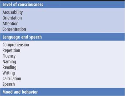

Table 5-4. Comprehensive mental status examination.
To determine the scope of the cognitive dysfunction (global or circumscribed), various spheres of cognition are tested in turn. These include memory, language, parietal lobe functions (pictorial construction, right–left discrimination, localization of objects in space), and frontal lobe or diffuse cerebral cortical functions (judgment, abstraction, thought content, the ability to perform previously learned acts). Multiple areas of cognitive function are impaired in dementia. The Minimental Status Examination (Table 5-5) provides a useful bedside screening test when dementia is suspected, but may not detect mild cognitive impairment.


Table 5-5. Minimental status examination.
Dementia from different causes may preferentially impair different spheres of cognition, and this can provide diagnostic clues. For example, Alzheimer disease affects memory disproportionately, whereas language function is often most impaired in frontotemporal dementia.
NEUROLOGIC EXAMINATION
Certain disorders that produce dementia also affect vision, coordination, or motor or sensory function. Detecting such associated neurologic abnormalities can help to establish an etiologic diagnosis. Neurologic signs suggesting causes of dementia are listed in Table 5-3.
LABORATORY INVESTIGATIONS
Laboratory studies that can help to identify the cause of dementia are listed in Table 5-6.
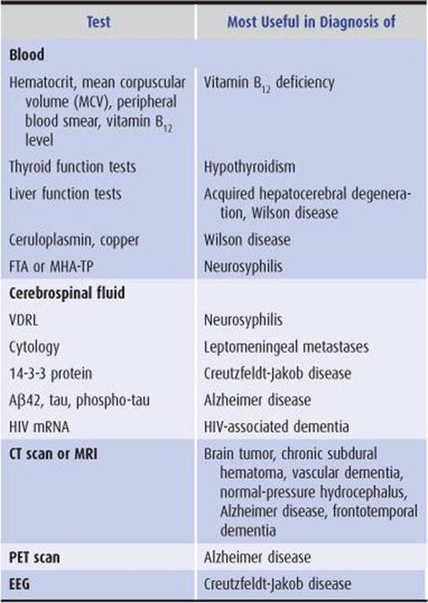
Table 5-6. Laboratory studies in dementia.
DEMENTIA
DIFFERENTIAL DIAGNOSIS
Common Causes of Dementia
A wide variety of diseases can produce dementia, but only a few do so commonly. In its most typical presentation—with gradual cognitive decline in an elderly (≥65 years) patient—the most common causes of dementia are Alzheimer disease, vascular (formerly “multi-infarct”) dementia, frontotemporal dementia, Lewy body disease, and Parkinson disease (Figure 5-3). In many patients, neurodegeneration and vascular disease coexist as causes of dementia in the same patient. Patients who present with dementia before age 45 years are much less likely to have Alzheimer disease or vascular dementia; in these patients, a wider range of neurodegenerative (Huntington disease, corticobasal degeneration), inflammatory (multiple sclerosis, systemic lupus erythematosus, vasculitis), and infective (prion disease) causes must be entertained. Dementia that progresses rapidly over weeks to months results most often from prion (Creutzfeldt-Jakob) disease.

Figure 5-3. Common causes of dementia based on age at presentation and rate of progression. “Mixed” refers to combined Alzheimer disease and vascular dementia. Totals of <100% reflect absence of an etiologic diagnosis in some cases. (Data from: Kelley BJ, Boeve BF, Josephs KA. Young-onset dementia: demographic and etiologic characteristics of 235 patients. Arch Neurol. 2008;65:1502-1508. Garre-Olmo J, Genís Batlle D, del Mar Fernández M, et al. Incidence and subtypes of early-onset dementia in a geographically defined general population. Neurology. 2010;75:1249-1255. Geschwind MD, Shu H, Haman A, Sejvar JJ, Miller BL. Rapidly progressive dementia. Ann Neurol. 2008;64:97-108.)
Other Causes of Dementia
Reversible causes of dementia, such as normal-pressure hydrocephalus, intracranial mass lesions, vitamin B12 deficiency, hypothyroidism, and neurosyphilis, are rare. However, they are important to diagnose because treatment can arrest or reverse the intellectual decline.
Diagnosing dementia caused by Huntington disease or other heritable disorders allows patients and their families to benefit from genetic counseling. If Creutzfeldt-Jakob disease or HIV-associated dementia is diagnosed, precautions can be instituted against transmission, and HIV disease can be treated with antiretroviral drugs. Progressive multifocal leukoencephalopathy may indicate underlying immunosuppression from HIV infection, lymphoma, or leukemia and may thereby bring these disorders to attention.
Approximately 15% of patients referred for evaluation of possible dementia instead have other disorders (pseudo-dementias), such as depression. Depression in this setting is important to identify because it is readily treatable. Drug intoxication, often cited as a cause of dementia in the elderly, actually produces an acute confusional state, rather than dementia.
NEURODEGENERATIVE PROTEINOPATHIES
In several neurodegenerative diseases, the production of misfolded proteins and their association to form insoluble aggregates appears to play an important role in pathogenesis (Table 5-7). These abnormal proteins can arise from either genetic or acquired modifications, and their pathologic effects may result from loss of normal protein function, gain of a toxic function, or a combination of these factors. Protein aggregation may be a mechanism for sequestering proteins that the cell’s proteolytic machinery cannot process, but protein aggregates may also exert adverse effects on the cell, such as by interfering with axonal transport.
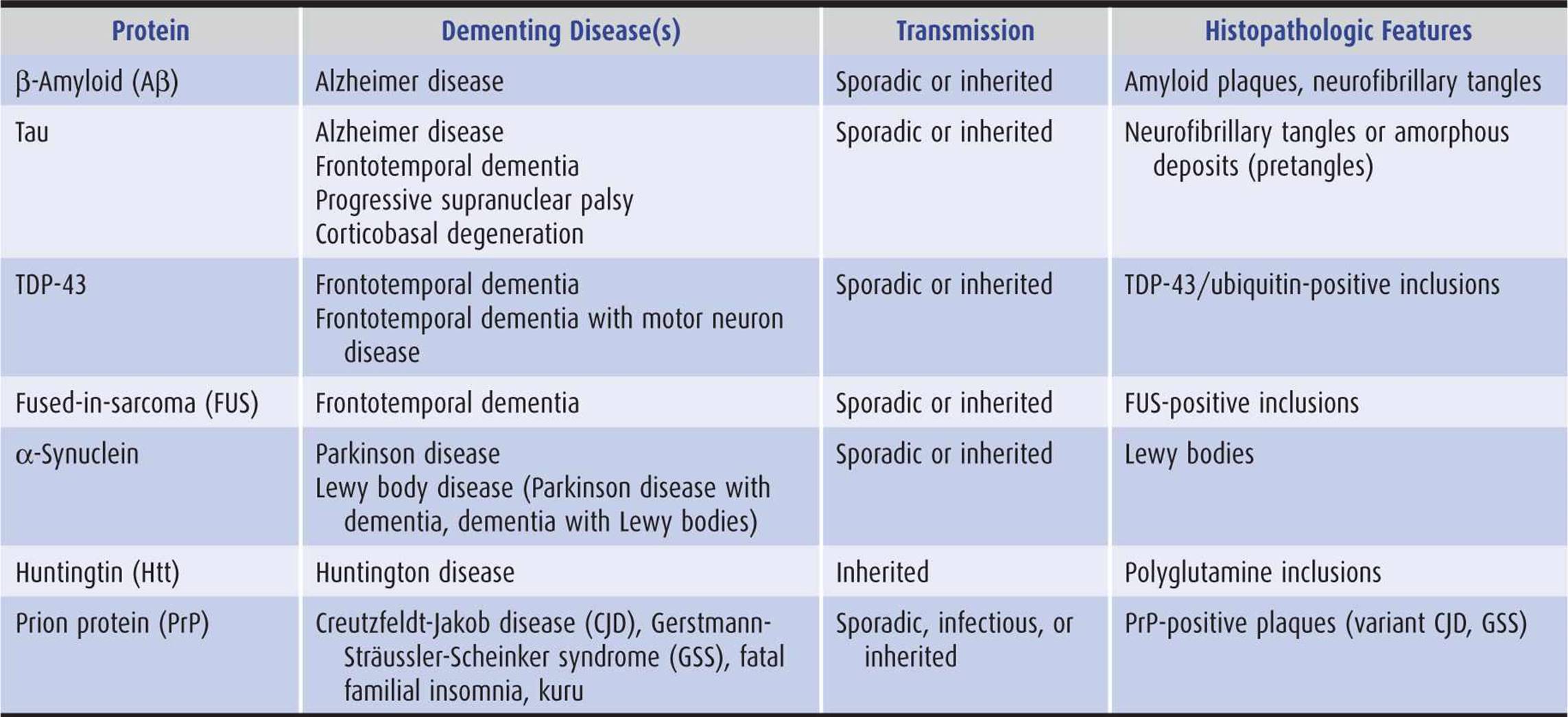
Table 5-7. Neurodegenerative proteinopathies.
Except in rare inherited or infectious cases, the underlying cause of neurodegenerative proteinopathies is unknown. However, these diseases share several features. In addition to protein misfolding and aggregation, which sometimes produces characteristic histopathologic findings (Table 5-7), these diseases may be associated with cell-to-cell (prionic) transmission (Figure 5-4), which allows them to spread through the nervous system to produce characteristic anatomic patterns of involvement. Evidence for this mechanism includes the finding that fetal tissue grafted into the brains of patients with Parkinson disease develops the same abnormal protein aggregates found in the recipient’s brain.
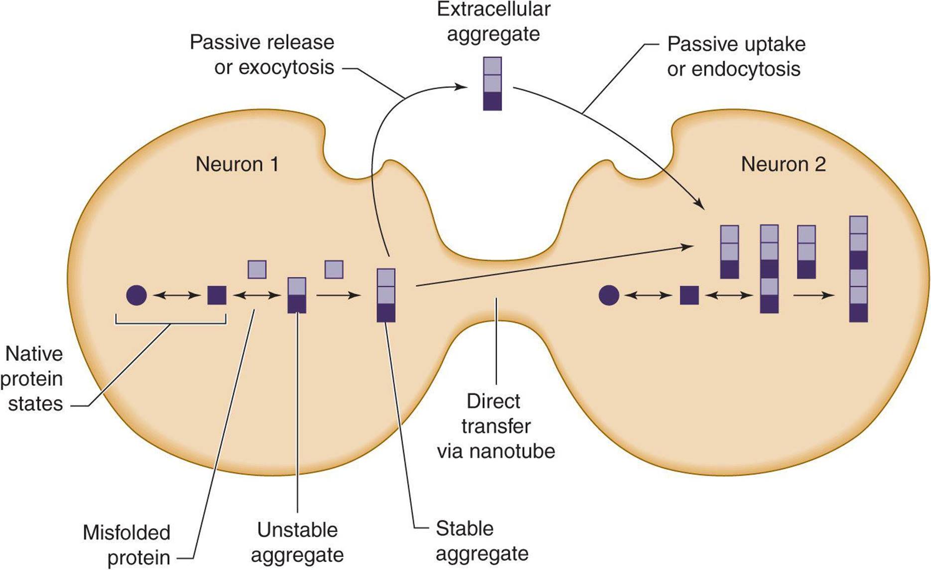
Figure 5-4. Cell-to-cell (prionic) transmission of neurodegenerative proteinopathies. Abnormal proteins associated with neurodegenerative disease may misfold, leading to the formation of protein aggregates; either mis-folded proteins, protein aggregates, or both may be toxic and contribute to neuronal dysfunction. In addition, toxic protein aggregates may be transferred between cells to propagate the disease.
ALZHEIMER DISEASE
Epidemiology
Alzheimer disease is the most common cause of dementia, accounting in whole or part for an estimated 60% to 70% of cases. Alzheimer disease affects approximately 15% of individuals age 65 years or older and approximately 45% of those age 85 years or over. Its prevalence is >5 million cases in the United States and approximately 30 million cases worldwide. Men and women are affected with equal frequency, when adjusted for age. However, because women live longer, they account for approximately two-thirds of Alzheimer patients.
Pathology
Alzheimer disease is defined by characteristic histopathologic features, especially neuritic (senile) plaques and neurofibrillary tangles. Neuritic plaques are extracellular deposits that contain β-amyloid (Aβ) and other proteins, including presenilin 1, presenilin 2, α1-antichymotrypsin, apolipoprotein E, α2-macroglobulin, and ubiquitin. Plaques may also be found in cerebral and meningeal blood-vessel walls, producing cerebral amyloid angiopathy.Neurofibrillary tangles are intracellular deposits containing hyperphosphorylated tau (a microtubule-associated protein) and ubiquitin.
Etiology
Alzheimer disease is a progressive, degenerative disorder that is caused by a genetic defect in rare cases (see later), but is usually sporadic and of unknown cause. Abnormal metabolism, deposition, or clearance of two proteins—Aβ and tau—appears to be closely linked to pathogenesis.
Pathogenesis
1. Genetics—In approximately 1% of patients, Alzheimer disease is a familial disorder that results from a mutation in one of three functionally related membrane proteins (Table 5-8): β-amyloid precursor protein (APP), presenilin 1 (PS1), or presenilin 2 (PS2). Onset of the disease in these patients is typically between the ages of 30 and 60 years. Patients with Down syndrome (trisomy 21) also develop early Alzheimer disease (mean onset at age 50 years), which is thought to be related to an extra copy of the APP gene, located on chromosome 21. Although the cause of sporadic Alzheimer disease is unknown, the gene defects in familial Alzheimer disease provide clues, implicating both APP, a protein with neurotrophic properties, and the presenilins, which are involved in APP metabolism.
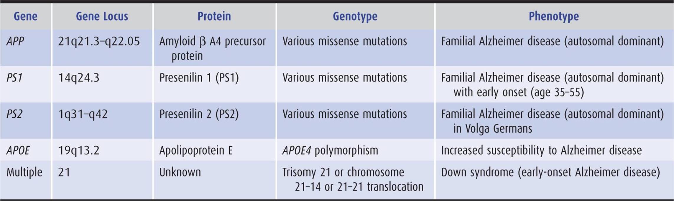
Table 5-8. Principal genes implicated in Alzheimer disease.
The risk of Alzheimer disease is also influenced by the inheritance pattern of apolipoprotein E (APOE) gene isoforms ε2, ε3, and ε4. Risk increases about threefold with a single apolipoprotein E ε4 (APOE4) allele and about 12-fold with two copies of APOE4; each copy of APOE4 also lowers the age at onset by about 5 years. In contrast to APOE4, APOE2 appears to confer relative protection from Alzheimer disease. The mechanism through which APOE genotype modifies susceptibility to Alzheimer disease is unknown, but may involve binding of the APOE protein to Aβ.
2. Aβ and neuritic plaques—Aβ is the principal constituent of neuritic plaques and is also deposited in cerebral and meningeal blood vessels in Alzheimer disease. Aβ is a 38- to 43-amino acid peptide produced by proteolytic cleavage of the transmembrane protein, APP (Figure 5-5). Normal processing of APP involves its cleavage by the enzyme α-secretase, which does not produce Aβ, and by β-secretase(BACE;β-site APP cleaving enzyme) and γ-secretase, yielding primarily a 40-amino acid fragment (Aγ40), which is secreted and cleared from the brain. In Alzheimer disease, a disproportionate amount of Aβ42, a longer form of the molecule with an increased tendency to aggregate, is produced. Presenilins 1 and 2 contribute to γ-secretase activity.

Figure 5-5. Normal and pathologic (amyloidogenic) processing of APP and Aβ. APP, a membrane-spanning protein, is normally cleaved by α-secretase (α), or by β-secretase (β) and then γ-secretase (γ, a protein complex that includes presenilin 1 or 2, nicastrin, APH1, and PEN2) to generate β-amyloid (Aβ), a secreted protein of unknown function. APP mutations associated with familial Alzheimer disease shift Aβ production from a nontoxic 40- to a toxic (amyloidogenic) 42-amino acid form, which has a greater tendency to form amyloid deposits. Aβ normally undergoes enzymatic breakdown (by neprilysin [NEP], insulin-degrading enzyme [IDE], or endothelin-converging enzyme [ECE-1]) and clearance from the brain. However, it can also aggregate to form oligomers of increasing size, which are thought to be neurotoxic. sAPP, soluble APP; C83 and C99, C-terminal fragments of APP; AICD, APP intracellular domain.
Evidence for a causal role of Aβ in Alzheimer disease includes the involvement of APP mutations in some familial cases and the neurotoxicity of Aβ under some circumstances. However, there is a poor correlation between the extent of amyloid plaque deposition in the brain and the severity of dementia in Alzheimer disease. One explanation for this disparity is that soluble Aβ oligomers, rather than insoluble plaques, may be the toxic agent. Another possibility is that Aβ aggregation produces Alzheimer disease indirectly, by promoting the formation of tau-containing neurofibrillary tangles.
3. Tau and neurofibrillary tangles—Tau is a cytoplasmic protein that binds to tubulin and stabilizes microtubules, cytoskeletal structures that help maintain cell structure and facilitate intracellular transport. In Alzheimer disease and other tauopathies, tau becomes hyperphosphorylated and dissociates from microtubules; the microtubules disassemble and hyperphosphorylated tau aggregates to form neurofibrillary tangles (Figure 5-6). How this leads to impaired neuronal function is unknown, but may involve a defect in axonal transport. A causal role for tau pathology in Alzheimer disease is supported by the observations that the abundance of neurofibrillary tangles correlates well with disease severity and that other tauopathies (eg, frontotemporal dementia), in which Aβ processing is normal, can also produce dementia.

Figure 5-6. Tau hyperphosphorylation and neurofibrillary tangle formation.
4. Synaptic dysfunction—Alzheimer disease is accompanied by early dysfunction and later loss of synapses, prominently affecting excitatory transmission in hippocampus and cerebral cortex. These changes may contribute to memory loss.
5. Neuronal loss and brain atrophy—Certain neuronal populations are preferentially lost in Alzheimer disease, including glutamatergic neurons in the entorhinal cortex and the CA1 sector of hippocampus, as well as cholinergic neurons in the basal forebrain. Focal brain atrophy is seen in the affected areas.
6. Vascular involvement—The extent to which vascular pathology may contribute to Alzheimer disease is controversial. Evidence for such a connection includes the overlap between risk factors for vascular disease and Alzheimer disease (including APOE genotype) and the involvement of blood vessels in amyloid pathology.
Risk Factors
The factors most conclusively associated with increased risk for Alzheimer disease are increasing age, female sex, and APOE4 genotype. Other factors that have been implicated in some studies include family history of Alzheimer disease, depression, low educational level, smoking, diabetes, hypertension, and fatty diet. Besides APOE4, several other genes that modify risk have been identified, but the magnitude of their effects is small.
Definitive evidence that the risk of Alzheimer disease can be reduced by diet, drugs, or lifestyle changes is lacking. However, cognitive engagement, physical activity, a low-fat and vegetable-rich diet, and light to moderate alcohol intake are all supported by some data.
Clinical Findings
The clinical progression of Alzheimer disease is thought to comprise a presymptomatic phase of up to about 10 years characterized by the deposition of amyloid plaques, followed by a symptomatic phase of up to about 10 years, during which tangle formation occurs (Figure 5-7).

Figure 5-7. Relationship between plaques, tangles, and clinical progression of Alzheimer disease (AD). MCI, mild cognitive impairment.
1. Early manifestations—The term mild cognitive impairment (MCI) is sometimes used to describe the early phase of cognitive decline observed in patients who later receive a diagnosis of Alzheimer disease. Impairment of recent memory is typically the first sign of Alzheimer disease and may be noticed only by family members. As the memory disorder progresses over months to several years, the patient becomes disoriented to time and then to place. Aphasia, anomia, and acalculia may develop, forcing the patient to leave work or give up the management of family finances. The depression apparent in the earlier stages of the disorder may give way to an agitated, restless state. Apraxias and visuospatial disorientation ensue, causing the patient to become lost easily. Primitive reflexes are commonly found. A frontal lobe gait disorder may become apparent, with short, slow, shuffling steps, flexed posture, wide base, and difficulty in initiating walking.
2. Late manifestations—In the late stages, previously preserved social graces are lost, and psychiatric symptoms, including psychosis with paranoia, hallucinations, or delusions, may be prominent. Seizures occur in some cases. Examination at this stage may show rigidity and bradykinesia. Rare and usually late features of the disease include myoclonus, incontinence, spasticity, extensor plantar responses, and hemiparesis. Mutism, incontinence, and a bedridden state are terminal manifestations. Eating problems, febrile episodes, dyspnea, pneumonia, and pain are frequent complications in the final months of life, and death typically occurs from 5 to 10 years after the onset of symptoms.
Investigative Studies
Laboratory investigations can exclude other disorders, including reversible or otherwise treatable conditions. Cognitive testing may also be useful in helping to distinguish between Alzheimer disease and other causes of dementia. In patients with Alzheimer disease, the CT scan or MRI often shows cortical (especially medial temporal lobe) atrophy and enlarged ventricles, but such changes are nonspecific. Positron emission tomography (PET) scanning may reveal hypometabolism and hypoperfusion in the temporal and parietal lobes. More specific PET tracers include [18F]FDDNP, which labels amyloid plaques and neurofibrillary tangles, and [11C] Pittsburgh compound B (PIB), which binds to amyloid plaques. Cerebrospinal fluid (CSF) levels of Aβ42, tau, and phospho-tau have also been found useful as biomarkers of Alzheimer disease.
Differential Diagnosis
Early Alzheimer disease may resemble depression or pure memory disorders such as the Korsakoff amnestic syndrome (see later discussion). More advanced Alzheimer disease must be distinguished from Lewy body dementia, vascular dementia, Creutzfeldt-Jakob disease, and other dementing disorders (see later discussion).
Treatment
No currently available treatment has been shown to reverse existing deficits or to arrest disease progression. However, memantine (Table 5-9), an NMDA-type glutamate receptor antagonist drug, may produce modest improvement in patients with moderate or severe Alzheimer disease.

Table 5-9. Drugs used in the treatment of Alzheimer disease.
Because cholinergic neuronal pathways degenerate and choline acetyltransferase is depleted in the brains of patients with Alzheimer disease, cholinergic replacement therapy has also been used for symptomatic treatment of cognitive dysfunction (Table 5-9). Acetylcholinesterase inhibitors, including tacrine, donepezil, rivastigmine, and galantamine, have all been shown to produce small improvements in tests of cognitive function. Side effects include nausea and vomiting, diarrhea, and dizziness; tacrine also elevates serum transaminase levels. The better side-effect profile of donepezil and its once-daily dosage schedule are advantageous.
Experimental treatments under investigation include vaccines directed against β-amyloid, secretase inhibitors, and metal chelators. Antipsychotic drugs, antidepressants, and anxiolytics may be useful in controlling behavioral disturbances associated with Alzheimer disease. However, evidence for their effectiveness is sparse, and in some cases (risperidone, olanzapine) their use is associated with an increased incidence of stroke in elderly patients.
Prognosis
Early in the course of the disease, patients can usually remain at home and continue social, recreational, and limited professional activities. Early diagnosis can allow patients time to plan orderly retirement from work, to arrange for management of their finances, and to discuss with physicians and family members the management of future medical problems. Patients in advanced stages of the disease may require care in a nursing facility and the use of psychoactive medications. These patients must be protected and prevented from injuring themselves and their families by injudicious actions or decisions. Death from inanition or infection generally occurs 5 to 10 years after the first symptoms.
FRONTOTEMPORAL DEMENTIA
Frontotemporal dementia (FTD) comprises a genetically and clinically heterogeneous group of dementing disorders that produce frontal and temporal lobe degeneration and affect behavior and language preferentially. FTD differs in these respects from Alzheimer disease, which involves primarily the temporal and parietal lobes and causes prominent memory disturbance. Both FTD and Alzheimer disease exhibit tau-containing inclusions, but abnormal amyloid processing and plaques are seen only in Alzheimer disease.
Epidemiology
FTD is thought to be the third most common cause of dementia, after Alzheimer disease and vascular dementia. The average age at onset is 50 to 60 years.
Pathology
Frontotemporal dementia is characterized by atrophy of the frontal and temporal lobes. Histopathologic findings include neuronal loss, gliosis, and intracellular inclusions containing either the microtubule-associated protein tau (MAPT); transactive response DNA-binding protein 43 (TDP-43) and ubiquitin; or fused-in-sarcoma (FUS). Tau inclusions in FTD differ from those found in Alzheimer disease: The former are twisted, ribbon-like structures rather than paired helical filaments and include neurofibrillary tangles, amorphous deposits (pretangles), and, in some cases, Pick bodies. Inclusions are also sometimes found in hippocampus, subcortical nuclei, brainstem, cerebellum, or spinal cord.
Etiology
In most cases, frontotemporal dementia is a sporadic neurodegenerative disease of unknown cause. However, 20% to 40% of patients report a family history of a neurodegenerative disorder, and approximately 10% appear to inherit frontotemporal dementia in an autosomal dominant fashion.
Pathogenesis
1. Genetics—Mutations in two genes—MAPT and progranulin (GRN)—appear to be responsible for approximately one-half of inherited cases. MAPT mutations are thought to produce disease largely by toxic gain of function, whereas GRN mutations cause loss of function through haploinsufficiency. Less common mutations producing frontotemporal dementia affect genes for valosin-containing protein (VCP), charged multivesicular body protein 2B (CHMP2B), TAR-DNA binding protein (TARDP), or FUS.
2. Tau and neurofibrillary tangles—Patients with MAPT mutations have tau-containing inclusions (neurofibrillary tangles or amorphous deposits). Disease-producing effects of mutant tau may include defective assembly and stability of microtubules, impaired axonal transport, increased aggregation to form toxic inclusions, and altered signal transduction. MAPT mutations and tau-positive inclusions are associated clinically with behavioral variant frontotemporal dementia, progressive nonfluent aphasia, corticobasal degeneration, and progressive supranuclear palsy (see later discussion).
3. Progranulin and TDP-43/ubiquitin-positive inclusions—Progranulin is a growth factor involved in neurite outgrowth and neuronal survival, and GRN mutations may deprive neurons of trophic support. These mutations are associated with inclusions that contain TDP-43, a transcriptional repressor and regulator of translation. TDP-43 is normally located in the nucleus, but it translocates to the cytoplasm and is ubiquitinated in frontotemporal dementia due to GRN mutations. GRN mutations and TDP-43/ubiquitin-containing inclusions can produce the clinical syndromes of behavioral variant frontotemporal dementia, semantic dementia, progressive nonfluent aphasia, corticobasal degeneration, and FTD with motor neuron disease (see later discussion).
4. FUS-positive inclusions—Some patients with FTD have tau- and TDP-43-negative, but ubiquitin-positive, inclusions that also stain for FUS, a nuclear protein involved in DNA repair, transcriptional regulation, and cytoplasmic localization. Patients with these inclusions have sporadic, behavioral variant FTD, with a tendency for early onset, usually before age 40 years.
5. Neuronal dysfunction, neuronal loss, and brain atrophy—It is unclear how much of the clinical picture in FTD results from abnormal neuronal function as opposed to neuronal loss. Eventually, however, there is marked brain atrophy affecting the frontal and anterior temporal lobes most prominently, together with neuronal loss and gliosis.
Clinical Findings
1. Behavioral variant frontotemporal dementia is characterized by prominent behavioral changes, including altered interpersonal interactions and personal conduct (eg, apathy and disinhibition), blunted emotions, and lack of insight. These behavioral abnormalities overshadow more modest cognitive defects, such as impaired judgment, inattention, or disorganization. This syndrome can be seen in patients with tau, TDP-43, or FUS pathology.
2. Semantic dementia produces fluent (receptive) aphasia (Chapter 1) with impaired comprehension and anomia and occurs with disease affecting the dominant temporal lobe. It is most common in patients with TDP-43 pathology.
3. Progressive nonfluent aphasia produces expressive aphasia (Chapter 1) with preserved comprehension and results from predominant involvement of the dominant frontal lobe. It is most common in patients with tau pathology.
4. Overlap syndromes occur in cases in which FTD is combined with features of parkinsonism (corticobasal degeneration [discussed later] or progressive supranuclear palsy [discussed later and in Chapter 11]) or motor neuron disease (amyotrophic lateral sclerosis [Chapter 9]). Parkinsonian syndromes are seen most often in patients with tau pathology, whereas motor neuron involvement is associated with TDP-43 pathology.
Investigative Studies
MRI shows frontal and temporal lobe atrophy (Figure 5-8), and PET may show hypometabolism in these regions. In both cases, the abnormalities are often asymmetric, with right-sided atrophy predominating in behavioral and left-sided atrophy predominating in language variants. Genetic screening can demonstrate mutations in MAPT or GRN in patients with a positive family history.
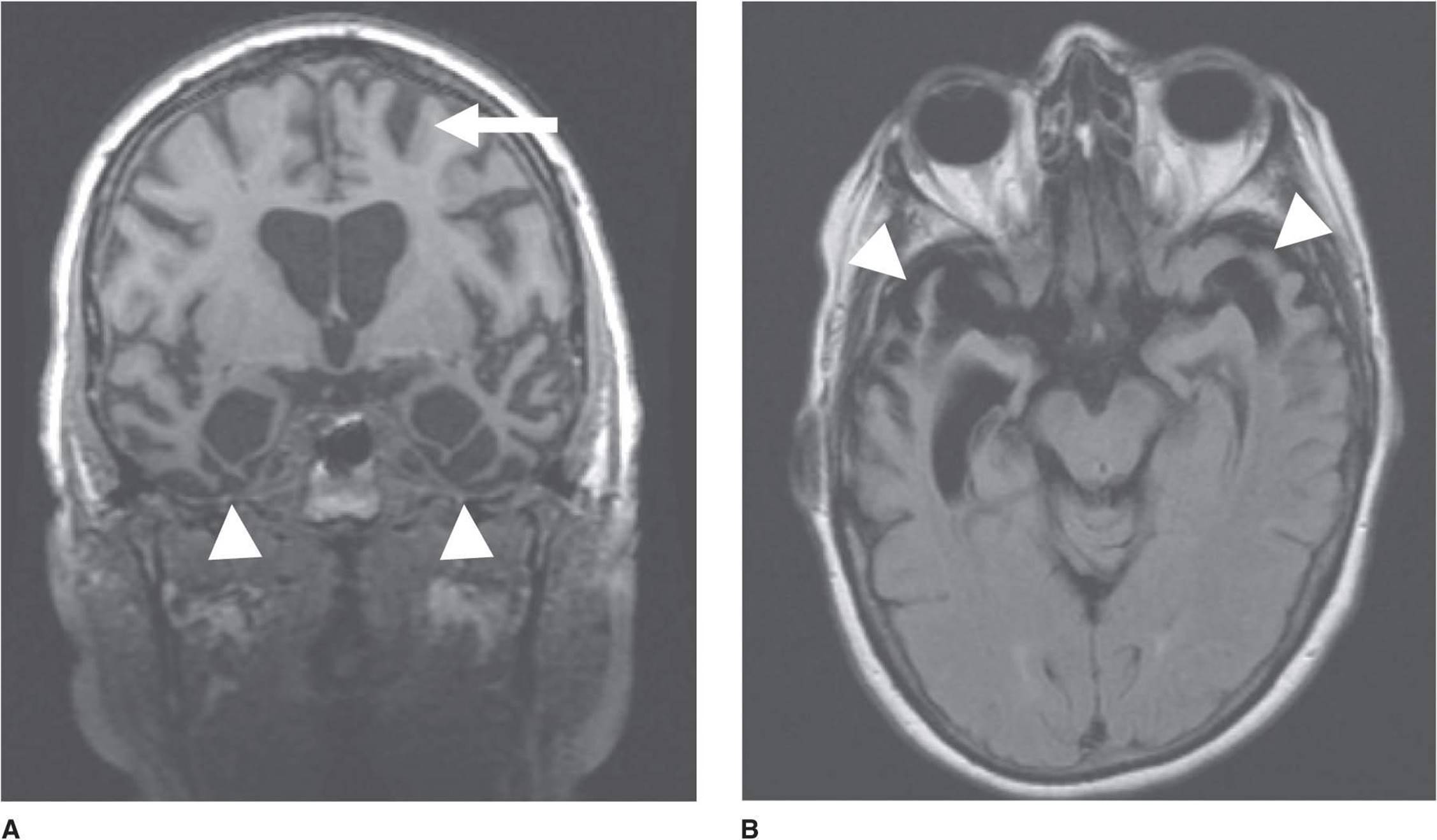
Figure 5-8. Axial (A) and coronal (B) FLAIR MRI in frontotemporal dementia showing regional atrophy of the frontal (arrow) and temporal (arrowheads) lobes. (Courtesy J. Handwerke.)
Differential Diagnosis
In contrast to Alzheimer disease, memory disturbance does not dominate the clinical picture in FTD, and onset typically occurs at an earlier age. The diagnosis is suggested by the onset of dementia before age 60 years, with behavioral disturbance or aphasia as the primary abnormality. FTD with altered behavior may be mistaken for a primary psychiatric disorder, and language variants can raise suspicion regarding stroke. FTD associated with parkinsonism or motor neuron disease must be distinguished from Parkinson disease or amyotrophic lateral sclerosis.
Treatment
Memantine and anticholinesterase drugs used to treat Alzheimer disease have not been shown to be effective in FTD. Antidepressants, especially selective serotonin reuptake inhibitors and trazodone, may be useful for managing behavioral symptoms. Patients with parkinsonian features (corticobasal degeneration or progressive supra-nuclear palsy) may benefit from levodopa/carbidopa or dopamine receptor agonists.
Prognosis
The duration of illness in FTD is highly variable (2-20 years), consistent with the clinical heterogeneity of the disorder.
CORTICOBASAL DEGENERATION
Corticobasal degeneration is a tauopathy related to FTD with tau pathology. It produces asymmetric frontoparietal cortical atrophy and depigmentation of the substantia nigra, with tau-positive neuronal and glial inclusions, ballooned neurons, and neuronal and glia cell loss. The classic corticobasal syndrome reflects involvement of both cerebral cortex and basal ganglia and consists of unilateral limb (usually arm) clumsiness and functional impairment due to some combination of apraxia, sensory loss, and myoclonus, together with extrapyramidal rigidity, bradykinesia, and postural tremor. Limb apraxia and sensory loss may produce the alien-hand sign, in which the limb moves seemingly of its own accord. In addition to corticobasal degeneration, the corticobasal syndrome can also be seen in progressive supranuclear palsy (see next section) and FTD. Rigidity and bradykinesia are typically unresponsive to antiparkinsonian medications.
PROGRESSIVE SUPRANUCLEAR PALSY
Progressive supranuclear palsy (PSP, or Steele-Richardson-Olszewski syndrome) is an idiopathic degenerative disorder that primarily affects the brainstem, subcortical gray matter, and cerebral cortex. Like tau-positive FTD and corticobasal degeneration, it is characterized pathologically by tau-positive intracellular inclusions. The classic clinical features are supranuclear ophthalmoplegia (especially affecting down-gaze), pseudobulbar palsy, axial dystonia with or without extrapyramidal rigidity of the limbs, and dementia. Because PSP usually presents as a movement disorder with parkinsonian features, it is discussed further in Chapter 11.
LEWY BODY DISEASE
Parkinson disease (Chapter 11), a sporadic or inherited neurodegenerative disorder characterized by tremor, rigidity, bradykinesia, and postural instability, is accompanied by dementia in approximately one-third of cases. Patients who develop dementia at least 1 year after the onset of motor symptoms are classified as having Parkinson disease with dementia, whereas those in whom dementia has its onset prior to or within 1 year of the first motor symptoms are given the diagnosis of dementia with Lewy bodies. However, these two diagnoses cannot be distinguished pathologically, and the term Lewy body diseaseis sometimes used to encompass both.
Lewy body disease is characterized histopathologically by round, eosinophilic, intracytoplasmic neuronal inclusions (Lewy bodies) in the brainstem and cerebral cortex. These inclusions contain α-synuclein, a protein that is also found in Lewy bodies in Parkinson disease without dementia, and both Lewy body disease and Parkinson disease are, therefore, classified as synucleinopathies.
Lewy body disease causes cognitive decline without prominent early memory impairment. Features include fluctuating cognitive ability, well-formed visual hallucinations, and signs of parkinsonism, especially rigidity and bradykinesia.
Motor manifestations of Lewy body disease are treated with antiparkinsonian medications (Chapter 11); some studies suggest that memantine or anticholinesterase drugs used to treat Alzheimer disease (Table 5-9) may also be beneficial in dementia associated with Lewy body disease.
HUNTINGTON DISEASE
Huntington disease is an autosomal dominant neurodegenerative disorder characterized by chorea, psychiatric symptoms, and dementia. The cause is an expanded CAG trinucleotide repeat coding for a polyglutamine tract in the huntingtin (Htt) gene. The brain shows atrophy affecting the caudate nucleus, putamen, and cerebral cortex, with Htt aggregation in cytoplasmic and nuclear inclusions. Dementia usually becomes apparent after chorea and psychiatric symptoms have been present for a few years, but precedes chorea in approximately one-fourth of cases. Impaired executivefunction (eg, judgment) and memory are prominent features, whereas language tends to be spared until late in the course. Huntington disease is discussed further in Chapter 11.
CREUTZFELDT-JAKOB (PRION) DISEASE
Creutzfeldt-Jakob disease (CJD) produces rapidly progressive dementia with variable focal degeneration of cerebral cortex, basal ganglia, cerebellum, brainstem, and spinal cord. It is caused by a proteinaceous infectious particle (prion) and may be sporadic (approximately 85% of cases), genetic, or infectious. In the latter case, CJD can be transmitted via prion-contaminated tissue or surgical instruments (iatrogenic CJD) or by consumption of contaminated beef (variant CJD). Documented human-to-human transmission (by corneal transplantation, cortical electrode implantation, or administration of human growth hormone) is rare. The infectious agent is present in the brain, spinal cord, eyes, lungs, lymph nodes, kidneys, spleen, liver, and CSF, but not other body fluids.
The annual incidence is approximately 1 case per 1 million population. The sporadic disease usually occurs in patients older than 40 years and has a mean age at onset of approximately 60 years; in contrast, the genetically acquired disease usually has its onset before age 55 years. More than one member of a family is affected in only 5% to 10% of cases, and conjugal cases are rare.
Pathogenesis
Familial CJD is an autosomal dominant disorder caused by a mutation in the PRNP gene, which codes for the prion protein cellular isoform (PrPC), a protein of unknown function. In sporadic CJD, PrPCundergoes a conformational change to produce an abnormal prion protein (scrapie isoform, or PrPSc). PrPSc then serves as a template on which PrPC is converted to additional PrPSc. In infectious CJD, PrPSc is introduced into the brain from an external source. In each case, the result is accumulation of abnormal PrPSc prions in brain tissue. The ability of PrPSc to induce the PrPSc conformation in PrPC prions enables it to replicate without nucleic acids.
Prions have also been implicated in diseases of animals and in three other rare human disorders (Table 5-10)—kuru, a dementing disease of Fore-speaking tribes of New Guinea (apparently spread by cannibalism); Gerstmann-Straüssler syndrome, a familial disorder characterized by dementia and ataxia; and fatal familial insomnia, which produces disturbances of sleep and of autonomic, motor, and endocrine function.

Table 5-10. Prion diseases.
Clinical Findings
The clinical picture may be that of a diffuse central nervous system (CNS) disorder or of more localized dysfunction (Table 5-11). Dementia is present in virtually all cases and may begin as mild global cognitive impairment or a focal cortical disorder such as aphasia, apraxia, or agnosia. Progression to akinetic mutism or coma typically ensues over a period of months. Psychiatric symptoms including anxiety, euphoria, depression, labile affect, delusions, hallucinations, and changes in personality or behavior may be prominent. Fever is absent.

Table 5-11. Clinical features of sporadic Creutzfeldt-Jakob disease.
Aside from cognitive abnormalities, the most frequent clinical manifestations are myoclonus (often induced by a startle), extrapyramidal signs (rigidity, bradykinesia, tremor, dystonia, chorea, or athetosis), cerebellar signs, and extrapyramidal signs. Visual field defects, cranial nerve palsies, and seizures occur less often.
A distinct variant of CJD results from the transmission of bovine spongiform encephalopathy (“mad cow disease”) to humans. This variant is characterized by earlier onset (typically in the teens or young adulthood), invariable cerebellar involvement, prominent early psychiatric abnormalities, and diffuse amyloid plaques.
Investigative Studies
The electroencephalogram (EEG) may show periodic sharp waves or spikes (Figure 5-9), which are absent in the variant form described previously. CSF protein may be elevated (≤100 mg/dL), and levels of 14–3–3 protein are increased. MRI scans may show hyperintense signals in the basal ganglia on T2-weighted images (Figure 5-9). Definitive diagnosis is by immunodetection of PrPSc in brain tissue obtained at biopsy or, in familial cases, by detection of mutant forms of PrPC in DNA from lymphocytes.


Figure 5-9. Diffusion-weighted image of the brain in Creutzfeldt-Jakob disease, showing characteristic hyperintensities (white) in the basal ganglia and cortical ribbon (A), and EEG with typical triphasic waves in all leads (B), which occur repetitively about once every second.
Differential Diagnosis
A variety of other disorders must be distinguished from CJD. Alzheimer disease is often a consideration, especially in patients with a less fulminant course and a paucity of cerebellar and extrapyramidal signs. Where subcortical involvement is prominent, Parkinson disease, cerebellar degeneration, or progressive supranuclear palsy may be suspected. Striking focal signs raise the possibility of an intracerebral mass lesion. Acute metabolic disorders that produce altered mentation and myoclonus (eg, sedative drug withdrawal) can mimic CJD.
Prognosis
No treatment is currently available. The disease is usually relentlessly progressive and invariably fatal. In most sporadic cases, death occurs within 1 year after the onset of symptoms. Depending on the specific mutation present, familial forms of the disease may have a longer course (1-5 years).
CEREBROVASCULAR DISEASE
VASCULAR DEMENTIA
Vascular disease is generally considered the second most common cause of dementia, after Alzheimer disease, and many patients have features of both diseases. Most patients with this diagnosis have either multiple large cortical infarcts from occlusion of major cerebral arteries or several smaller infarcts (lacunar state; Chapter 13) affecting subcortical white matter, basal ganglia, or thalamus.
The relationship between cerebral vascular disease and dementia is poorly characterized. For example, the number of strokes, their locations, and the total infarct volume required for strokes to produce dementia are uncertain, making it often difficult to determine whether strokes are the cause of dementia in a given patient. The absence of neuroradiologic signs of cerebrovascular disease argues strongly against a vascular basis for dementia, but the presence of vascular lesions does not prove that they are causal. This is especially true when another cause of dementia, such as Alzheimer disease, coexists with cerebrovascular disease.
Clinical Findings
As classically described, patients with vascular dementia have a history of hypertension, a stepwise progression of deficits, a more or less abrupt onset of dementia, and focal neurologic symptoms or signs. Because extensive pathologic changes may already exist at presentation, it is assumed that patients can remain functionally well compensated until a new and perhaps otherwise innocuous infarct tips the balance.
The neurologic examination commonly shows pseudo-bulbar palsy with dysarthria, dysphagia, and pathologic emotionality (pseudobulbar affect); focal motor and sensory deficits; ataxia; gait apraxia; hyperreflexia; and extensor plantar responses. Memory disturbance is typically less prominent than in Alzheimer disease.
Investigative Studies
The MRI (Figure 5-10) may show multiple large infarcts, multiple small (lacunar) infarcts, areas of low density in subcortical white matter (Binswanger disease, or subcortical arteriosclerotic encephalopathy), or combinations of these findings and is more sensitive than CT scan for detecting these abnormalities.

Figure 5-10. T2-weighted MRI in vascular dementia, showing foci of abnormal high signal intensity adjacent to the lateral ventricles (arrows) and within the basal ganglia (arrowheads).
Additional laboratory studies should be performed to exclude cardiac emboli, polycythemia, thrombocytosis, cerebral vasculitis, and meningovascular syphilis as causes of multiple infarctions, particularly in younger patients or those without a history of hypertension.
Treatment
Hypertension, when present, should be treated to reduce the incidence of subsequent infarction and to prevent other end-organ diseases. Antiplatelet agents (Chapter 13) may help to reduce the risk of future strokes.
CHRONIC SUBDURAL HEMATOMA
Chronic subdural hematoma usually affects patients aged 50 to 70 years, often after minor head trauma. Other risk factors include alcoholism, cerebral atrophy, epilepsy, anticoagulation, ventricular shunts, and long-term hemo-dialysis. The onset of symptoms may be delayed for months after trauma. Hematomas are bilateral in approximately one-sixth of cases.
Clinical Findings
Headache is the initial symptom in most patients. Confusion, dementia, hemiparesis, and vomiting may ensue. The most frequent signs are cognitive disturbance, hemiparesis, papilledema, and extensor plantar responses. Aphasia, visual field defects, and seizures are uncommon, but can occur.
The hematoma can usually be seen on CT scan or MRI (Figure 5-11) as an extra-axial crescent-shaped area of decreased density, with ipsilateral obliteration of cortical sulci and often ventricular compression. The scan should be carefully reviewed for evidence of bilateral subdural collections. Isodense collections may become more apparent after contrast infusion. In a few cases, demonstration of the hematoma may require cerebral arteriography, which should always be undertaken bilaterally.
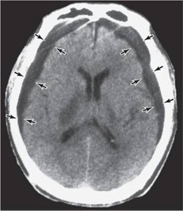
Figure 5-11. CT scan in chronic subdural hematoma, showing bilateral low-density collections between the inner table of the skull and the cerebral hemispheres (arrows).
Treatment
Unless contraindicated by medical problems or spontaneous improvement, symptomatic hematomas should be surgically evacuated.
OTHER CEREBRAL DISORDERS
NORMAL-PRESSURE HYDROCEPHALUS
Normal-pressure hydrocephalus, a potentially reversible cause of dementia, is characterized by the clinical triad of dementia, gait apraxia, and incontinence. It may be idiopathic or secondary to conditions that interfere with CSF absorption, such as meningitis or subarachnoid hemorrhage. The dementia is often mild and insidious in onset and is typically preceded by gait disorder and incontinence. It is characterized initially by mental slowness and apathy and later by global cognitive dysfunction. Deterioration of memory is common, but aphasia and agnosia are rare.
Pathophysiology
Normal-pressure hydrocephalus is sometimes called communicating hydrocephalus (because the lateral, third, and fourth ventricles remain in communication) or nonobstructive hydrocephalus (because the flow of CSF between the ventricles is not impaired). It is presumed to be caused by impaired CSF absorption from arachnoid granulations in the subarachnoid space over the convexity of the hemispheres (Figure 5-12), for example, from meningeal fibrosis and adhesions after meningitis or subarachnoid hemorrhage. In contrast, noncommunicating or obstructive hydrocephalus is caused by a blockade of CSF circulation within the ventricular system (eg, by an intraventricular cyst or tumor) and is associated with increased CSF pressure and often with headache and papilledema.
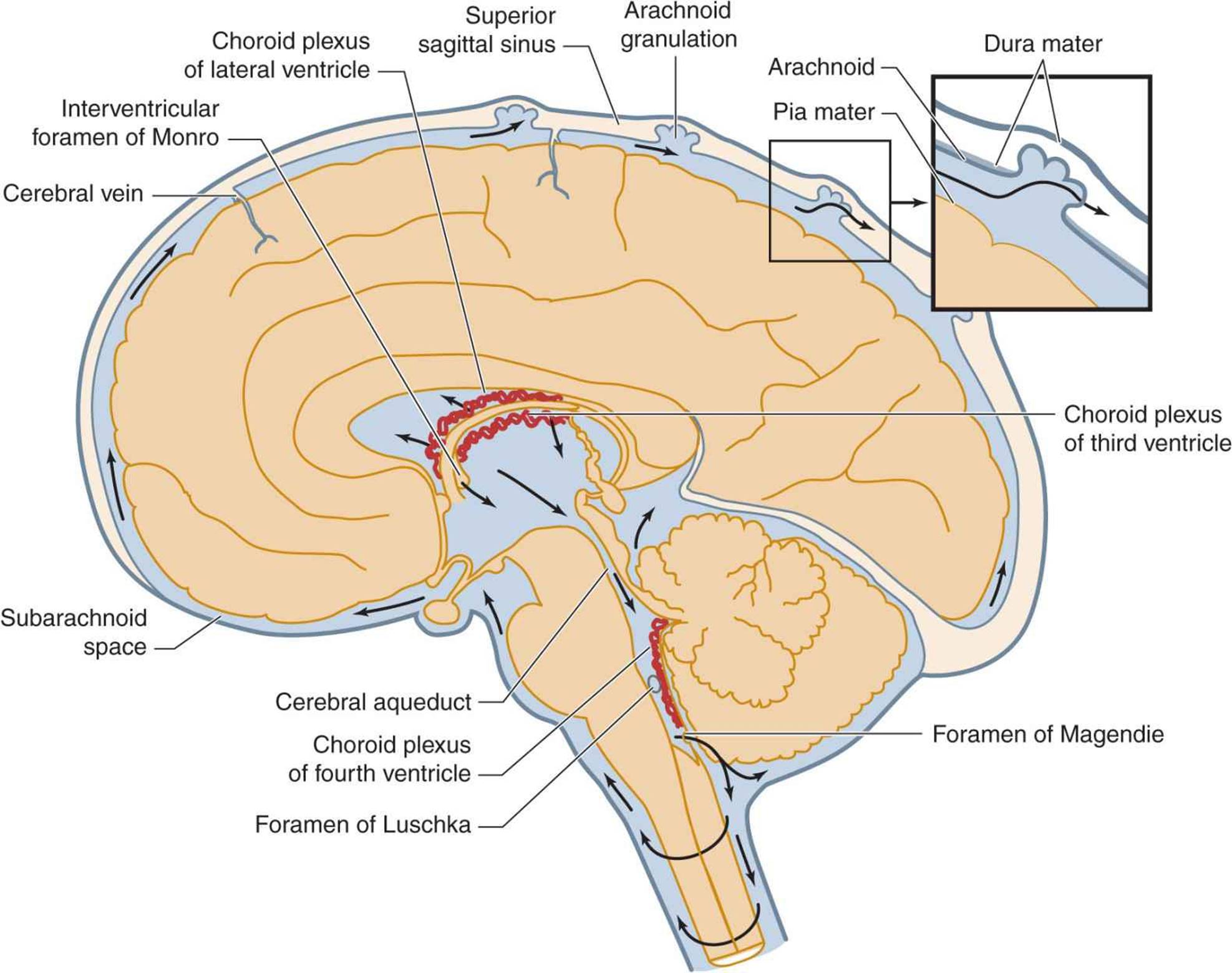
Figure 5-12. Circulation of cerebrospinal fluid (CSF). CSF is produced by the choroid plexus, which consists of specialized secretory tissue located within the cerebral ventricles. It flows from the lateral and third ventricles through the cerebral aqueduct and fourth ventricle and exits the ventricular system through two laterally situated foramina of Luschka and a single, medially located foramen of Magendie. CSF then enters and circulates through the subarachnoid space surrounding the brain and spinal cord. It is ultimately absorbed through arachnoid granulations into the venous circulation.
Clinical Findings
Normal-pressure hydrocephalus usually develops over a period of months; a gait disorder is often the initial manifestation. This typically takes the form of gait apraxia, characterized by unsteadiness on standing and difficulty in initiating walking (magnetic gait), even though there is no weakness or ataxia. The patient can perform the leg movements associated with walking, bicycling, or kicking a ball and can trace figures with the feet while lying or sitting but is unable to do so when the legs are bearing weight. The patient typically appears to be glued to the floor, and walking, once underway, is slow and shuffling. Pyramidal signs, including spasticity, hyperreflexia, and extensor plantar responses, are sometimes present. Motor perseveration (the inappropriate repetition of motor activity) and grasp reflexes in the hands and feet may occur. Dementia is manifested by psychomotor slowing, inattention, and impaired decision making and spatial recognition. Urinary incontinence is a later development, and patients may be unaware of it; fecal incontinence is uncommon.
Investigative Studies
Lumbar puncture reveals normal or low opening pressure. The CT scan or MRI typically shows enlarged lateral ventricles without increased prominence of cortical sulci (Figure 5-13). Radionuclide cisternography classically shows isotope accumulation in the ventricles, delayed clearance, and failure of ascent over the cerebral convexities. This pattern is not necessarily present in patients who respond to shunting, however. Transient improvement in gait, cognitive testing, or sphincteric function after the removal of 30 to 50 mL of CSF by lumbar puncture (tap test) or external lumbar drainage of 750 to 1,000 mL of CSF over 72 hours is probably the best predictor of a favorable clinical response to shunting (see Treatment).
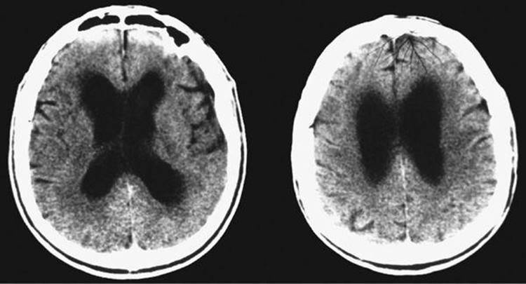
Figure 5-13. CT scan at two transverse levels in normal-pressure hydrocephalus, showing enlarged lateral ventricles without enlargement of the cortical sulci.
Differential Diagnosis
A variety of conditions that produce dementia must be considered in the differential diagnosis. Alzheimer disease tends to follow a longer course, often with prominent focal cortical dysfunction and enlarged cortical sulci shown in a CT scan or MRI. Parkinsonism may be simulated by the gait disorder but can be distinguished by extrapyramidal rigidity, tremor, and response to antiparkinsonian medications. Vascular dementia should be suspected if the disorder follows a stepwise course, or pseudobulbar palsy, focal sensorimotor signs, or a history of stroke is encountered. In such cases, CT scan or MRI should demonstrate vascular lesions.
Treatment
Some patients, especially those with hydrocephalus from meningitis or subarachnoid hemorrhage, recover or improve after ventriculoatrial, ventriculoperitoneal, or lumboperitoneal shunting. In idiopathic normal-pressure hydrocephalus, approximately 60% of patients respond and approximately 40% show sustained improvement after shunting. Complications of shunting occur in approximately 40% of patients and include shunt infection, subdural hematoma, and shunt malfunction that necessitates replacement.
BRAIN TUMOR
Brain tumors (Chapter 6) produce dementia and related syndromes by a combination of local and diffuse effects, including edema, compression of adjacent brain structures, increased intracranial pressure, and impairment of cerebral blood flow. Cognitive function in patients with brain tumor can also be impaired by radiotherapy or chemotherapy. The tumors most likely to produce generalized cerebral syndromes are gliomas arising in the frontal or temporal lobes or the corpus callosum. Although such lesions tend to infiltrate subcortical white matter extensively, they initially give rise to few focal neurologic signs.
The dementia associated with brain tumor is characterized by prominent mental slowness, apathy, impaired concentration, and subtle alterations in personality. Depending on the areas of involvement, memory disorder, aphasia, or agnosia may be seen early. Brain tumors ultimately produce headache, seizures, or focal sensorimotor disturbances.
Meningeal neoplasia, discussed in Chapter 4 as a cause of confusional states, may also produce dementia that is commonly associated with headache, as well as symptoms and signs of dysfunction at multiple sites in the nervous system. The diagnosis is established by cytologic studies of the CSF.
CHRONIC TRAUMATIC ENCEPHALOPATHY
Severe or repetitive head injury may cause a progressive syndrome manifested by impaired memory and concentration, personality changes, headache, tremor, rigidity, bradykinesia, dysarthria, cerebellar ataxia, pyramidal signs, and seizures. Although classically described in boxers (dementia pugilistica), this condition is recognized increasingly in athletes subject to more subtle forms of head trauma, including concussion. Neuroimaging studies may show cortical atrophy and cavum septi pellucidum. Histopathologic features include neurofibrillary tangles consistent with a progressive tauopathy.
SYSTEMIC DISORDERS
INFECTION
HIV-Associated Dementia
Human immunodeficiency virus (HIV-1) infection of the brain can produce a range of HIV-associated neurocognitive disorders. These include asymptomatic neurocognitive impairment (demonstrable only by cognitive testing), minor neurocognitive disorder (symptomatic but of little functional impact), and HIV-associated dementia (also termed AIDS dementia complex or HIV encephalopathy). Although the incidence and severity of HIV-associated dementia have declined with highly active antiretroviral therapy, it still affects 2% to 8% of patients with treated HIV infection. Risk factors include intravenous drug use, female sex, and increased age.
A. Pathogenesis
HIV-associated dementia results from invasion of the brain by blood-borne macrophage-tropic retrovirus, which infects macrophages, microglia, and astrocytes, but not neurons. The virus appears to reach the CNS early in the course of systemic HIV-1 infection, but may remain latent and asymptomatic for years. Alternatively it can produce transient symptomatic HIV-1 meningitis (Chapter 4). HIV-associated neurocognitive disorders are thought to result from the release of proinflammatory cytokines and toxic viral gene products, astrocyte activation, loss of synapses, dendritic degeneration, and neuronal death. Hepatitis C virus coinfection, which is present in a large proportion of patients, may contribute to cognitive impairment.
B. Pathology
The brain in HIV-associated dementia shows perivascular infiltration by macrophages, multinucleated giant cells, and astrogliosis, which affect the basal ganglia, subcortical white matter, thalamus, and brainstem. Neuronal loss may be noted in basal ganglia and frontal and temporal cortex. The spinal cord may also be affected by a vacuolar myelopathy.
C. Clinical Findings
The onset is usually insidious and is associated with cognitive, behavioral, and motor deficits. These include memory loss, apathy, difficulty with reading and writing, gait disorder, and tremor. Examination may show primitive reflexes, hyperreflexia, extensor plantar responses, and cerebellar ataxia.
D. Investigative Studies
There is no definitive laboratory test for HIV-associated dementia, but CSF levels of HIV mRNA are elevated in untreated patients. CSF also usually shows mild to moderate elevation of protein (≤200 mg/dL), modest, usually mononuclear pleocytosis (≤50 cells/μL), and oligoclonal bands. MRI shows cortical and subcortical atrophy with diffuse signal abnormalities in subcortical white matter (Figure 5-14) and is useful for excluding other HIV-related neuropathologic processes, such as opportunistic infection.

Figure 5-14. T2-weighted MRI in HIV-associated dementia, showing increased signal intensity (white) in subcortical white matter.
E. Treatment
Patients with HIV-associated dementia should receive highly active antiretroviral therapy with at least three drugs. Those drugs with the best brain penetration in each class are zidovudine (nucleoside reverse transcriptase inhibitor), nevirapine (non-nucleoside reverse transcriptase inhibitor), indinavir (protease inhibitor), vicriviroc (viral entry inhibitor), and raltegravir (integrase inhibitor). Side effects include fatty liver and myopathy (nucleosides), skin rash (non-nucleosides), and gastrointestinal disturbances (protease inhibitors).
F. Prognosis
The course may be relatively static (especially in treated patients), steadily progressive, or acutely exacerbated by concurrent pulmonary infection. With highly active antiretroviral treatment, survival has been extended from months to years.
Neurosyphilis
Neurosyphilis was a common cause of dementia before the widespread use of penicillin permitted effective treatment of early syphilis. Dementia from neurosyphilis is now rare, but the resurgence of syphilis in recent years suggests that it may become more common.
A. Clinical Findings
1. Early syphilis—Syphilis is caused by Treponema pallidum transmitted by sexual contact, which results in infection in approximately one-third of encounters with infected individuals. Primary syphilis is characterized by local skin lesions (chancres) that usually appear within 1 month of exposure. There are no neurologic symptoms. Hematogenous spread of T. pallidum produces symptoms and signs of secondary syphilis within 1 to 6 months. These include fever, skin rash, alopecia, anogenital skin lesions, and ulceration of mucous membranes; neurologic symptoms are still uncommon at this stage. Meningeal syphilis, the earliest form of symptomatic neurosyphilis, is most often seen 2 to 12 months after primary infection. Clinical features include headache, stiff neck, nausea and vomiting, and cranial nerve (especially II, VII, or VIII) involvement.
2. Meningovascular syphilis—This delayed manifestation of neurosyphilis occurs 4 to 7 years into the course of the disease and usually presents with transient ischemic attacks or stroke (Chapter 13).
3. Late (parenchymatous) neurosyphilis—This produces the syndromes of general paresis and tabes dorsalis, which can occur separately or together (taboparesis); either one can occur in combination with optic atrophy.
A. GENERAL PARESIS—A chronic meningoencephalitis caused by active spirochetal infection, this was the usual cause of dementia and psychiatric disorders related to neurosyphilis in the pre-penicillin era. Onset is with gradual memory loss or altered affect, personality, or behavior. This is followed by global intellectual deterioration with grandiosity, depression, psychosis, and focal weakness. Terminal features include incontinence, seizures, or strokes. Neurologic examination may show tremor of the face and tongue, paucity of facial expression, dysarthria, and pyramidal signs.
B. TABOPARESIS—In taboparesis, the coexistence of tabes dorsalis (Chapter 10) with general paresis, signs and symptoms include Argyll Robertson pupils (Chapter 7), lancinating (stabbing) pains, areflexia, posterior column sensory deficits with sensory ataxia and Romberg sign, incontinence, impotence, Charcot (hypertrophic) joints, and genu recur-vatum (hyperextended knees). Optic atrophy may also be present.
B. Investigative Studies
Treponemal serologic blood tests (fluorescent treponemal antibody absorbed [FTA-ABS] or microhemagglutination-Treponema pallidum [MHATP]) are reactive in almost all patients with active neurosyphilis, but non-treponemal blood tests (Venereal Disease Research Laboratory [VDRL] or rapid plasma reagin [RPR]) can be negative; therefore, a treponemal blood test should be obtained in all suspected cases. If this is nonreactive, neurosyphilis is effectively excluded; if it is reactive, lumbar puncture should be performed to confirm the diagnosis of neurosyphilis and provide a baseline CSF profile against which to gauge the efficacy of subsequent treatment. The CSF in active neurosyphilis shows a lymphocytic pleocytosis and reactive non-treponemal CSF serology in almost all cases. The exceptions are acute syphilitic meningitis and meningovascular syphilis, in which pleocytosis may precede seroconversion so that non-treponemal CSF tests are falsely negative early on, and end-stage tabes dorsalis, in which the CSF can be normal. Other CSF abnormalities include protein elevation, increased γ-globulin, and the presence of oligoclonal bands. The MRI in syphilitic dementia may show unilateral or bilateral, medial temporal lobe T2 high-intensity abnormalities with or without associated atrophy.
C. Treatment
Neurosyphilis is treated with a 10-day course of aqueous penicillin G, 2 to 4 × 10⁶ units intravenously every 4 hours. Tetracycline or erythromycin can be used for patients allergic to penicillin. Fever and leukocytosis may occur shortly after therapy is started (Herxheimer reaction) but are transient. Failure of the CSF to return to normal within 6 months requires re-treatment. Neither failure of treatment nor relapse is convincingly more common in HIV-1–infected patients.
D. Prognosis
After penicillin (or other antibiotic) treatment for general paresis, the clinical condition may improve or stabilize; in some cases it continues to deteriorate. Patients with persistent CSF abnormalities or symptomatic progression despite therapy should be re-treated. Patients with reactive CSF serologic tests but no pleocytosis are unlikely to respond to penicillin therapy but are usually treated nevertheless.
Progressive Multifocal Leukoencephalopathy
Progressive multifocal leukoencephalopathy (PML) results from infection with a papovavirus called JC virus. Antibodies are present in most adults, but symptomatic infection is rare. It is most common in patients with HIV infection, lymphoma or leukemia, carcinoma, sarcoidosis, tuberculosis, or pharmacologic immunosuppression after organ transplantation. Progressive multifocal leukoencephalopathy also occurs as a side effect of natalizumab treatment for multiple sclerosis, but is generally rare in those with normal immune function. The virus infects oligodendrocytes, leading to diffuse and patchy demyelination that primarily affects white matter of the cerebral hemispheres, but also involves the brainstem and cerebellum.
The course is typically subacute and progressive, leading to death in approximately 50% of patients within 3 to 6 months, although mortality is lower (approximately 20%) in patients on natalizumab. Fever and systemic symptoms are absent. Dementia and focal cortical dysfunction are prominent. Signs of the latter include hemiparesis, visual deficits, aphasia, dysarthria, and sensory impairment. Ataxia and headache are uncommon, and seizures do not occur.
The CSF is usually normal but may show a mild increase in pressure, white cell count, or protein. The CT scan or MRI shows multifocal white matter abnormalities (Figure 5-15). When the diagnosis is in doubt, it can be established by brain biopsy.

Figure 5-15. Axial FLAIR MRI in progressive multifocal leukoencephalopathy, showing abnormally high signal intensity (arrows) in white matter of the right parietal and occipital lobes.
Treatment is with highly active antiretroviral therapy or, in patients on natalizumab, discontinuation of the offending drug and plasmapheresis.
METABOLIC DISORDERS
Alcoholism
Certain complications of alcoholism can cause dementia. These include acquired hepatocerebral degeneration from alcoholic liver disease, chronic subdural hematoma from head trauma, and nutritional deficiency states.
1. Pellagra, caused by deficiency of nicotinic acid (niacin), affects neurons in the cerebral cortex, basal ganglia, brainstem, cerebellum, and anterior horns of the spinal cord. Systemic involvement is manifested by diarrhea, glossitis, anemia, and erythematous skin lesions. Neurologic involvement may produce dementia; psychosis; confusional states; pyramidal, extrapyramidal, and cerebellar signs; polyneuropathy; and optic neuropathy. Treatment is with nicotinamide, but the neurologic deficits may persist despite treatment.
2. Marchiafava-Bignami syndrome is characterized by necrosis of the corpus callosum and subcortical white matter and occurs most often in malnourished alcoholics. The course can be acute, subacute, or chronic. Clinical features include dementia, spasticity, dysarthria, gait disorder, and coma. The diagnosis can sometimes be made by CT scan or MRI. No specific treatment is available, but cessation of drinking and improvement of nutrition are advised. The outcome is variable: patients may die, survive with dementia, or recover.
3. Alcoholic dementia caused by direct toxic effects of ethanol on the brain has been proposed to occur, but no distinctive abnormalities have been identified in the brains of demented alcoholics. Dementia in alcoholics is more likely to result from one or more of the metabolic and traumatic disorders mentioned previously.
Hypothyroidism
Hypothyroidism (myxedema), which is discussed in Chapter 4 as a cause of acute confusional states, can also produce a reversible dementia or chronic organic psychosis. The dementia is a global disorder characterized by mental slowness, memory loss, and irritability. Focal cortical deficits do not occur. Psychiatric manifestations are typically prominent and include depression, paranoia, visual and auditory hallucinations, mania, and suicidal behavior.
Patients with myxedema may complain of headache, hearing loss, tinnitus, vertigo, weakness, or paresthesia. Examination may show deafness, dysarthria, or cerebellar ataxia. The most suggestive finding is delayed relaxation of the tendon reflexes. Diagnosis and treatment are discussed in Chapter 4. Cognitive dysfunction is usually reversible with treatment.
Vitamin B12 Deficiency
Vitamin B12 deficiency is a rare cause of reversible dementia and organic psychosis. Like the acute confusional state associated with vitamin B12 deficiency (Chapter 4), dementia can occur with or without hematologic and other neurologic manifestations (although diminished vibratory sensation and proprioception in the lower extremities are common). The dementia consists of global cognitive dysfunction with mental slowness, impaired concentration, and memory disturbance; aphasia and other focal cortical disorders do not occur. Psychiatric manifestations are often prominent and include depression, mania, and paranoid psychosis with visual and auditory hallucinations. Laboratory findings, CNS imaging, and treatment are discussed in Chapter 4.
ORGAN FAILURE
Dialysis Dementia
This is a rare disorder in patients receiving chronic hemo-dialysis. Clinical features include dysarthria, myoclonus, and seizures. These are initially intermittent, but later become permanent, and dementia supervenes. The EEG shows paroxysmal high-voltage slowing with intermixed spikes and slow waves; these abnormalities can be reversed by diazepam. Aluminum in the dialysate is a major etio-logic suspect, and removing trace metals from the dialysate has decreased the syndrome’s incidence. Mean survival is 6 months.
Non-Wilsonian Hepatocerebral Degeneration
Acquired (non-Wilsonian) hepatocerebral degeneration is an uncommon complication of chronic hepatic cirrhosis with spontaneous or surgical portosystemic shunting. Symptoms may be related to failure of the liver to detoxify ammonia. Neurologic symptoms precede hepatic symptoms in approximately one-sixth of patients.
A. Clinical Findings
Systemic manifestations of chronic liver disease are usually present. The neurologic syndrome is fluctuating but progressive over 1 to 9 years and may be punctuated by episodes of acute hepatic encephalopathy. Dementia, dysarthria, and cerebellar, extrapyramidal, and pyramidal signs are the most common features. Dementia is marked by mental slowness, apathy, impaired attention and concentration, and memory disturbance. Cerebellar signs include gait and limb ataxia and dysarthria; nystagmus is rare. Extrapyramidal involvement may produce rigidity, resting tremor, dystonia, chorea, or athetosis. Asterixis, myoclonus, hyperreflexia, and extensor plantar responses are common; paraparesis is rare.
Laboratory studies show abnormal hepatic blood chemistries and elevated blood ammonia, but the degree of abnormality bears no direct relationship to the severity of neurologic symptoms. The CSF is normal, except for increased glutamine and occasional mild elevation of protein.
B. Differential Diagnosis
Wilson disease can be distinguished by its earlier onset, Kayser-Fleischer rings and abnormal copper metabolism. Alcoholic cerebellar degeneration primarily affects gait and is not accompanied by extrapyramidal or pyramidal signs.
C. Treatment & Prognosis
Patients may benefit from a low-protein diet, lactulose, neomycin, liver transplantation, or portosystemic shunting, and improvement after levodopa or bromocriptine therapy has been described. Death results from progressive liver failure or variceal bleeding.
Wilson Disease
Wilson disease (hepatolenticular degeneration) is a rare but treatable autosomal recessive hereditary disorder of copper metabolism that produces dementia and extrapyramidal symptoms. The disease results from mutations in the ATP7B gene, which codes for the β polypeptide of a copper-transporting ATPase. Wilson disease is discussed further in Chapter 11.
PSEUDODEMENTIA
Depression is the disorder most commonly mistaken for dementia. Because depression is common and treatable, distinguishing between the two conditions is important. Both dementia and depression can be characterized by mental slowness, apathy, self-neglect, withdrawal, irritability, difficulty with memory and concentration, and changes in behavior and personality. Moreover, depression can be a feature of dementing illnesses, depression and dementia may coexist as independent disorders, and late-life depression may be a harbinger of subsequent dementia. Clinical features that help in the differentiation are listed in Table 5-12. When depression is being considered, psychiatric consultation should be obtained. If depression is identified as a significant problem and is not correctable by treatment of an underlying disease or by a change in medication, it should be treated directly. Modes of treatment include psychotherapy, tricyclic and related antidepressants, selective serotonin reuptake inhibitors, monoamine oxidase inhibitors, and electro-convulsive therapy.
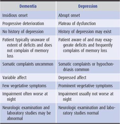
Table 5-12. Dementia versus pseudodementia of depression: distinguishing features.
AMNESTIC SYNDROMES
A disorder of memory (amnestic syndrome) may occur as one feature of an acute confusional state or dementia, or as an isolated abnormality. The latter condition is discussed in this section.
Memory is a complex function that can be viewed as having different components. Declarative (conscious) memory includes working memory, which permits acute manipulation of newly presented information, as well as longer-term semantic (factual) and episodic (personal) memory. Nondeclarative (unconscious) memory includes procedural memory required to carry out well-learned and seemingly automatic tasks, and emotional memory that attaches affective significance to objects or events.
Memory can also be seen as comprising the phases of registration, storage, and retrieval of information. Autopsy and imaging studies suggest that the hippocampus, parahippocampal region of the medial temporal lobe, and neocortical association areas are important in memory processing. Bilateral damage to these regions results in impairment of short-term memory, which is manifested clinically by the inability to form new memories. Long-term memory, which involves retrieval of previously learned information, is relatively preserved, perhaps because well-established memories are stored diffusely in the cerebral cortex. Some patients with amnestic syndromes may attempt to fill in gaps in memory with false recollections (confabulation), which can take the form of elaborate contrivances or of genuine memories misplaced in time. The longest-standing and most deeply ingrained memories, however, such as one’s own name, are almost always spared in organic memory disturbances. In contrast, such personal memories may be prominently or exclusively impaired in dissociative (psychogenic) amnesia.
The cellular basis of memory is poorly understood, but repetitive neuronal firing produces lasting pre- and post-synaptic changes (plasticity) that facilitate neurotransmission at hippocampal synapses (long-term potentiation). These changes appear to involve the release of glutamate, which stimulates the entry of calcium into postsynaptic neurons, and the production of retrograde signals (eg, nitric oxide) that act on presynaptic nerve terminals to increase transmitter release upon subsequent firing.
ACUTE AMNESIA
HEAD TRAUMA
Head injuries resulting in loss of consciousness are invariably associated with an amnestic syndrome. Patients seen shortly after such an injury exhibit a confusional state in which they are unable to incorporate new memories (anterograde, or posttraumatic amnesia; Figure 5-16), although they may behave in an apparently normal automatic fashion. In addition, retrograde amnesia is present, covering a variable period prior to the trauma. Features characteristic of transient global amnesia (see later) may be seen.

Figure 5-16. Retrograde and anterograde amnesia in posttraumatic memory disorders. Head trauma may produce transient coma, followed by a confusional state during which the patient is unable to form new memories. With recovery, this ability is restored, but there is persistent amnesia for the period of coma and confusion (anterograde amnesia) and for a variable period preceding the trauma (retrograde amnesia); the latter deficit may improve with time.
As full consciousness returns, the ability to form new memories is restored. Events occurring in the confusional interval tend to be permanently lost to memory, however. Exceptions are islands of memory for a lucid interval between trauma and unconsciousness, or for periods of lesser impairment in the course of a fluctuating posttraumatic confusional state. The period of retrograde amnesia begins to shrink, with the most remote memories being the first to return. The severity of the injury tends to correlate with the duration of confusion and with the extent of permanent retrograde and posttraumatic amnesia.
HYPOXIA OR ISCHEMIA
Because of the selective vulnerability of pyramidal neurons in the Sommer sector (h1 sector of Scholz) of the hippocampus, conditions resulting in cerebral hypoxia or ischemia, such as cardiac arrest or carbon monoxide poisoning, can produce amnestic syndromes. Amnesia tends to occur in patients in whom coma has lasted at least 12 hours. There is severe impairment of the ability to incorporate new memories, with relative preservation of registration and remote memory; patients typically appear to have an isolated disorder of short-term memory. A period of retrograde amnesia preceding the insult may occur. Patients exhibit a lack of concern about their impairment and sometimes confabulate. Amnesia after cardiac arrest may be the sole manifestation of neurologic dysfunction, or it may coexist with other cerebral watershed syndromes, such as bibrachial paresis, cortical blindness, or visual agnosia (Chapter 13). Recovery often occurs within several days, although deficits may persist.
Amnestic syndromes from carbon monoxide poisoning are frequently associated with affective disturbances. Other abnormalities include focal cortical and extrapyramidal dysfunction. Acute carbon monoxide poisoning is suggested by cherry-red coloration of the skin and mucous membranes, elevated carboxyhemoglobin levels, or cardiac arrhythmia. The CT brain scan may show lucencies in the basal ganglia and dentate nuclei. Treatment consists of the administration of normo- or hyperbaric oxygen.
BILATERAL POSTERIOR CEREBRAL ARTERY OCCLUSION
The posterior cerebral artery supplies the medial temporal lobe, thalamus, posterior internal capsule, and occipital cortex (Figure 5-17). Ischemia or infarction in this territory, typically when bilateral, may produce a transient or permanent amnestic syndrome. Emboli in the vertebrobasilar system (Chapter 13) are frequent causes of such disorders.

Figure 5-17. T1-weighted MRI in a patient with an old left posterior cerebral artery occlusion, showing tissue loss in the medial temporal (small arrows) and occipital (large arrows) lobes and associated dilation of the temporal and occipital horns of the lateral ventricle. (Courtesy of A. Gean.)
The amnestic syndrome is usually associated with unilateral or bilateral hemianopia and sometimes with visual agnosia, alexia without agraphia, anomia, sensory disturbances, or signs of upper midbrain dysfunction (especially impaired pupillary light reflex). Recent memory tends to be selectively impaired, with relative preservation of remote memory and registration.
The CT scan shows lucencies, which may or may not be enhanced by use of contrast material, in any combination of the previously mentioned regions. Evaluation and treatment are described in Chapter 13.
TRANSIENT GLOBAL AMNESIA
Transient global amnesia is a syndrome of acute memory loss that tends to occur in middle-aged or elderly patients. Transient cerebral ischemia, spreading depression (waves of reduced electrical activity in the cerebral cortex), and physical or emotional stress have all been suggested as the cause. The disorder is recurrent in fewer than 10% of patients.
Transient global amnesia affects short-term memory and lasts for up to 24 (typically 6-10) hours. Patients appear agitated and perplexed and may repeatedly inquire about their whereabouts, the time, and the nature of what they are experiencing. Knowledge of personal identity is preserved, as are remote memories and registration. New memories cannot be formed, however, which accounts for the patient’s repetitive questions. Retrograde amnesia for a variable period preceding the episode may be present, but this period shrinks as the episode resolves. Associated symptoms may include headache, nausea, and dizziness.
The patient’s obvious concern about the condition distinguishes transient global amnesia from most other organically based amnestic syndromes and may give rise to the suspicion that amnesia is psychogenic. High-resolution MRI may show signal abnormalities affecting the CA1 field of hippocampus.
ALCOHOLIC BLACKOUTS
Short-term consumption of large amounts of ethanol by alcoholic or nonalcoholic individuals may lead to “blackouts”—transient amnestic episodes that are not caused by global confusion, seizures, head trauma, or the Wernicke-Korsakoff syndrome. These spells are characterized by an inability to form new memories, without impairment of long-term memory or immediate recall. Although the cause is unknown, alcoholic blackouts may result from ethanol-induced depression of synaptic (especially serotonin- or glutamate-mediated) neurotransmission. The disorder is self-limited, and no specific treatment is required, but reduction of the ethanol intake should be counseled, and thiamine should be given to treat possible Wernicke encephalopathy (Chapter 4).
WERNICKE ENCEPHALOPATHY
Wernicke encephalopathy is caused by thiamine deficiency and classically produces an acute confusional state, ataxia, and ophthalmoplegia. Amnesia may be the major or sole cognitive disturbance, however, especially after thiamine treatment is begun and other cognitive abnormalities improve. Because patients with Wernicke encephalopathy usually present with global confusion rather than isolated amnesia, the disorder is discussed more fully in Chapter 4.
DISSOCIATIVE (PSYCHOGENIC) AMNESIA
Amnesia may be a manifestation of a dissociative disorder or of malingering. In such patients a prior psychiatric history, additional psychiatric symptoms, or a precipitating emotional stress can often be identified. Dissociative amnesia is characterized by an isolated or a disproportionate loss of traumatic or stressful personal memories. Dissociative amnesia is usually localized in time to the immediate aftermath of a traumatic experience or selective for some but not other events during such a period. Less frequent patterns include systematized amnesia restricted to certain categories of information, continuous amnesia for events from some time in the past up to and including the present, and generalized amnesia. In some cases, patients may be unable to remember even their own name, an exceedingly rare finding in organic amnesia. Despite such disorientation to person, orientation to place and time may be preserved. In addition, recent memories may be less affected than remote memories, the reverse of the pattern customarily seen in amnesia is caused by organic disease. Examination under hypnosis or after administration of amobarbital sodium may be helpful in establishing that amnesia is of psychogenic origin.
CHRONIC AMNESIA
ALCOHOLIC KORSAKOFF AMNESTIC SYNDROME
The Korsakoff amnestic syndrome, which occurs in chronic alcoholism and other malnutrition states, is thought to be caused by thiamine deficiency. It is usually preceded by one or more episodes of Wernicke encephalopathy, but such a history may be lacking. The memory disorder may be related to bilateral degeneration of the dorsomedial thalamic nuclei.
An amnestic syndrome of variable severity follows recovery from Wernicke encephalopathy in approximately three-fourths of cases and is often associated with polyneuropathy and other residua such as nystagmus or gait ataxia. The essential defect is an inability to form new memories, resulting in significant impairment of short-term memory. Long-term memory is also frequently affected, although to a lesser extent. Registration is intact. Patients are typically apathetic and lack insight into their disorder. They may attempt to reassure the physician that no impairment exists and try to explain away their obvious inability to remember. Confabulation is often, but not invariably, a feature.
Korsakoff syndrome can be prevented or its severity decreased by prompt administration of thiamine to patients with Wernicke encephalopathy. Patients with established Korsakoff syndrome should also receive thiamine to prevent the progression of deficits, although existing deficits are unlikely to be reversed.
POSTENCEPHALITIC AMNESIA
Patients who recover from acute viral encephalitis (Chapter 4), particularly that caused by herpes simplex virus, may be left with a permanent and static amnestic syndrome. The syndrome is similar to that produced by chronic alcoholism in that an inability to form new memories is its outstanding feature. Remote memories are affected to a lesser extent than are recent ones, and registration is intact. Confabulation may occur. Often there is total amnesia for the period of the acute encephalitis.
Patients may also exhibit other symptoms of limbic system disease. These include docility, indifference, flatness of mood and affect, inappropriate jocularity and sexual allusions, hyperphagia, impotence, repetitive stereotyped motor activity, and the absence of goal-oriented activity. Complex partial seizures, with or without secondary generalization, may occur.
BRAIN TUMOR
Brain tumor is a rare cause of amnestic syndrome. Tumors that can present in this manner include those that are located in the third ventricle or that compress its floor or walls from without. The amnestic syndrome closely resembles Korsakoff syndrome and may be accompanied by lethargy, headache, endocrine disturbances, visual field deficits, or papilledema. Cranial irradiation or chemotherapy may also impair memory by inhibiting neurogenesis in the hippocampus.
The diagnosis of brain tumor is made by CT scan or MRI. Treatment consists of surgery or irradiation or both, depending on the type of tumor and its location.
PARANEOPLASTIC LIMBIC ENCEPHALITIS
An inflammatory and degenerative disorder of gray matter regions of the CNS can occur as a remote effect of systemic cancer. When limbic structures are primarily affected, an amnestic syndrome is a prominent clinical feature. The disorder is thought to be autoimmune in origin because, as in other paraneoplastic neurologic syndromes, antineuronal autoantibodies can be detected.
Paraneoplastic limbic encephalitis is most often associated with small-cell cancer of the lung, and symptoms typically precede diagnosis of the underlying cancer. Histopathologic findings include neuronal loss, reactive gliosis, microglial proliferation, and perivascular lymphocytic cuffing. Gray matter of the hippocampus, cingulum, piriform cortex, inferior frontal lobes, insula, and amygdala is characteristically affected. Symptoms develop over several weeks. The disorder is characterized by profound impairment of recent memory, corresponding to the inability to learn new material. Remote memory is less impaired, and registration is unaffected; confabulation occurs in some cases. Mood disorders, delusions, hallucinations, sleep disturbance, complex partial or generalized seizures, and dementia may develop. Depending on the extent to which gray matter regions outside the limbic system are involved, cerebellar, pyramidal, bulbar, and peripheral nerve disturbances may also occur.
The CSF may also show a modest mononuclear pleocytosis and mildly elevated protein. Diffuse slowing or bitemporal slow waves and spikes are sometimes seen on EEG. An MRI may reveal abnormal signal intensity in the medial temporal lobes (Figure 5-18). A majority of patients have increased CSF protein and mononuclear cell pleocytosis, as well as antineuronal antibodies in serum or CSF. Anti-Hu antibodies are most common and are usually associated with small-cell lung cancer, but a wide range of autoantibodies and underlying malignancies are reported.

Figure 5-18. FLAIR MRI with axial (A) and coronal (B) views in limbic encephalitis, showing increased signal (white arrows) in the medial temporal lobes.
The paraneoplastic amnestic syndrome can be static, progressive, or remitting. Excluding other, especially treatable, disorders (eg, herpes simplex virus encephalitis) is of primary importance. Korsakoff syndrome caused by thiamine deficiency should also be considered, because patients with cancer are susceptible to nutritional deficiency, and thiamine administration may prevent these symptoms from worsening. Treatment of paraneoplastic limbic encephalitis includes administration of corticosteroids and therapy directed at the associated malignancy.
REFERENCES
Dementia (General)
Brundin P, Melki R, Kopito R. Prion-like transmission of protein aggregates in neurodegenerative diseases. Nat Rev Mol Cell Biol. 2010;11:301-307.
Dickson DW. Neuropathology of non-Alzheimer degenerative disorders. Int J Clin Exp Pathol. 2010;3:1-23.
Frost B, Diamond MI. Prion-like mechanisms in neurodegenerative diseases. Nat Rev Neurosci. 2010;155-159.
Garre-Olmo J, Genís Batlle D, del Mar Fernández M, et al. Incidence and subtypes of early-onset dementia in a geographically defined general population. Neurology. 2010;75:1249-1255.
Geschwind MD, Shu H, Haman A, Sejvar JJ, Miller BL. Rapidly progressive dementia. Ann Neurol. 2008;64:97-108.
Goedert M, Clavaguera F, Tolnay M. The propagation of prion-like protein inclusions in neurodegenerative diseases. Trends Neurosci. 2010;33:317-325.
Kelley BJ, Boeve BF, Josephs KA. Young-onset dementia: demographic and etiologic characteristics of 235 patients. Arch Neurol. 2008;65:1502-1508.
Kordower JH, Chu Y, Hauser RA, Freeman TB, Olanow CW. Lewy body-like pathology in long-term embryonic nigral transplants in Parkinson’s disease. Nat Med. 2008;14:504-506.
Li J, Englund E, Holton JL, et al. Lewy bodies in grafted neurons in subjects with Parkinson’s disease suggest host-to-graft disease propagation. Nat Med. 2008;14: 501-503.
Mitchell SL, Teno JM, Kiely DK, et al. The clinical course of advanced dementia. N Engl J Med. 2009;361:1529-1528.
Morris M, Maeda S, Vossel K, Mucke L. The many faces of tau. Neuron. 2011;70:410-426.
Petersen RC. Mild cognitive impairment. N Engl J Med. 2011;364:2227-2234.
Rabinovici GD, Jagust WJ. Amyloid imaging in aging and dementia: Testing the amyloid hypothesis in vivo. Behav Neurol. 2009;21:117-128.
Savva GM, Wharton SB, Ince PG, Forster G, Matthews FE, Brayne C. Age, neuropathology, and dementia. N Engl J Med. 2009;360:2302-2309.
van der Flier WM, Scheltens P. Use of laboratory and imaging investigations in dementia. J Neurol Neurosurg Psychiatry. 2005;76(Suppl V):v45-v52.
Alzheimer Disease
Anonymous. Drugs for cognitive loss and dementia. Treat Guide Med Lett. 2010;8:19-24.
Ballard C, Gauthier S, Corbett A, Brayne C, Aarsland D, Jones E. Alzheimer’s disease. Lancet. 2011;377:1019-1031.
Bettens K, Sleegers K, Van Broeckhoven C. Current status on Alzheimer disease molecular genetics: from past, to present, to future. Hum Mol Genet. 2010;19:R4-R11.
Blennow K, Hampel H, Weiner M, Zetterberg H. Cerebrospinal fluid and plasma biomarkers in Alzheimer disease. Nat Rev Neurol. 2010;6:131-144.
Braak H, Del Tredici K. Alzheimer’s pathogenesis: is there neuron-to-neuron propagation? Acta Neuropathol. 2011;121:589-595.
Caselli RJ, Dueck AC, Locke DE, et al. Longitudinal modeling of age-related memory decline and the APOE ε4 effect. N Engl J Med. 2009;361:255-263.
Daviglus ML, Bell CC, Berrettini W, et al. National Institutes of Health State-of-the-Science Conference statement: preventing Alzheimer disease and cognitive decline. Ann Int Med. 2010;153:176-181.
De Strooper B. Proteases and proteolysis in Alzheimer disease: a multifactorial view in the disease process. Physiol Rev. 2010;90:465-494.
Dolan D, Troncoso J, Resnick SM, Crain BJ, Zonderman AB, O’Brien RJ. Age, Alzheimer’s disease and dementia in the Baltimore longitudinal study of ageing. Brain. 2010;133:2225-2231.
Duyckaerts C, Delatour B, Potier MC. Classification and basic pathology of Alzheimer disease. Acta Neuropathol. 2009;118:5-36.
Ewers M, Sperling RA, Klunk WE, Weiner MW, Hampel H. Neuroimaging markers for the prediction and early diagnosis of Alzheimer’s disease dementia. Trends Neurosci. 2011;34: 430-442.
Green RC, Roberts JS, Cupples LA, et al. Disclosure of APOE genotype for risk of Alzheimer’s disease. N Engl J Med. 2009;361:245-254.
Holtzman DM, Morris JC, Goate AM. Alzheimer’s disease: the challenge of the second century. Science Transl Med. 2011;3:77sr1.
Ittner LM, Gotz J. Amyloid-β and tau—a toxic pas de deux in Alzheimer’s disease. Nat Rev Neurosci. 2011;12:67-72.
Jalbert JJ, Daiello LA, Lapane KL. Dementia of the Alzheimer type. Epidemiol Rev. 2008;30:15-34.
Nelson PT, Head E, Schmitt FA, et al. Alzheimer’s disease is not “brain aging”: neuropathological, genetic, and epidemiological human studies. Acta Neuropathol. 2011;121:571-587.
Querfurth HW, LaFerla FM. Alzheimer’s disease. N Engl J Med. 2010;362:329-344.
Rabinovici GD, Jagust WJ. Amyloid imaging in aging and dementia: testing the amyloid hypothesis in vivo. Behav Neurol. 2009;21:117-128.
Salloway S, Correia S. Alzheimer disease: time to improve its diagnosis and treatment. Cleve Clin J Med. 2009;76:49-58.
Scarmeas N, Honig LS, Choi H, et al. Seizures in Alzheimer disease: who, when, and how common? Arch Neurol. 2009; 66:992-997.
Smith EE, Greenberg SM. β-Amyloid, blood vessels, and brain function. Stroke. 2009;40:2601-2606.
Williamson J, Goldman J, Marder KS. Genetic aspects of Alzheimer disease. Neurologist. 2009;15:80-86.
Frontotemporal Dementia
Arvanitakis Z. Update on frontotemporal dementia. Neurologist. 2010;16:16-22.
Josephs KA. Frontotemporal dementia and related disorders: deciphering the enigma. Ann Neurol. 2008;64:4-14.
Kaye ED, Petrovic-Poljak A, Verhoeff NP, Freedman M. Frontotemporal dementia and pharmacologic interventions. J Neuropsychiatry Clin Neurosci. 2010;22:19-29.
Rabinovici GD, Miller BL. Frontotemporal lobar degeneration: epidemiology, pathophysiology, diagnosis and management. CNS Drugs. 2010;24:375-398.
Seelaar H, Rohrer JD, Pijnenburg YA, Fox NC, van Swieten JC. Clinical, genetic and pathological heterogeneity of frontotemporal dementia: a review. J Neurol Neurosurg Psychiatry. 2011;82:476-486.
Snowden JS, Hu Q, Rollinson S, et al. The most common type of FTLD-FUS (aFTLD-U) is associated with a distinct clinical form of frontotemporal dementia but is not related to mutations in the FUS gene. Acta Neuropathol. 2011;122:99-110.
Van der Zee J, Sleegers K, Van Broeckhoven C. The Alzheimer-frontotemporal lobar degeneration spectrum. Neurology. 2008;71:1191-1197.
Yu C-E, Bird TD, Bekris LM, et al. The spectrum of mutations in progranulin. A collaborative study screening 545 cases of neurodegeneration. Arch Neurol. 2010;67:161-170.
Lewy Body Disease
Johansen KK, White LR, Sando SB, Aasly JO. Biomarkers: Parkinson disease with dementia and dementia with Lewy bodies. Parkinsonism Relat Disord. 2010;16:307-315.
Kovari E, Horvath J, Bouras C. Neuropathology of Lewy body disorders. Brain Res Bull. 2009;80:203-210.
Sonnen JA, Postupna N, Larson EB, et al. Pathologic correlates of dementia in individuals with Lewy body disease. Brain Pathol. 2010;20:654-659.
Corticobasal Degeneration
Ling H, O’Sullivan SS, Holton JL, et al. Does corticobasal degeneration exist? A clinicopathological re-evaluation. Brain. 2010; 133:2045-2057.
Wadia PM, Lang AE. The many faces of corticobasal degeneration. Parkinsonism Relat Disord. 2007;13(Suppl 3):S336-S340.
Huntington Disease
Roos RA. Huntington’s disease: a clinical review. Orphanet J Rare Dis. 2010;5:40.
Progressive Supranuclear Palsy
Lubarsky M, Juncos JL. Progressive supranuclear palsy: a current review. Neurologist. 2008;14:79-88.
Stamelou M, de Silva R, Arias-Carrión O, et al. Rational therapeutic approaches to progressive supranuclear palsy. Brain. 2010;133:1578-1590.
Prion Disease
Brown K, Mastrianni JK. The prion diseases. J Geriat Psychiatr Neurol. 2010;23:277-298.
Colby DW, Prusiner SB. Prions. Cold Spring Harb Perspect Biol. 2011;3:a006833.
Vascular Dementia
Dolan H, Crain B, Troncoso J, Resnick SM, Zonderman AB, O’Brien RJ. Atherosclerosis, dementia, and Alzheimer disease in the Baltimore Longitudinal Study of Aging cohort. Ann Neurol. 2010;68:231-240.
Jellinger KA. Morphologic diagnosis of “vascular dementia”—a critical update. J Neurol Sci. 2008;270:1-12.
Lee JH, Kim SH, Kim GH, et al. Identification of pure subcortical vascular dementia using 11C-Pittsburgh compound B. Neurology. 2011;77:18-25.
Nagata K, Saito H, Ueno T, et al. Clinical diagnosis of vascular dementia. J Neurol Sci. 2007;257:44-48.
Schneck MJ. Vascular dementia. Top Stroke Rehabil. 2008;15:22-26.
Strozyk D, Dickson DW, Lipton RB, et al. Contribution of vascular pathology to the clinical expression of dementia. Neurobiol Aging. 2010;31:1710-1720.
Tomimoto H. Subcortical vascular dementia. Neurosci Res. 2011; 71:193-199.
Normal-Pressure Hydrocephalus
Shprecher D, Schwalb J, Kurlan R. Normal pressure hydrocephalus: diagnosis and treatment. Curr Neurol Neurosci Rep. 2008;8:371-376.
Brain Tumor
Janelsins MC, Kohli S, Mohile SG, Usuki K, Ahles TA, Morrow GR. An update on cancer- and chemotherapy-related cognitive dysfunction: current status. Semin Oncol. 2011;38: 431-438.
Chronic Traumatic Encephalopathy
McKee AC, Cantu RC, Nowinski CJ, et al. Chronic traumatic encephalopathy in athletes: progressive tauopathy after repetitive head injury. J Neuropathol Exp Neurol. 2009;68:709-735.
Infection
Hellwig K, Gold R. Progressive multifocal leukoencephalopathy and natalizumab. J Neurol. 2011;258:1920-1928.
Luo W, Ouyang Z, Xu H, Chen J, Ding M, Zhang B. The clinical analysis of general paresis with 5 cases. J Neuropsychiatry Clin Neurosci. 2008;20:490-493.
Mateen FJ, Muralidharan R, Carone M, et al. Progressive multi-focal leukoencephalopathy in transplant recipients. Ann Neurol. 2011;70:305-322.
McArthur JC, Steiner J, Sacktor N, Nath A. Human immunodeficiency virus-associated neurocognitive disorders mind the gap. Ann Neurol. 2010;67:699-714.
Metabolic Disorders
Langan RC, Zawistoski KJ. Update on vitamin B12 deficiency. Am Fam Physician. 2011;83:1425-1430.
Roberts EA, Schilsky ML. Diagnosis and treatment of Wilson disease: an update. Hepatology. 2008;47:2089-2111.
Pseudodementia
Saez-Fonseca JA, Lee L, Walker Z. Long-term outcome of depressive pseudodementia in the elderly. J Affect Disord. 2007;101:123-129.
Amnestic Syndromes & Memory (General)
Blundon JA, Zakharenko SS. Dissecting the components of long-term potentiation. Neuroscientist. 2008;14:598-608.
Dickerson BC, Eichenbaum H. The episodic memory system: neurocircuitry and disorders. Neuropsychopharmacology. 2010; 35:86-104.
Morgado-Bernal I. Learning and memory consolidation: linking molecular and behavioral data. Neuroscience. 2011;176:12-19.
Mozzachiodi R, Byrne JH. More than synaptic plasticity: role of nonsynaptic plasticity in learning and memory. Trends Neurosci. 2010;33:17-26.
Peng S, Zhang Y, Zhang J, Wang H, Ren B. Glutamate receptors and signal transduction in learning and memory. Mol Biol Rep. 2011;38:453-460.
Perouansky M, Pearce RA. How we recall (or don’t): the hippocampal memory machine and anesthetic amnesia. Can J Anesth. 2011;58:157-166.
Hypoxia or Ischemia
Weaver LK. Clinical practice. Carbon monoxide poisoning. N Engl J Med. 2009;360:1217-1225.
Bilateral Posterior Cerebral Artery Occlusion
Szabo K, Förster A, Jäger T, et al. Hippocampal lesion patterns in acute posterior cerebral artery stroke: clinical and MRI findings. Stroke. 2009;40:2042-2045.
Transient Global Amnesia
Bartsch T, Schönfeld R, Müller FJ, et al. Focal lesions of human hippocampal CA1 neurons in transient global amnesia impair place memory. Science. 2010;328:1412-1415.
Alcoholic Blackouts
Lee H, Roh S, Kim DJ. Alcohol-induced blackout. Int J Environ Res Public Health. 2009;6:2783-2792.
Wernicke Encephalopathy
Galvin R, Bråthen G, Ivashynka A, Hillbom M, Tanasescu R, Leone MA. EFNS guidelines for diagnosis, therapy and prevention of Wernicke encephalopathy. Eur J Neurol. 2010; 17:1408-1418.
Dissociative (Psychogenic) Amnesia
Schmidtke K, Pohlmann S, Metternich B. The syndrome of functional memory disorder: definition, etiology, and natural course. Am J Geriatr Psychiatry. 2008;16:981-988.
Alcoholic Korsakoff Amnestic Syndrome
Kopelman MD, Thomson AD, Guerrini I, Marshall EJ. The Korsakoff syndrome: clinical aspects, psychology and treatment. Alcohol. 2009;44:148-154.
Postencephalitic Amnesia
Grydeland H, Walhovd KB, Westlye LT, et al. Amnesia following herpes simplex encephalitis: diffusion-tensor imaging uncovers reduced integrity of normal-appearing white matter. Radiology. 2010;257:774-781.
Brain Tumor
Monje ML, Vogel H, Masek M, Ligon KL, Fisher PG, Palmer TD, et al. Impaired human hippocampal neurogenesis after treatment for central nervous system malignancies. Ann Neurol. 2007;62:515-520.
Paraneoplastic Limbic Encephalitis
Foster AR, Caplan JP. Paraneoplastic limbic encephalitis. Psychosomatics. 2009;50:108-113.