
Approach to Diagnosis
Functional Anatomy Of The Visual System
Visual Input
Peripheral Visual Pathways
Central Visual Pathways
Vascular Supply Of Visual Pathways
Functional Anatomy of the Ocular Motor System
Extraocular Muscles
Cranial Nerves
Cranial Nerve Nuclei
Supranuclear Control of Eye Movements
History
Nature of Complaint
Temporal Pattern of Symptoms
Associated Neurologic Abnormalities
Medical History
Neuro-Ophthalmic Examination
Visual Acuity
Visual Fields
Ophthalmoscopy
Pupils
Optokinetic Response
Eyelids
Exophthalmos
Eye Movements
Disorders of the Visual System
Monocular Disorders
Transient Monocular Blindness
Optic Neuritis
Nonarteritic Anterior Ischemic Optic Neuropathy
Giant Cell (Temporal) Arteritis
Binocular Disorders
Papilledema
Chiasmal Lesions
Retrochiasmal Lesions
Disorders of Eye Movement
Gaze Palsies
Hemispheric Lesions
Midbrain Lesions
Pontine Lesions
Internuclear Ophthalmoplegia
Ocular Nerve Palsies
Oculomotor (III) Nerve Lesions
Trochlear (IV) Nerve Lesions
Abducens (VI) Nerve Lesions
Diabetic Ophthalmoplegias
Painful Ophthalmoplegias
Myasthenia Gravis
Ocular Myopathies
References
APPROACH TO DIAGNOSIS
Disorders that affect the ocular muscles, ocular motor (III, IV, and VI) cranial nerves, or visual or ocular motor pathways in the brain produce a wide variety of neuro-ophthalmic disturbances. Because the anatomic pathways of the visual and ocular motor systems traverse major portions of the brainstem and cerebral hemispheres, neuro-ophthalmic symptoms and signs are often of great value in the anatomic localization and diagnosis of neurologic disease.
FUNCTIONAL ANATOMY OF THE VISUAL SYSTEM
VISUAL INPUT
Visual information enters the nervous system when light, refracted and focused by the lens, creates a visual image on the retina at the posterior pole of the eye (Figure 7-1). The action of the lens causes this image to be reversed in the horizontal and vertical planes. Thus the superior portion of the visual image falls on the inferior retina and vice versa, and the temporal (lateral) and nasal (medial) fields are likewise reversed (Figure 7-2). The center of the visual field is focused at the fovea, where the retina’s perceptual sensitivity is greatest. Within the retina, photoreceptor cells (rods and cones) transduce incident light into neuronal impulses, which are transmitted by retinal neurons to the optic (II) nerve. At this and all other levels of the visual system, the topographic relations of the visual field are preserved.

Figure 7-1. Representation of the visual field at the level of the retina. The point of fixation is focused on the fovea, the physiologic blind spot on the optic disk, the temporal half of the visual field on the nasal side of the retina, and the nasal half of the visual field on the temporal side of the retina.
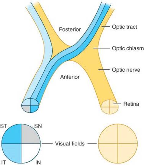
Figure 7-2. Representation of the visual field at the level of the optic nerve, chiasm, and tract. Quadrants of the visual field are designated ST (superior temporal), IT (inferior temporal), SN (superior nasal), and IN (inferior nasal).
PERIPHERAL VISUAL PATHWAYS
Each optic nerve contains fibers from one eye, but as shown in Figure 7-2, the nasal (medial) fibers, conveying information from the temporal (lateral) visual fields, cross in the optic chiasm. As a result, each optic tract contains fibers not from one eye, but from one-half of the visual field. Because of this arrangement, prechiasmal lesions affect vision in the ipsilateral eye and retrochiasmal lesions produce defects in the contralateral half of the visual field of both eyes.
CENTRAL VISUAL PATHWAYS
The optic tracts terminate in the lateral geniculate nuclei, where their neurons synapse on neurons that project through the optic radiations to the primary visual or calcarine cortex (area 17), located near the posterior poles of the occipital lobes, and visual association areas (areas 18 and 19). Here, too, the visual image is represented in such a way that its topographic organization is preserved (Figure 7-3). The central region of the visual field (macula) is represented in the most posterior portion of the visual cortex, whereas the inferior and superior parts of the field (superior and inferior retina) are represented above and below the calcarine fissure, respectively.
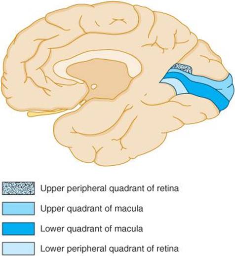
Figure 7-3. Representation of the visual field at the level of the primary visual cortex, midsagittal view, shows the medial surface of the right occipital lobe, which receives visual input from the left side of the visual field of both eyes.
VASCULAR SUPPLY OF VISUAL PATHWAYS
The vascular supply of the visual system is derived from the ophthalmic, middle cerebral, and posterior cerebral arteries (Figure 7-4); thus ischemia or infarction in the territory of any of these vessels can produce visual field defects.

Figure 7-4. Arterial supply of the visual system, viewed from below.
1. Retina—The retina is supplied by the central retinal artery, a branch of the ophthalmic artery, which is itself a branch of the internal carotid artery. Because the central retinal artery subsequently divides into superior and inferior retinal branches, vascular disease of the retina tends to produce altitudinal (ie, superior or inferior) visual field deficits.
2. Optic nerve—The optic nerve receives arterial blood primarily from the ophthalmic artery and its branches.
3. Optic radiations—As the optic radiations course backward toward the visual cortex, they are supplied by branches of the middle cerebral artery. Ischemia or infarction in the distribution of the middle cerebral artery may thus cause loss of vision in the contralateral visual field.
4. Primary visual cortex—The principal source of arterial blood for the primary visual cortex is the posterior cerebral artery. Occlusion of one posterior cerebral artery produces blindness in the contralateral visual field, although the dual (middle and posterior cerebral) arterial supply to the macular region of the visual cortex may spare central (macular) vision. Because the posterior cerebral arteries arise together from the basilar artery, occlusion at the tip of the basilar artery can cause bilateral occipital infarction and complete cortical blindness—although, in some cases, macular vision is spared.
FUNCTIONAL ANATOMY OF THE OCULAR MOTOR SYSTEM
EXTRAOCULAR MUSCLES
Movement of the eyes is accomplished by the action of six muscles attached to each globe (Figure 7-5). These muscles act to move the eye into each of six cardinal positions of gaze. Equal and opposed actions of these six muscles in the resting state place the eye in mid- or primary position, that is, looking directly forward. When the function of one extraocular muscle is disrupted, the eye is unable to move in the direction of action of the affected muscle (ophthalmoplegia) and may deviate in the opposite direction because of the unopposed action of other extraocular muscles. When the eyes are thus misaligned, visual images of perceived objects fall on a different region of each retina, creating the illusion of double vision, or diplopia.
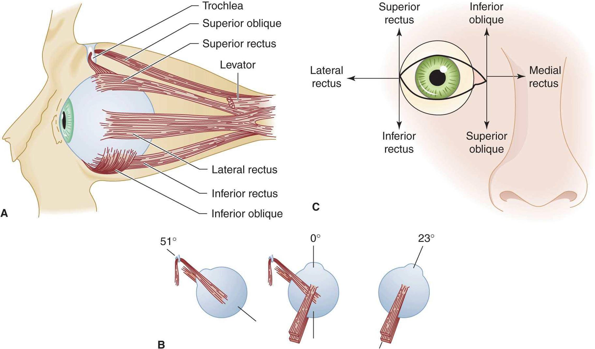
Figure 7-5. Anatomy and function of extraocular muscles. A: Extraocular muscles in the right orbit (lateral view). B: An illustration of the right eye viewed from above in the primary position (center figure) showing the angle of attachment of the superior and inferior rectus muscles and the superior and inferior oblique muscles. With the eye directed to the right, the superior and inferior rectus muscles can now be examined as pure elevators and depressors of the globe (right image), and with the eye deviated to the left, the oblique muscles can now be examined as pure elevators and depressors of the globe as illustrated in part C. C: The six cardinal positions of gaze for testing eye movement. The eye is adducted by the medial rectus and abducted by the lateral rectus. The adducted eye is elevated by the inferior oblique and depressed by the superior oblique; the abducted eye is elevated by the superior rectus and depressed by the inferior rectus.
CRANIAL NERVES
The extraocular muscles are innervated by the oculomotor (III), trochlear (IV), and abducens (VI) nerves. Because of this differential innervation of the ocular muscles, the pattern of their involvement in pathologic conditions can help to distinguish a disorder of the ocular muscles per se from a disorder that affects a cranial nerve. Cranial nerves that control eye movement traverse long distances to pass from the brainstem to the eye; they are thereby rendered vulnerable to injury by a variety of pathologic processes.
1. Oculomotor (III) nerve—The oculomotor nerve supplies the medial rectus, superior and inferior rectus, and inferior oblique muscles and carries fibers to the levator palpebrae (which raises the eyelid). It also supplies the parasympathetic fibers responsible for pupillary constriction. With a complete nerve III lesion, the eye is partially abducted, and there is an inability to adduct, elevate, and depress the eye; the eyelid droops (ptosis), and the pupil is nonreactive.
2. Trochlear (IV) nerve—The trochlear nerve innervates the superior oblique muscle. Lesions of this nerve result in defective depression of the adducted eye.
3. Abducens (VI) nerve—Lesions of the abducens nerve cause lateral rectus palsy, with impaired abduction of the affected eye.
CRANIAL NERVE NUCLEI
The nuclei of the oculomotor (III) and trochlear (IV) nerves are located in the dorsal midbrain, ventral to the cerebral aqueduct (of Sylvius), whereas the abducens (VI) nerve nucleus occupies a similarly dorsal and periventricular position in the pons. Lesions involving these nuclei give rise to clinical abnormalities similar to those produced by involvement of their respective cranial nerves; in some cases, nuclear and nerve lesions can be distinguished.
1. Oculomotor (III) nerve nucleus—Although each oculomotor nerve supplies muscles of the ipsilateral eye only, fibers to the superior rectus originate in the contralateral oculomotor nerve nucleus, and the levator palpebrae receives bilateral nuclear innervation. Thus ophthalmoplegia affecting only one eye with ipsilateral ptosis or superior rectus palsy suggests oculomotor nerve disease, whereas ophthalmoplegia with by bilateral ptosis or a contralateral superior rectus palsy is probably due to a nuclear lesion.
2. Trochlear (IV) nerve nucleus—It is not possible to distinguish clinically between lesions of the trochlear nerve (see earlier) and those of its nucleus.
3. Abducens (VI) nerve nucleus—In disorders affecting the abducens nerve nucleus rather than the nerve itself, lateral rectus paresis is often associated with facial weakness, paresis of ipsilateral conjugate gaze, or a depressed level of consciousness. This is because of the proximity of the abducens nerve nucleus to the facial (VII) nerve fasciculus, pontine lateral gaze center, and ascending reticular activating system, respectively. When a Horner syndrome (miosis of the pupil, ptosis, and sometimes segmental anhidrosis) accompanies an abducens nerve palsy, this indicates that the lesion is in the cavernous sinus.
SUPRANUCLEAR CONTROL OF EYE MOVEMENTS
Supranuclear control of eye movements enables the two eyes to act in concert to produce version (conjugate gaze) or vergence (convergence and divergence) movements.
1. Brainstem gaze centers—Centers that control horizontal (lateral) and vertical gaze are located in the pons and in the pretectal region of the midbrain, respectively, and receive descending inputs from the cerebral cortex that allow voluntary control of gaze (Figure 7-6). Each lateral gaze center, located in the paramedian pontine reticular formation (PPRF) adjacent to the abducens nerve nucleus, mediates ipsilateral, conjugate, horizontal gaze via its connections to the ipsilateral abducens (VI) and contralateral oculomotor (III) nerve nucleus. A lesion in the pons affecting the PPRF therefore produces a gaze preference away from the side of the lesion and toward the side of an associated hemiparesis, if present. Disorders of vertical gaze, typically impaired upgaze, may result from mass lesions that exert downward pressure on the dorsal mid-brain, such as pineal tumors (Parinaud syndrome).
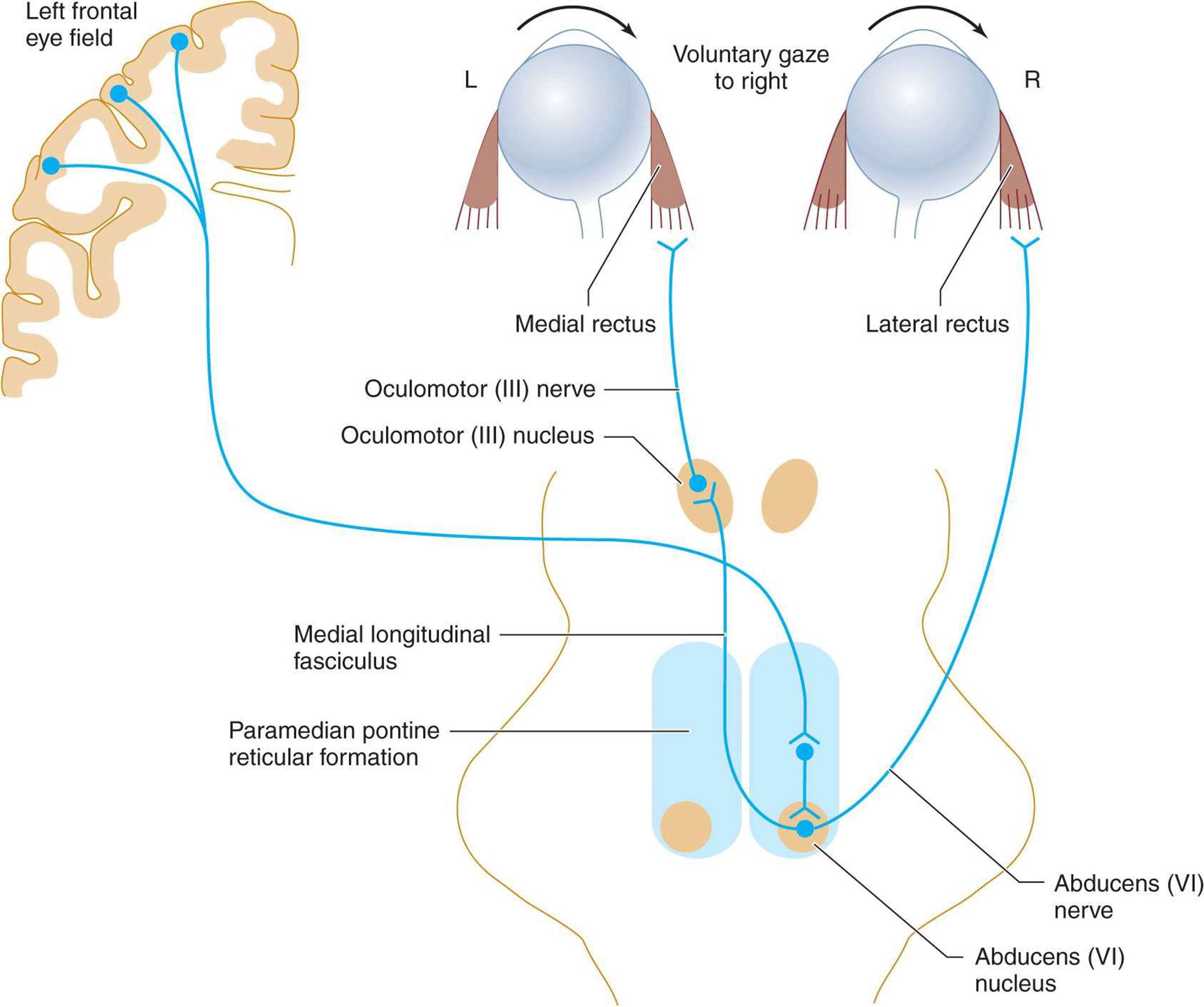
Figure 7-6. Neuronal pathways involved in horizontal gaze.
2. Cortical input—The PPRF receives cortical input from the contralateral frontal lobe, which regulates rapid eye movements (saccades), and from the ipsilateral parietooccipital lobe, which regulates slow eye movements (pursuits). Therefore, a destructive lesion affecting the frontal cortex interferes with the mechanism for contralateral horizontal gaze and may result in a gaze preference toward the side of the lesion (and away from the side of associated hemiparesis). By contrast, an irritative (seizure) focus in the frontal lobe may cause gaze away from the side of the focus.
HISTORY
NATURE OF COMPLAINT
The first step in evaluating a neuro-ophthalmic disorder is to obtain a clear description of the complaint. Patients may only complain of vague symptoms, such as blurred vision, which provide little diagnostic information. Therefore, it is important to determine whether the patient means to describe decreased visual acuity in one or both eyes, loss of vision in part of the visual field, diplopia, an unstable visual image, pain in or about the eye, or some other problem.
TEMPORAL PATTERN OF SYMPTOMS
Once the nature of the complaint has been established, inquiries regarding its temporal pattern can provide clues to the underlying pathologic process.
1. Sudden onset—Vascular disorders that affect the eye or its connections in the brain tend to produce symptoms of sudden onset.
2. Slow onset—With inflammatory or neoplastic disease, symptoms usually evolve over a longer period.
3. Transient, recurrent symptoms—Symptoms that are transient and recurrent suggest a select group of pathologic processes, including intermittent ischemia, multiple sclerosis, and myasthenia gravis.
ASSOCIATED NEUROLOGIC ABNORMALITIES
The nature of any associated neurologic abnormalities, such as impaired facial sensation, weakness, ataxia, or aphasia, can be valuable in localizing the anatomic site of involvement.
MEDICAL HISTORY
The history should be scrutinized for conditions that predispose the patient to neuro-ophthalmic problems.
1. Multiple sclerosis often involves the optic nerve or brainstem, leading to a variety of neuro-ophthalmic disorders. A history of disturbances that also involve other parts of the central nervous system should suggest this diagnosis.
2. Atherosclerosis, hypertension, and diabetes can be complicated by vascular disorders of the eye, cranial nerves, or visual or ocular motor pathways in the brain.
3. Endocrine disorders (eg, hyperthyroidism) can cause ocular myopathy.
4. Connective tissue disease and systemic cancer can affect the visual and ocular motor systems at a variety of sites in the brain or subarachnoid space.
5. Nutritional deficiencies may present with neuroophthalmic symptoms, as in the amblyopia (decreased visual acuity) associated with malnutrition and the ophthalmoplegia of Wernicke encephalopathy.
6. Drugs (eg, ethambutol, isoniazid, digitalis, clioquinol) are known to be toxic to the visual system, and others (sedative drugs, anticonvulsants) commonly produce ocular motor disorders.
NEURO-OPHTHALMIC EXAMINATION
VISUAL ACUITY
Assessment
To identify neuro-ophthalmic problems, vision should be tested under conditions that eliminate refractive errors. Therefore, patients who wear glasses should be examined while wearing them (a pinhole can be substituted if the corrective lenses usually worn are not available at the time of testing). Visual acuity must be assessed for each eye separately. Distant vision is tested using a Snellen eye chart, with the patient 6 m (20 ft) away. Near vision is tested with a Rosenbaum pocket eye chart held approximately 36 cm (14 in) from the patient. In each case, the smallest line of print that can be read is noted.
Recording
Visual acuity is expressed as a fraction (eg, 20/20, 20/40, 20/200). The numerator is the distance (in feet) from the test figures at which the examination is performed, and the denominator is the distance (in feet) at which figures of a given size can be correctly identified by persons with normal vision. For example, if a patient standing 20 ft away from the eye chart is unable to identify figures that can normally be seen from that distance but can identify the larger figures that would be visible 40 ft away with normal acuity, the visual acuity is recorded as 20/40. If the patient can read most of a given line but makes some errors, acuity may be recorded as 20/40–1, for example, indicating that all but one letter on the 20/40 line were correctly identified. When visual acuity is markedly reduced, it can still be quantified, though less precisely, in terms of the distance at which the patient can count fingers (CF), discern hand movement (HM), or perceive light. If an eye is totally blind, the examination will reveal no light perception (NLP).
Red–Green Color Vision
Red–green color vision is often disproportionately impaired in optic nerve lesions and can be tested with colored objects such as pens or hatpins or with color vision plates.
VISUAL FIELDS
Evaluating the visual fields can be a lengthy and tedious procedure if conducted in an undirected fashion. Familiarity with the common types of visual field defects is important if testing is to be reasonably rapid and yield useful information. The most common visual field abnormalities are illustrated in Figure 7-7.

Figure 7-7. Common visual field defects and their anatomic bases. 1. Central scotoma caused by inflammation of the optic disk (optic neuritis) or optic nerve (retrobulbar neuritis). 2. Total blindness of the right eye from a complete lesion of the right optic nerve. 3. Bitemporal hemianopia caused by pressure exerted on the optic chiasm by a pituitary tumor. 4. Right nasal hemianopia caused by a perichiasmal lesion (eg, calcified internal carotid artery). 5. Right homonymous hemianopia from a lesion of the left optic tract. 6. Right homonymous superior quadrantanopia caused by partial involvement of the optic radiation by a lesion in the left temporal lobe (Meyer loop). 7. Right homonymous inferior quadrantanopia caused by partial involvement of the optic radiation by a lesion in the left parietal lobe. 8. Right homonymous hemianopia from a complete lesion of the left optic radiation. (A similar defect may also result from lesion 9.) 9. Right homonymous hemianopia (with macular sparing) resulting from posterior cerebral artery occlusion.
Extent of Visual Fields
The normal monocular visual field subtends an angle of approximately 160 degrees in the horizontal plane and approximately 135 degrees in the vertical plane. With binocular vision, the horizontal range of vision exceeds 180 degrees.
Physiologic Blind Spot
Within the normal field of each eye is a 5-degree blind spot, corresponding to the optic disk, which lacks receptor cells. The blind spot is located 15 degrees temporal to fixation in each eye.
Measurement Techniques
Like visual acuity, the visual field must be examined separately for each eye.
1. Confrontation (Figure 7-8) is the simplest method for visual field testing. The examiner stands at about arm’s length from the patient, with the eyes of both patient and examiner aligned in the horizontal plane. The eye not being tested is covered by the patient’s hand or an eye patch. The examiner closes the eye opposite the patient’s covered eye, and the patient is instructed to fix on the examiner’s open eye. Now the monocular fields of patient and examiner are superimposed, which allows comparison of the patient’s field with the examiner’s presumably normal field. The examiner uses the index fingers of either hand to locate the boundaries of the patient’s field, moving them slowly inward from the periphery in all directions until the patient detects them. The boundaries are then defined more carefully by determining the farthest peripheral sites at which the patient can detect slight movements of the fingertips or the white head of a pin. The patient’s blind spot can be located in the region of the examiner’s own blind spot, and the sizes of these spots can be compared using a pin with a white head as the target. The procedure is then repeated for the other eye.

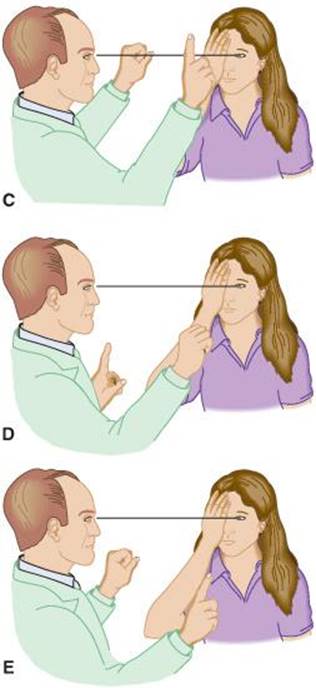
Figure 7-8. Confrontation testing of the visual field. A: The left eye of the patient and the right eye of the examiner are aligned. B: Testing the superior nasal quadrant. C: Testing the superior temporal quadrant. D: Testing the inferior nasal quadrant. The procedure is then repeated for the patient’s other eye. E: Testing the inferior temporal quadrant.
2. Subtle field defects may be detected by asking the patient to compare the brightness of colored objects presented at different sites in the field or by measuring the fields using a pin with a red head as the target.
3. In young children, the fields may be assessed by standing behind the child and bringing an attention-getting object, such as a toy, forward around the child’s head in various directions until it is first noticed.
4. A gross indication of visual field abnormalities may be obtained in obtunded patients by determining whether they blink in response to a visual threat—typically the examiner’s finger—brought toward the patient’s eye in various regions of the field.
5. Although many visual field deficits are detectable by these screening procedures, more precise mapping of the fields requires the use of standard tangent screen testing or automated perimetry techniques.
OPHTHALMOSCOPY
Preparation of the Patient
Ophthalmoscopy of the optic fundus is particularly important for evaluating disorders that affect the retina or optic disk and examining patients with a suspected increase in intracranial pressure. The examination should be conducted in a dark room so that the pupils are dilated; in some patients, the use of mydriatic (sympathomimetic or anticholinergic) eye drops is necessary. In the latter case, visual acuity and pupillary reflexes should always be assessed before instilling the drops. Mydriatic agents should be avoided in patients with untreated closed angle glaucoma, and in situations—such as impending or ongoing transtentorial herniation—in which the state of pupillary reactivity is an important guide to management.
Examination of the Fundus
Familiarity with the normal appearance of the optic fundus (Figure 1-10) is necessary if abnormalities are to be appreciated.
A. Optic Disk
1. Normal appearance—The optic disk is usually easily recognizable as a yellowish, slightly oval structure situated nasally at the posterior pole of the eye. The temporal side of the disk is often paler than the nasal side. The disk margins should be sharply demarcated, though the nasal edge is commonly somewhat less distinct than the temporal edge. The disk is normally in the same plane as the surrounding retina. Blood vessels crossing the border of the optic disk are distinct and become obscured when the disk swells.
2. Optic disk swelling—The ophthalmoscopic abnormality that most often requires prompt interpretation and attention is optic nerve swelling due to papilledema. This condition implies increased intracranial pressure and must be differentiated from swelling that is due to other causes, such as local inflammation (papillitis) and ischemic optic neuropathy. In making this distinction, it is helpful to bear in mind that papilledema is almost always bilateral, does not typically impair vision (except for enlargement of the blind spot), and is not associated with eye pain. Papilledema can be simulated by disk abnormalities such as drusen (colloid or hyaline bodies).
Increased intracranial pressure is thought to cause papilledema by blocking axonal transport in the optic nerve. Because the optic nerve sheath communicates with the subarachnoid space, disorders associated with increased intracranial pressure that also obstruct the subarachnoid space, such as meningitis, are less likely to cause papilledema. The ophthalmoscopic changes in papilledema typically develop over days or weeks but may become apparent within hours after a sudden increase in intracranial pressure—as, for example, after intracranial hemorrhage. In early papilledema (Figure 1-11), the retinal veins appear engorged and spontaneous venous pulsations are absent. The disk may be hyperemic, and linear hemorrhages may be seen at its borders. The disk margins become blurred, with the temporal edge last to be affected. In fully developed papilledema, the optic disk is elevated above the plane of the retina, and blood vessels crossing the border of the disk become obscured.
3. Optic disk pallor—Optic disk pallor with impaired visual acuity, visual field defects, or loss of pupillary reactivity is associated with a wide variety of disorders that affect the optic nerve, including inflammatory conditions, nutritional deficiencies, and degenerative diseases. A pale optic disk with normal visual function can also occur as a congenital variant, and an optic disk may appear artificially pale if a cataract has been removed.
B. Arteries & Veins
The caliber of the retinal arteries and veins should be observed where they arise from the disk and pass over its edges onto the retina. Features to note include whether these vessels are easily visible throughout their course, whether they appear engorged, and whether spontaneous venous pulsations (which indicate normal intracranial pressure) are present. The remainder of the visible retina is inspected, noting the presence of hemorrhages, exudates, or other abnormalities.
C. Macula
The macula, a somewhat paler area than the rest of the retina, is located approximately two disk diameters temporal to the temporal margin of the optic disk. It can be visualized quickly by having the patient look at the light from the ophthalmoscope. Ophthalmoscopic examination of the macula can reveal abnormalities related to visual loss from age-related macular degeneration, macular holes, or hereditary cerebromacular degenerations.
PUPILS
Size
The size and reactivity of the pupils reflect the integrity of neuronal pathways from the optic nerve to the midbrain (Figure 7-9). The normal pupil is round, regular, and centered within the iris; its size varies with age and with the intensity of ambient light. In a brightly illuminated room, the diameter of normal pupils is approximately 3 mm in adults, smaller in the elderly, and ≥5 mm in children. Pupil size may be asymmetric in up to 20% of people (physiologic anisocoria), but the difference is ≤1 mm. Symmetrically rapid constriction of the pupils in bright light indicates that pupillary function is normal and excludes oculomotor (III) nerve compression.
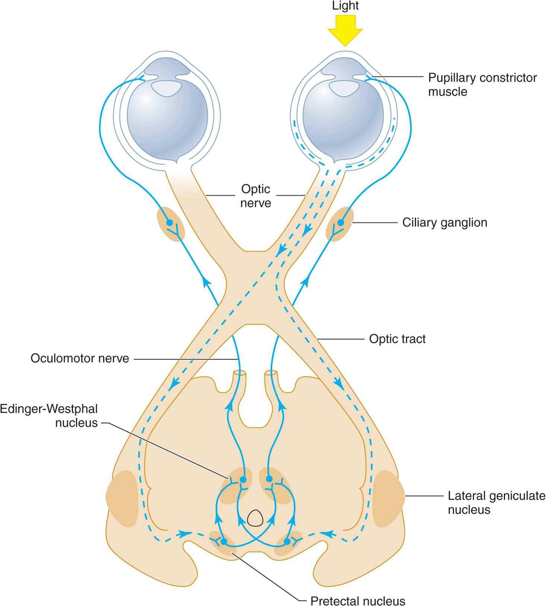
Figure 7-9. Anatomic basis of the pupillary light reflex. The afferent visual pathways from the retina to the pretectal nuclei of the midbrain are represented by dashed lines and the efferent pupilloconstrictor pathways from the mid-brain to the pupillary constrictor muscles by solid lines. Note that illumination of one eye results in bilateral pupillary constriction.
Reaction to Light
Direct (ipsilateral) and consensual (contralateral) pupillary constriction in response to a bright light shone in one eye demonstrates the integrity of the pathways shown in Figure 7-9. Normally, the direct response to light is slightly brisker and more pronounced than the consensual response.
Reaction to Accommodation
When the eyes converge to focus on a nearer object (accommodation), the pupils normally constrict. This reaction is tested by having the patient focus alternately on a distant object and a finger held just in front of his or her nose.
Pupillary Abnormalities
A. Nonreactive Pupils
Unilateral disorders of pupillary constriction are seen with local disease of the iris (trauma, iritis, glaucoma), oculomotor (III) nerve compression (tumor, aneurysm), administration of a mydriatic agent, and optic nerve disorders (optic neuritis, multiple sclerosis).
B. Light-Near Dissociation
Impaired pupillary reactivity to light with preserved constriction during accommodation (light-near dissociation) is usually bilateral and may result from neurosyphilis, diabetes, optic nerve disorders, and tumors compressing the midbrain tectum.
C. Argyll Robertson Pupils
These pupils are small, poorly reactive to light, often irregular in shape, and frequently unequal in size; they show light-near dissociation. Neurosyphilis is the classic cause, but other lesions in the region of the Edinger-Westphal nucleus (eg, multiple sclerosis) are now more common (Table 7-1).

Table 7-1. Common pupillary abnormalities.
D. Tonic Pupil
The tonic (Adie) pupil (Table 7-1) is larger than the contralateral unaffected pupil and reacts sluggishly to changes in both illumination and accommodation. Because the tonic pupil does eventually react, anisocoria becomes less marked during the examination. This abnormality is most commonly a manifestation of a benign, often familial disorder that frequently affects young women (Holmes-Adie syndrome) and may be associated with depressed deep tendon reflexes (especially in the legs), segmental anhidrosis (localized lack of sweating), orthostatic hypotension, or cardiovascular autonomic instability. The condition may be bilateral. The pupillary abnormality may be caused by degeneration of the ciliary ganglion, followed by aberrant reinnervation of the pupilloconstrictor muscles.
E. Horner Syndrome
Horner syndrome (Tables 7-1 and 7-2) results from a lesion of the central or peripheral sympathetic nervous system and consists of a small (miotic) pupil associated with mild ptosis (Figure 7-10) and sometimes loss of sweating (anhidrosis).
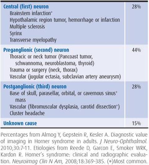
Table 7-2. Causes of Horner syndrome.


Figure 7-10. A: Right Horner syndrome after direct carotid puncture for arterial injection. B: Oculosympathetic pathway involved in Horner syndrome. This three-neuron pathway projects from the hypothalamus to the intermediolateral column of the spinal cord, then to the superior cervical (sympathetic) ganglion, and finally to the pupil, smooth muscle of the eyelid, and sweat glands of the forehead and face.
1. Oculosympathetic pathways—The sympathetic pathway controlling pupillary dilation consists of an uncrossed three-neuron arc: hypothalamic neurons, which descend through the brainstem to the intermediolateral column of the spinal cord at the T1 level, preganglionic sympathetic neurons projecting from the spinal cord to the superior cervical ganglion, and postganglionic sympathetic neuronsthat originate in the superior cervical ganglion, ascend in the neck along the internal carotid artery, and enter the orbit with the first (ophthalmic) division of the trigeminal (V) nerve. Horner syndrome is caused by interruption of these pathways at any site.
2. Clinical features—The lesions and resulting pupillary abnormality are usually unilateral. The pupil diameter on the involved side is typically reduced by 0.5 to 1 mm compared with the normal side. This inequality is most marked in dim illumination. The pupillary abnormality is accompanied by mild to moderate ptosis (see later) of the upper lid (as opposed to the pronounced ptosis seen with oculomotor nerve lesions), often associated with elevation of the lower lid (lower lid ptosis). When Horner syndrome has been present since infancy, the ipsilateral iris is lighter and blue (heterochromia iridis).
Deficits in the pattern of sweating, which are most prominent in acute-onset Horner syndrome, can help localize the lesion. If sweating is decreased on an entire half of the body and face, the lesion is in the central nervous system. Cervical lesions produce anhidrosis of the face, neck, and arm only. Sweating is unimpaired if the lesion is above the bifurcation of the carotid artery. The differential diagnosis of Horner syndrome is presented in Table 7-2.
F. Relative Afferent Pupillary Defect (Marcus Gunn Pupil)
In this condition, one pupil constricts less markedly in response to direct illumination than to illumination of the contralateral pupil, whereas normally the direct response is greater than the consensual response. The abnormality is detected by rapidly moving a bright flashlight back and forth between the eyes while continuously observing the suspect pupil (Gunn pupillary test). Relative afferent pupillary defect is commonly associated with disorders of the ipsilateral optic nerve, which interrupt the afferent limb and affect the pupillary light reflex (Figure 7-9). Such disorders also commonly impair vision (especially color vision) in the involved eye.
OPTOKINETIC RESPONSE
Optokinetic nystagmus consists of eye movements elicited by sequential fixation on a series of targets passing in front of a patient’s eyes, such as telephone poles seen from a moving train. For clinical testing, a revolving drum with vertical stripes or a vertically striped strip of cloth is moved across the visual field to generate these movements. Testing produces a slow following phase in the direction of the target’s movement, followed by a rapid return jerk in the opposite direction. The slow (pursuit) phase tests parieto-occipital and the rapid (saccadic) movement tests frontal lobe function in the hemisphere toward which the stimulus is moved. The presence of an optokinetic response reflects the ability to perceive movement or contour and is sometimes useful for documenting visual perception in newborns or in psychogenic blindness. Visual acuity required to produce the optokinetic response is minimal, however (20/400, or finger counting at 3-5 ft). Unilateral impairment of the optokinetic response may be found when targets are moved toward the side of a parietal lobe lesion.
EYELIDS
The eyelids (palpebrae) should be examined with the patient’s eyes open. The distance between the upper and lower lids (interpalpebral fissure) is usually approximately 10 mm and equal in both eyes, though physiologic asymmetries do occur. The position of the inferior margin of the upper lid relative to the superior border of the iris should be noted in order to detect drooping (ptosis) or abnormal elevation of the eyelid (lid retraction). The upper lid normally covers 1 to 2 mm of the iris.
Unilateral ptosis is seen with paralysis of the levator palpebrae muscle itself, lesions of the oculomotor (III) nerve or its superior branch, and Horner syndrome. In the last condition, ptosis is customarily associated with miosis and may be momentarily overcome by effortful eye opening.
Bilateral ptosis suggests a disorder affecting the oculomotor (III) nerve nucleus, neuromuscular junction (eg, myasthenia gravis), or muscle (eg, myotonic, ocular, or oculopharyngeal dystrophy).
Lid retraction (abnormal elevation of the upper lid) is seen in hyperthyroidism and in Parinaud syndrome caused by tumors in the pineal region.
EXOPHTHALMOS
Abnormal protrusion of the eye from the orbit (exophthalmos or proptosis) is best detected by standing behind the seated patient and looking down at his or her eyes from above. Causes include hyperthyroidism (Graves disease, Figure 7-11), orbital tumor or pseudotumor, and carotid artery–cavernous sinus fistula. A bruit may be audible on auscultation over the proptotic eye in patients with carotid artery–cavernous sinus fistula or other vascular anomalies.

Figure 7-11. Graves ophthalmopathy in a 41-year-old woman. Note the protrusion of the right globe.
EYE MOVEMENTS
Ocular Excursion & Gaze
Ocular palsies and gaze palsies are detected by having the patient gaze in each of the six cardinal positions (Figure 7-5). If voluntary eye movement is impaired or the patient is unable to cooperate with the examination (eg, is comatose), reflex eye movements can be induced by doll’s head (oculocephalic) or cold-water caloric (oculovestibular) testing (Chapter 3). If limitations in movement are observed, the muscles involved are noted, and the nature of the abnormality is determined according to the following scheme.
A. Ocular Palsy
Weakness of one or more eye muscles results from nuclear or infranuclear (nerve, neuromuscular junction, or muscle) lesions. An ocular palsy cannot be overcome by caloric stimulation of reflex eye movement. Nerve lesions produce distinctive patterns of ocular muscle involvement.
1. Oculomotor (III) nerve palsy—A complete lesion of the oculomotor nerve produces closure of the affected eye because of impaired levator function. Passively elevating the paralyzed lid (Figure 7-12) shows the involved eye to be laterally deviated because of the unopposed action of the lateral rectus muscle, which is not innervated by the oculomotor nerve. Diplopia is present in all directions of gaze except for lateral gaze toward the side of involvement. Pupil function may be normal (pupillary sparing) or impaired.

Figure 7-12. Clinical findings with oculomotor (III) nerve lesion. With the ptotic lid passively elevated, the affected (right) eye is abducted; it cannot adduct. On attempted downgaze, the unaffected superior oblique muscle, which is innervated by the trochlear (IV) nerve, causes the eye to turn inward.
2. Trochlear (IV) nerve palsy—With trochlear nerve lesions, which paralyze the superior oblique muscle, the involved eye is elevated during primary (forward) gaze; the extent of elevation increases during adduction and decreases during abduction. Elevation is greatest when the head is tilted toward the side of the involved eye and abolished by tilt in the opposite direction (Bielschowsky head-tilt test; Figure 7-13). Diplopia is most pronounced when the patient looks downward with the affected eye adducted (as in looking at the end of one’s nose). Spontaneous head tilting, intended to decrease or correct the diplopia, is present in approximately one-half of patients with unilateral palsies and in a greater number with bilateral palsies.
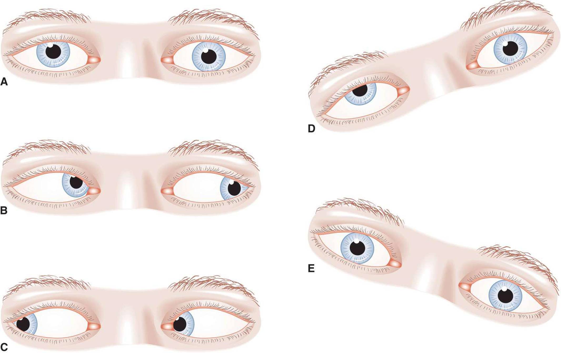
Figure 7-13. Clinical findings with trochlear (IV) nerve lesion. The affected (right) eye is elevated on forward gaze (A). The extent of elevation is increased with adduction (B) and decreased with abduction (C). Elevation increases with head tilting to the affected side (D) and decreases with head tilting in the opposite direction (E).
3. Abducens (VI) nerve palsy—An abducens nerve lesion causes paralysis of the lateral rectus muscle, resulting in adduction of the involved eye at rest and failure of attempted abduction (Figure 7-14). Diplopia occurs on lateral gaze to the side of the affected eye.

Figure 7-14. Clinical findings with abducens (VI) nerve lesion. The affected (right) eye is adducted at rest (A) and cannot be abducted (B).
B. Gaze Palsy
Gaze palsy is the diminished ability of a pair of yoked muscles (muscles that operate in concert to move the two eyes in a given direction) to move the eyes in voluntary gaze; it is caused by supranuclear lesions in the brainstem or cerebral hemisphere. Gaze palsy, unlike ocular palsies, affects both eyes and usually can be overcome by caloric stimulation. Its pathophysiology and causes are discussed more fully in the section that follows on binocular disorders of eye movement. Mild impairment of upgaze is not uncommon in normal elderly subjects.
C. Internuclear Ophthalmoplegia
Internuclear ophthalmoplegia (INO) results from a lesion of the medial longitudinal fasciculus, an ascending pathway in the brainstem that projects from the abducens (VI) to the contralateral oculomotor (III) nerve nucleus. As a consequence, the actions of the abducens (VI) and oculomotor (III) nerves during voluntary gaze or caloric-induced movement are uncoupled. Excursion of the abducting eye is full, but adduction of the contralateral eye is impaired (Figure 7-15). INO cannot be overcome by caloric stimulation, but can be distinguished from oculomotor (III) nerve palsy by noting preserved adduction with convergence. It is usually caused by multiple sclerosis or brainstem stroke.

Figure 7-15. Eye movements in internuclear ophthalmoplegia (INO) resulting from a bilateral lesion of the medial longitudinal fasciculus.
D. One-and-a-Half Syndrome
A pontine lesion affecting both the medial longitudinal fasciculus and the ipsilateral paramedian pontine reticular formation (lateral gaze center) produces a syndrome that combines internuclear ophthalmoplegia with an inability to gaze toward the side of the lesion (Figure 7-16). The ipsilateral eye is immobile in the horizontal plane, and movement of the contralateral eye is restricted to abduction, which may be associated with nystagmus. The causes include pontine infarct, multiple sclerosis, and pontine hemorrhage.

Figure 7-16. One-and-a-half syndrome. This results from a pontine lesion (shaded area) involving the paramedian pontine reticular formation (lateral gaze center) and medial longitudinal fasciculus, and sometimes also the abducens (VI) nucleus, and affecting the neuronal pathways indicated by dotted lines. Attempted gaze away from the lesion (A) activates the uninvolved right lateral gaze center and abducens (VI) nucleus; the right lateral rectus muscle contracts, and the right eye abducts normally. Involvement of the medial longitudinal fasciculus interrupts the pathway to the left oculomotor (III) nucleus, and the left eye fails to adduct. On attempted gaze toward the lesion (B), the left lateral gaze center cannot be activated, and the eyes do not move. There is a complete (bilateral) gaze palsy in one direction (toward the lesion) and one-half (unilateral) gaze palsy in the other direction (away from the lesion), accounting for the name of the syndrome.
Diplopia Testing
When the patient complains of double vision (diplopia), testing of eye movements should be used to determine its anatomic basis. The patient is asked to fix his or her vision on an object, such as a flashlight, in each of the six cardinal positions of gaze (Figure 7-5). With normal conjugate gaze, light from the flashlight falls at the same spot on both corneas; a lack of such congruency confirms that gaze is disconjugate. When the patient notes diplopia in a given direction of gaze, each eye should be covered in turn and the patient asked to report which of the two images disappears. The image displaced farther in the direction of gaze is always referable to the weak eye, because that image will not fall on the fovea. A variation of this procedure is the red glass test, in which one eye is covered with translucent red glass, plastic, or cellophane; this allows the eye responsible for each image to be identified.
Nystagmus
Nystagmus is rhythmic oscillation of the eyes. Pendular nystagmus, which usually has its onset in infancy, occurs with equal velocity in both directions. Jerk nystagmus is characterized by a slow phase of movement followed by a fast phase in the opposite direction; the direction of jerk nystagmus is specified by stating the direction of the fast phase (eg, leftward-beating nystagmus). Jerk nystagmus usually increases in amplitude with gaze in the direction of the fast phase (the Alexander law).
Nystagmus can occur at the extremes of voluntary gaze in normal subjects and is also a normal component of both the optokinetic response and the response to caloric stimulation of reflex eye movements. In other settings, however, it may be due to anticonvulsant or sedative drugs or disease in the peripheral vestibular apparatus, central vestibular pathways, or cerebellum.
To detect nystagmus, the eyes should be observed in the primary position and in each of the cardinal positions of gaze (Figure 7-5). Nystagmus is described in terms of the position of gaze in which it occurs, its direction and amplitude, precipitating factors such as changes in head position, and associated symptoms, such as vertigo.
Many forms of nystagmus and related ocular oscillations have been described, but two syndromes of acquired pathologic jerk nystagmus are by far the most common.
A. Gaze-Evoked Nystagmus
Gaze-evoked nystagmus occurs when the patient attempts to gaze in one or more directions away from the primary position. The fast phase of nystagmus is in the direction of gaze. Nystagmus evoked by gaze in a single direction is a common sign of early or mild residual ocular palsy. Multidirectional gaze-evoked nystagmus is most often an effect of anticonvulsant or sedative drugs, but can also result from cerebellar or central vestibular dysfunction.
B. Vestibular Nystagmus
Peripheral vestibular nystagmus caused by a lesion of the peripheral vestibular apparatus is characteristically unidirectional, horizontal or both horizontal and rotatory, and associated with severe vertigo. Its amplitude increases with gaze toward the fast phase. In contrast, central vestibular nystagmus may be bidirectional and purely horizontal, vertical, or rotatory, and the accompanying vertigo is typically mild. Positional nystagmus, elicited by changes in head position, can occur with either peripheral or central vestibular lesions. The most helpful distinguishing features are the presence of hearing loss or tinnitus with peripheral lesions and of corticospinal tract or additional cranial nerve abnormalities with central lesions.
DISORDERS OF THE VISUAL SYSTEM
MONOCULAR DISORDERS
Common syndromes of monocular visual loss include two reversible and two irreversible disorders. Transient monocular blindness caused by optic nerve or retinal ischemia is sudden in onset and resolves rapidly. Optic neuritisproduces subacute, painful, unilateral visual loss with partial resolution. Less reversible visual loss of sudden onset occurs in idiopathic ischemic optic neuropathy and in giant cell(temporal) arteritis.
TRANSIENT MONOCULAR BLINDNESS
This condition, sometimes called amaurosis fugax, is characterized by unilateral transient diminution or loss of vision that develops over seconds, remains maximal for 1 to 5 minutes, and resolves over 10 to 20 minutes. Although the cause of these episodes often remains uncertain, the presence of what appears to be embolic material in retinal arteries during episodes suggests that these emboli are the cause. Their major site of origin appears to be atherosclerotic lesions at the carotid bifurcation, but mitral valve prolapse and other cardiac sources of emboli can produce a similar syndrome. The risk for subsequent hemispheric infarction is increased (14% within 7 years) in patients with a history of transient monocular blindness, but is only approximately one-half of that in patients with hemispheric transient ischemic attacks (TIAs).
Diagnostic evaluation and treatment of patients with transient monocular blindness resemble that recommended for patients with hemispheric TIAs (Chapter 13). In patients with both transient monocular blindness or TIAs and high-grade (>70%) stenosis of the carotid artery at angiography (but not with transient monocular blindness alone), treatment with a combination of aspirin plus surgical removal of plaque (endarterectomy) is superior to aspirin alone.
OPTIC NEURITIS
Inflammation of the optic nerve produces the syndrome of optic neuritis. The most common cause is demyelination (acute demyelinating optic neuritis). Less common causes include parameningeal, meningeal, or intraocular inflammation associated with viral infections or postviral syndromes. Rare causes include toxins (eg, methanol, ethambutol), neurosyphilis, and vitamin B12 deficiency. Unilateral impairment of visual acuity occurs over hours to days, becoming maximal within 1 week. Visual loss is associated with headache, globe tenderness, or eye pain in more than 90% of patients; the pain is typically exacerbated by eye movement.
On visual field testing, there is usually a central scotoma (blind spot) associated with decreased visual acuity. Examination of the fundus shows unilateral disk swelling when the nerve head is involved, but is normal when the inflammatory process is posterior to the optic disk (retrobulbar neuritis), as is most common in demyelinating disease. The pupils are equal in size but show less pronounced constriction in response to illumination of the affected eye (relative afferent pupillary defect; discussed earlier).
Diffuse gadolinium enhancement of the optic nerve on MRI scans is typical of acute demyelinating optic neuritis, and T2-hyperintense lesions are also seen in the brain in 50% to 70% of these patients. With 10-year follow-up, approximately 55% of patients with one or more T2 lesions but only approximately 20% without T2 lesions develop multiple sclerosis.
In acute demyelinating optic neuritis, visual acuity usually but not invariably improves by 2 to 3 weeks, with eventual return to normal or near normal vision. Intravenous methylprednisolone, 1 g/d for 3 to 5 days, with or without an oral prednisone taper, from 1 mg/kg/d over 11 days, can hasten recovery but does not alter the final outcome or the likelihood of developing multiple sclerosis. Evolving data support immunomodulatory treatment for optic neuritis as presumed multiple sclerosis if demyelinating lesions are seen on a brain magnetic resonance imaging (MRI) scan (Figure 9-4).
NONARTERITIC ANTERIOR ISCHEMIC OPTIC NEUROPATHY
Idiopathic infarction of the anterior portion of the optic nerve is termed anterior ischemic optic neuropathy. It occurs after the age of 50 years. Visual loss is sudden in onset, usually painless, always monocular, and without premonitory ocular symptoms. The visual deficit is usually maximal at onset and frequently subtotal; an altitudinal (superior or inferior) field defect is typical. In some cases the course is stuttering or progressive.
Examination reveals ipsilateral disk swelling, often with peripapillary hemorrhages. In the absence of this finding, the diagnosis is tenuous, and other causes, such as a rapidly expanding intracranial mass or neoplastic meningitis, should be sought.
Although ischemic optic neuropathy is often assumed to be atherosclerotic in origin, there is no consistent association with other risk factors for cerebrovascular disease, such as hypertension, diabetes, or atherosclerotic carotid artery disease. Patients with anterior ischemic optic neuropathy have a structurally smaller than normal disk; 25% will go on to have the other eye affected within 2 to 4 years.
Attempts at treatment have been uniformly unsuccessful. As disk swelling resolves, ophthalmoscopic evaluation shows optic atrophy.
GIANT CELL (TEMPORAL) ARTERITIS
Arteritic infarction of the anterior portion of the optic nerve is the most devastating complication of giant cell, or temporal, arteritis. This disorder is usually accompanied by systemic symptoms such as fever, malaise, night sweats, weight loss, and headache (Chapter 6) and often by polymyalgia rheumatica (Chapter 9). Scalp tenderness and jaw claudication may occur. The visual loss is usually sudden and often total, but transient retinal ischemia, mimicking embolic events, may precede optic nerve infarction. On examination, the optic disk appears swollen and pale. The erythrocyte sedimentation rate and C-reactive protein are typically increased. Definitive diagnosis is by temporal artery biopsy.
Patients should be treated immediately with corticosteroids (methylprednisolone 1,000 mg/d intravenously for at least 3 days, followed by prednisone 60-80 mg/d orally) to protect what vision remains. Prednisone may be gradually reduced over many months while monitoring the erythrocyte sedimentation rate.
Because giant cell arteritis is treatable, it is most important to distinguish it from idiopathic or nonarteritic anterior ischemic optic neuropathy as the cause of monocular visual loss. Patients with giant cell arteritis tend to be older (aged 70-80 years) and may have premonitory symptoms. The most helpful differential features are the erythrocyte sedimentation rate, which is greater than 50 mm/h (Westergren) in most patients with giant cell arteritis, and elevated C-reactive protein.
BINOCULAR DISORDERS
PAPILLEDEMA
Papilledema is painless, passive bilateral disk swelling associated with increased intracranial pressure. Associated nonspecific symptoms of raised intracranial pressure include headache, nausea, vomiting, and diplopia from abducens (VI) nerve palsy.
The speed with which papilledema develops is dictated by the underlying cause. When intracranial pressure increases suddenly, as in subarachnoid or intracerebral hemorrhage, disk swelling may be seen within hours, but it most often evolves over days. Papilledema may require 2 to 3 months to resolve after restoration of normal intracranial pressure.
Funduscopic examination (Figure 1-11) reveals (in order of onset) blurring of the nerve fiber layer, absence of venous pulsations (signifying intracranial pressure greater than approximately 200 mm Hg), hemorrhages in the nerve fiber layer, elevation of the disk surface with blurring of the margins, and disk hyperemia.
Papilledema requires urgent evaluation to search for an intracranial mass and to exclude papillitis from meningeal carcinoma, sarcoidosis, or syphilis, which may produce a similar ophthalmoscopic appearance. If an intracranial mass lesion and other disorders associated with intracranial hypertension listed in Table 6-5 (eg, venous sinus thrombosis, polycythemia, endocrinopathy, hypervitaminosis A) are excluded by the history, examination, and computed tomography (CT) scanning or MRI, meningeal inflammation is excluded by CSF examination, and CSF pressure is elevated, a diagnosis of idiopathic intracranial hypertension (pseudotumor cerebri) is established by exclusion. The idiopathic form, which is the most common, occurs most often in obese women during the childbearing years. Although this disorder is usually self-limited, prolonged elevation of intracranial pressure with papilledema can lead to permanent visual loss.
Less common causes of papilledema include congenital cyanotic heart disease and disorders associated with increased cerebrospinal fluid (CSF) protein content, such as spinal cord tumor and idiopathic inflammatory polyneuropathy (Guillain-Barré syndrome).
CHIASMAL LESIONS
The major lesions that produce visual impairment at the level of the optic chiasm are tumors, especially those of pituitary origin. Other causes include trauma, multiple sclerosis, and berry aneurysms. The classic pattern of visual deficit caused by lesions of the optic chiasm is bitemporal hemianopia (Figure 7-7). Except in pituitary apoplexy due to acute intrapituitary hemorrhage, chiasmal visual loss is gradual in onset, and the resulting impairment in depth perception or in the lateral visual fields may not be noted for some time. Associated involvement of the oculomotor (III), trochlear (IV), trigeminal (V), or abducens (VI) nerve suggests tumor expansion laterally into the cavernous sinus. Nonophthalmic manifestations of pituitary tumors include headache, acromegaly, amenorrhea, galactorrhea, and Cushing syndrome.
Headache, endocrine abnormalities, and occasionally blurred or double vision may occur in patients with an enlarged sella turcica, but in whom neither tumor nor increased intracranial pressure is found. This empty sella syndrome is most common in women and occurs mainly between the fourth and seventh decades of life. Treatment is symptomatic.
RETROCHIASMAL LESIONS
Optic Tract & Lateral Geniculate Body
Lesions of the optic tract and lateral geniculate body are usually due to infarction. The resulting visual field abnormality is typically a noncongruous homonymous hemianopia; that is, the field defect is not the same in the two eyes. Associated hemisensory loss may occur with thalamic lesions.
Optic Radiations
Lesions of the optic radiations produce a congruous (bilaterally symmetric) homonymous hemianopia. Visual acuity is normal in the unaffected portion of the field. With lesions in the temporal lobe, where tumors are the most common cause, the field deficit is denser superiorly than inferiorly, resulting in a superior quadrantanopia (“pie in the sky” deficit; Figure 7-7).
Lesions affecting the optic radiations in the parietal-lobe may be due to tumor or vascular disease and are usually associated with contralateral weakness and sensory loss. A gaze preference is common in the acute phase, with the eyes conjugately deviated to the side of the parietal lesion. The visual field abnormality is either complete homonymous hemianopia or inferior quadrantanopia (Figure 7-7). The optokinetic response to a visual stimulus moved toward the side of the lesion is impaired, which is not the case with pure temporal or occipital lobe lesions.
Occipital Cortex
Lesions in the occipital cortex usually produce homonymous hemianopia affecting the contralateral visual field. The patient may be unaware of the visual deficit. Because the region of the occipital cortex in which the macula is represented is often supplied by branches of both the posterior and middle cerebral arteries (Figure 7-4), visual field abnormalities caused by vascular lesions in the occipital lobe may show sparing of macular vision (Figure 7-7). Macular sparing may also result from bilateral cortical representation of the macular region of the visual field.
By far the most common cause of visual impairment in the occipital lobe is infarction in the posterior cerebral artery territory. Occipital lobe arteriovenous malformations (AVMs), vertebral angiography, and watershed infarction after cardiac arrest are less common causes. Additional symptoms and signs of basilar artery ischemia may occur. Tumors and occipital lobe AVMs are often associated with unformed visual hallucinations that are typically unilateral, stationary or moving, and brief or flickering; they can be colored or not colored.
Bilateral occipital lobe involvement produces cortical blindness. Pupillary reactions are normal, and bilateral macular sparing may preserve central (tunnel) vision. With more extensive lesions, denial of blindness may occur (Anton syndrome).
DISORDERS OF EYE MOVEMENT
GAZE PALSIES
Lesions in the cortex or brainstem above the level of the oculomotor (III) nuclei may impair conjugate (yoked) movement of the eyes, producing gaze disorders. In milder gaze palsies, the eyes may move fully, but the speed or the amplitude of the fast eye movements is reduced.
HEMISPHERIC LESIONS
Acute hemispheric lesions produce tonic deviation of both eyes toward the side of the lesion and away from the side of the hemiparesis (Figure 7-17). This gaze deviation may last for several days in alert patients (somewhat longer in comatose patients). Seizure discharges involving the frontal gaze centers can also produce gaze deviation by driving the eyes away from the discharging focus. When the ipsilateral motor cortex is also involved, producing focal motor seizures, the patient gazes toward the side of the motor activity (Figure 7-17).
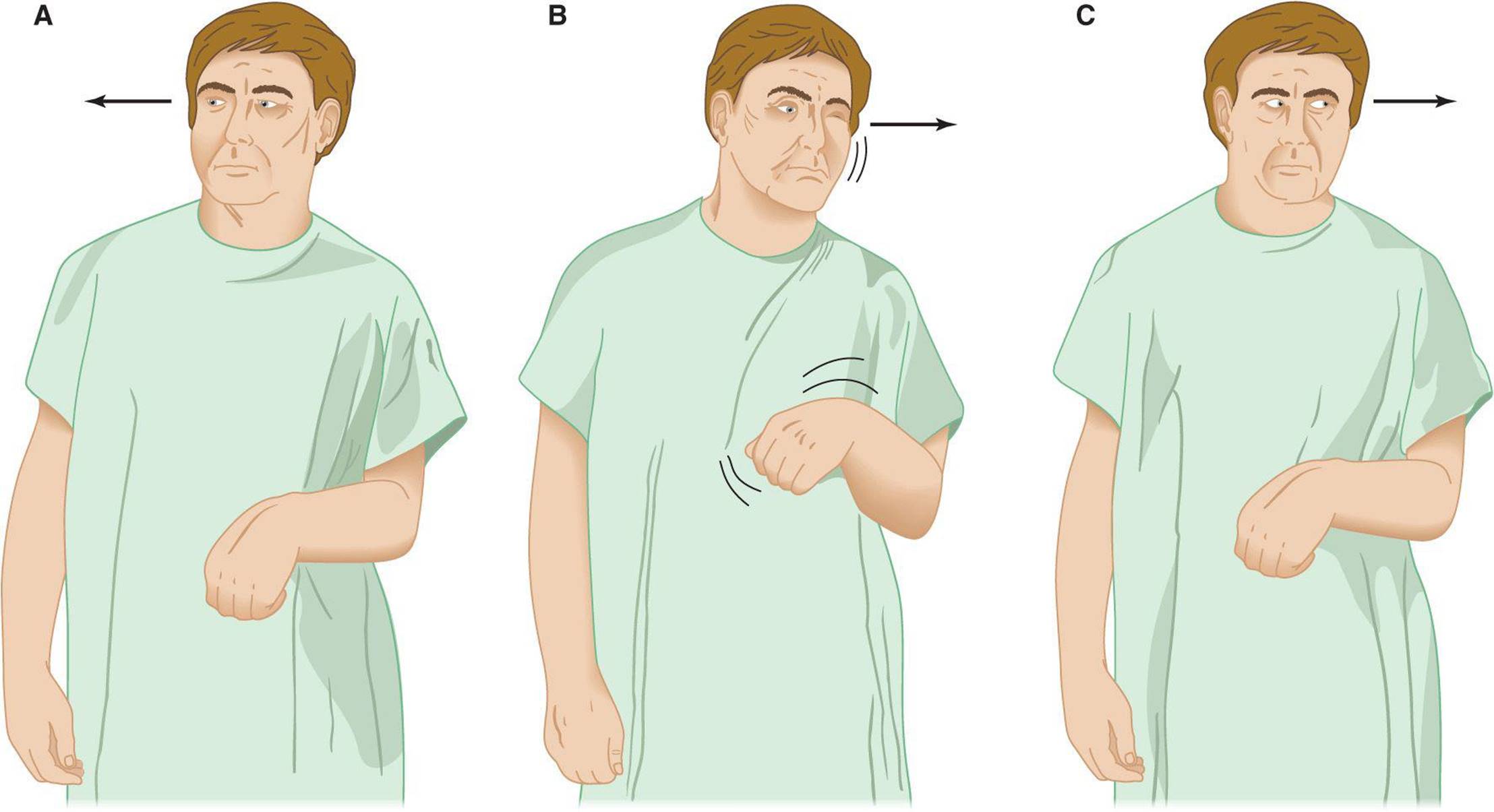
Figure 7-17. Disorders of gaze associated with hemispheric and brainstem lesions. A: Destructive lesion in the frontal lobe of the right cerebral hemisphere. B: Seizure arising from the frontal lobe of the right cerebral hemisphere. C: Destructive lesion in the right pons. Arrows indicate the direction of gaze preference (away from the hemiparetic side in A and toward the convulsing or hemiparetic side in B and C).
MIDBRAIN LESIONS
Lesions of the dorsal midbrain affect the center responsible for voluntary upward gaze and may therefore produce upgaze paralysis. In addition, all or some of the features of Parinaud syndrome may occur: preserved reflex vertical eye movements with the doll’s head maneuver or Bell phenomenon (elevation of the eye upon eyelid closure), nystagmus (especially on downward gaze and typically associated with retraction of the eyes), paralysis of accommodation, midsized pupils, and light-near dissociation.
PONTINE LESIONS
Brainstem lesions at the level of the pontine gaze centers produce disorders of conjugate horizontal gaze. Gaze palsies from pontine involvement (unlike those from hemispheric lesions) cause eye deviation toward, rather than away from, the side of the hemiparesis (Figure 7-17), because the corticobulbar pathways that regulate gaze have crossed at this level, but the descending motor pathways have not. Brainstem gaze palsies are characteristically far more resistant to attempts to move the eyes (via the doll’s head maneuver or caloric stimulation) than are hemispheric gaze pareses. In addition, gaze palsies from brainstem lesions are commonly associated with abducens (VI) nerve palsies because these lesions often also involve the abducens (VI) nerve nucleus.
INTERNUCLEAR OPHTHALMOPLEGIA
INO results from lesions of the medial longitudinal fasciculus between the midpons and the oculomotor nerve nucleus that disconnect the abducens (VI) nucleus from the contralateral oculomotor (III) nucleus (Figure 7-6). The site of the INO is named according to the side on which oculomotor (III) nerve function is impaired. There is a characteristic abnormality consisting of disconjugate gaze with impaired adduction and nystagmus of the abducting eye (Figure 7-15).
An INO usually implies intrinsic brainstem disease. The most common cause, especially in young adults and patients with bilateral involvement, is multiple sclerosis. In older patients and those with unilateral involvement, stroke is likely. These two causes encompass 80% or more of all cases. Rarer causes include brainstem encephalitis, intrinsic brainstem tumors, syringobulbia, sedative drug intoxication, and Wernicke encephalopathy. Because myasthenia gravis can closely mimic a lesion of the medial longitudinal fasciculus, myasthenia must be ruled out in patients with isolated INO.
OCULAR NERVE PALSIES
OCULOMOTOR (III) NERVE LESIONS
Lesions of the oculomotor (III) nerve can occur at any of several levels. The most common causes are listed in Table 7-3; oculomotor disorders resulting from diabetes are discussed separately later.
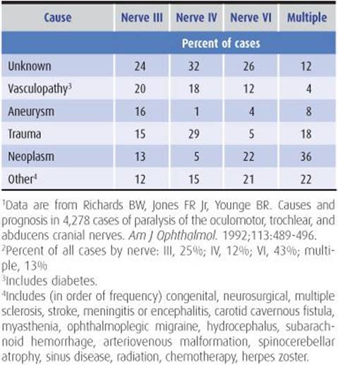
Table 7-3. Causes of oculomotor (III), trochlear (IV), and abducens (VI) nerve lesions.1,2
Brainstem
Within the brainstem, other neurologic signs permit localization of the lesion; associated contralateral hemiplegia (Weber syndrome) and contralateral ataxia (Benedikt syndrome) are the most common vascular syndromes.
Subarachnoid Space
As the oculomotor (III) nerve exits the brainstem in the interpeduncular space, it is susceptible to injury from trauma and from aneurysms of the posterior communicating artery. Such compressive lesions typically impair the pupillary light reflex, which tends to be spared with ischemic (eg, diabetic) lesions..
Cavernous Sinus
In the cavernous sinus (Figure 7-18), the oculomotor (III) nerve is usually involved together with the trochlear (IV) and abducens (VI) nerves and the first (V1) and sometimes the second (V2) division of the trigeminal nerve. Horner syndrome may occur. Oculomotor (III) nerve lesions in the cavernous sinus tend to produce partial deficits that may or may not spare the pupil.


Figure 7-18. Position of cranial nerves in the cavernous sinus and adjacent structures. A: Coronal view through the cavernous sinus, with the midline at left and the temporal lobe at right. B: Location of cranial nerves as they course anteriorly (left to right) in relation to the cavernous sinus, superior orbital fissure, and orbital apex. Note that a lesion in the cavernous sinus spares the optic (II) and mandibular (V3) nerves, a lesion in the superior orbital fissure additionally spares the maxillary (V2) nerve, and a lesion in the orbital apex spares both V2 and V3 but may involve II.
Orbit
Unlike cavernous sinus lesions, orbital lesions that affect the oculomotor (III) nerve are often associated with optic (II) nerve involvement and exophthalmos; however, disorders of the orbit and cavernous sinus may be clinically indistinguishable except by CT scanning or MRI.
TROCHLEAR (IV) NERVE LESIONS
Head trauma, often minor, is a common cause of an isolated trochlear (IV) nerve palsy (Table 7-3). Although trochlear palsies in middle-aged and elderly patients are frequently attributed to vascular disease or diabetes, they often occur without obvious cause. For patients with isolated trochlear (IV) nerve palsies without a history of trauma, and in whom diabetes, myasthenia, thyroid disease, and orbital mass lesions have been excluded, observation is the appropriate clinical approach.
ABDUCENS (VI) NERVE LESIONS
Patients with abducens (VI) nerve lesions complain of horizontal diplopia due to weakness of the lateral rectus muscle. Lateral rectus palsies can occur as a result of disorders of either the muscle itself or the abducens (VI) nerve, and each of these possibilities should be investigated in turn. The causes of abducens (VI) nerve lesions are summarized in Table 7-3. In elderly patients, abducens (VI) nerve involvement is most often idiopathic or caused by vascular disease or diabetes, but the erythrocyte sedimentation rate should be determined to exclude a rare presentation of giant cell arteritis. Radiographic investigation of the base of the skull is indicated to exclude nasopharyngeal carcinoma or other tumors. In painless abducens palsy, when the above studies are normal, other systemic and neurologic symptoms are absent, and intracranial pressure is not elevated, patients can be followed conservatively. A trial of prednisone (60 mg/d orally for 5 days) may produce dramatic relief in painful abducens (VI) nerve palsy, giving support to a tentative diagnosis of idiopathic inflammation of the superior orbital fissure (superior orbital fissure syndrome) or cavernous sinus (Tolosa-Hunt syndrome). Persistent pain despite treatment with steroids should prompt investigation of the cavernous sinus by CT scanning or MRI, followed, in some cases, by angiography. The presence of a concurrent Horner syndrome localizes the underlying lesion to the cavernous sinus.
DIABETIC OPHTHALMOPLEGIAS
An isolated oculomotor (III), trochlear (IV), or abducens (VI) nerve lesion may occur in patients with diabetes mellitus; noninvasive imaging procedures (CT scanning or MRI) reveal no abnormality. Diabetic oculomotor (III) nerve lesions are characterized by pupillary sparing, which is commonly attributed to infarction of the central portion of the nerve with sparing of the more peripherally situated fibers that mediate pupillary constriction. Pupil-sparing oculomotor palsies also can be seen with compressive, infiltrative, or inflammatory lesions of the oculomotor (III) nerve or with infarcts, hemorrhages, or tumors that affect the oculomotor (III) nucleus or fascicle within the midbrain. Pain, when present, may be severe enough to suggest aneurysmal expansion as a likely diagnosis.
In known diabetics, painful ophthalmoplegia with exophthalmos and metabolic acidosis requires urgent attention to determine the possibility of fungal infection in the paranasal sinus, orbit, or cavernous sinus by mucormycosis.The diagnosis is usually made by biopsy of the nasal mucosa. Urgent treatment with amphotericin B and surgical debridement of necrotic tissue is required.
PAINFUL OPHTHALMOPLEGIAS
Dysfunction of one or more of the ocular motor nerves with accompanying pain may be produced by lesions located anywhere from the posterior fossa to the orbit (Table 7-4). The evaluation should consist of careful documentation of the clinical course, inspection and palpation of the globe for proptosis (localizing the process to the orbit or anterior cavernous sinus), auscultation over the globe to detect a bruit (supporting a diagnosis of carotid artery–cavernous sinus fistula or another vascular anomaly), and evaluation for diabetes. Useful laboratory studies include blood glucose, orbital CT scan or MRI, carotid arteriography, and orbital venography.

Table 7-4. Causes of painful ophthalmoplegia.
Therapy for these disorders is dictated by the specific diagnosis. Idiopathic inflammation of the orbit (orbital pseudotumor) or cavernous sinus (Tolosa-Hunt syndrome) responds dramatically to corticosteroids (prednisone 60-100 mg/d orally). However, the pain and ocular signs associated with some neoplasms may also improve transiently during corticosteroid therapy so that a specific etio-logic diagnosis may depend on biopsy.
MYASTHENIA GRAVIS
Myasthenia eventually involves the ocular muscles in approximately 90% of patients; more than 60% present with ocular muscle involvement. The syndrome is painless, pupillary responses are always normal, and there are no sensory abnormalities. The diagnosis is confirmed by a positive response to intravenous edrophonium (Tensilon). Details of this disorder are discussed in Chapter 9. The classic disorder is associated with observable fatigue of normal eyelid position, and the eyelid may show a characteristic twitch.
OCULAR MYOPATHIES
Ocular myopathies are painless syndromes that spare pupillary function and are usually bilateral. The most common is the myopathy of hyperthyroidism, a cause of double vision beginning in midlife or later. Many patients are otherwise clinically euthyroid at the time of diagnosis. Double vision on attempted elevation of the globe is the most common symptom, but in mild cases there is lid retraction during staring or lid lag during rapid up-and-down movements of the eye. Exophthalmos is a characteristic finding, especially in advanced cases. The diagnosis can be confirmed by the forced duction test, which detects mechanical resistance to forced movement of the anesthetized globe in the orbit. This restrictive ocular myopathy is usually self-limited. The patient should be referred for testing of thyroid function and treated for hyperthyroidism as appropriate.
The progressive external ophthalmoplegias are a group of syndromes characterized by slowly progressive, symmetric impairment of ocular movement that cannot be overcome by caloric stimulation. Pupillary function is spared, and there is no pain; ptosis may be prominent. This clinical picture can be produced by ocular or oculopharyngeal muscular dystrophy. Progressive external ophthalmoplegia associated with myotonic contraction on percussion of muscle groups (classically, the thenar group in the palm) suggests the diagnosis of myotonic dystrophy. In Kearns-Sayre-Daroff syndrome, which has been associated with deletions in muscle mitochondrial DNA, progressive external ophthalmoplegia is accompanied by pigmentary degeneration of the retina, cardiac conduction defects, cerebellar ataxia, and elevated CSF protein. The muscle biopsy shows ragged red fibers that reflect the presence of abnormal mitochondria. Disorders that simulate progressive external ophthalmoplegia include progressive supranuclear palsy and Parkinson disease, but in these conditions the impairment of (usually vertical) eye movements can be overcome by oculocephalic or caloric stimulation.
REFERENCES
Antonio-Santos AA, Eggenberger ER. Medical treatment options for ocular myasthenia gravis. Curr Opin Ophthalmol. 2008; 19:468-478.
Atkins EJ, Bruce BB, Newman NJ, Biousse V. Treatment of nonarteritic anterior ischemic optic neuropathy. Surv Ophthalmol. 2010;55:47-63.
Bartalena L, Tanda ML. Graves’ ophthalmopathy. N Engl J Med. 2009;360:994-1001.
Beck RW, Cleary PA, Anderson MM Jr, et al. A randomized, controlled trial of corticosteroids in the treatment of acute optic neuritis. N Engl J Med. 1992;326: 581-588.
Beck RW, Cleary PA, Trobe JD, et al. The effect of corticosteroids for acute optic neuritis on the subsequent development of multiple sclerosis. The Optic Neuritis Study Group. N Engl J Med. 1993;329:1764-1769.
Bird HA. Criteria for polymyalgia rheumatica. Tale without end. J Rheumatol. 2008;35:188-189.
Brass SD, Zivadinov R, Bakshi R. Acute demyelinating optic neuritis: a review. Front Biosci. 2008;13:2376-2390.
Caballero PEJ, Candela MS, Alvarez CIC, Tejerina AA. Chronic progressive external ophthalmoplegia: a report of 6 cases and a review of the literature. Neurologist. 2007;13:33-36.
De Schryver EL, Algra A, Donders RC, van Gijn J, Kappelle LJ. Type of stroke after transient monocular blindness or retinal infarction of presumed arterial origin. J Neurol Neurosurg Psychiatry. 2006;77:734-738.
Fernández S, Godino O, Martínez-Yélamos S, et al. Cavernous sinus syndrome: a series of 126 patients. Medicine (Baltimore). 2007;86:278-281.
Fisher CM. Some neuroophthalmological observations. J Neurol Neurosurg Psychiatry. 1967;30:383-392.
Friedman DI. Pearls: diplopia. Semin Neurol. 2010;30:54-65.
Graves J, Balcer LJ. Eye disorders in patients with multiple sclerosis: natural history and management. Clin Ophthalmol. 2010;4:1409-1422.
Hayreh SS. Acute retinal arterial occlusive disorders. Prog Retin Eye Res. 2011;30:359-394.
Hernández-Rodríguez J, Font C, García-Martínez A, et al. Development of ischemic complications in patients with giant cell arteritis presenting with apparently isolated polymyalgia rheumatica: study of a series of 100 patients. Medicine (Baltimore). 2007;86:233-241.
Kale N, Eggenberger E. Diagnosis and management of giant cell arteritis: a review. Curr Opin Ophthalmol. 2010;21:417-422.
Keane JR. The pretectal syndrome: 206 patients. Neurology. 1990;40:684-690.
Keane JR. Fourth nerve palsy: historical review and study of 215 inpatients. Neurology. 1993;43:2439-2443.
Keane JR. Cavernous sinus syndrome. Analysis of 151 cases. Arch Neurol. 1996;53:967-971.
Keane JR. Internuclear ophthalmoplegia: unusual causes in 114 of 410 patients. Arch Neurol. 2005;62:714-717.
Keane JR. Multiple cranial nerve palsies: analysis of 979 cases. Arch Neurol. 2005;62:1714-1717.
Keane JR. Triplopia: thirteen patients from a neurology inpatient service. Arch Neurol. 2006;63:388-389.
Keane JR. Bilateral ocular paralysis: analysis of 31 inpatients. Arch Neurol. 2007;64:178-180.
Lutt JR, Lim LL, Phal PM, Rosenbaum JT. Orbital inflammatory disease. Semin Arthritis Rheum. 2008;37:207-222.
Melson MR, Weyand CM, Newman NJ, Biousse V. The diagnosis of giant cell arteritis. Rev Neurol Dis. 2007;4:128-142.
Plant GT. Optic neuritis and multiple sclerosis. Curr Opin Neurol. 2008;21:16-21.
Prasad S, Volpe NJ. Paralytic strabismus: third, fourth, and sixth nerve palsy. Neurol Clin. 2010;28:803-833.
Reede DL, Garcon E, Smoker WR, Kardon R. Horner’s syndrome: clinical and radiographic evaluation. Neuroimaging Clin North Am. 2008;18:369-385.
Rucker JC. An update on acquired nystagmus. Semin Ophthalmol. 2008;23:91-97.