
Approach to Diagnosis
Vertigo
Peripheral Vertigo
Central Vertigo
Ataxia
Vestibular Ataxia
Cerebellar Ataxia
Sensory Ataxia
History
Symptoms & Signs
Onset & Time Course
Medical History
Family History
General Physical Examination
Neurologic Examination
Mental Status Examination
Stance & Gait
Cranial Nerves
Motor System
Sensory System
Reflexes
Investigative Studies
Blood Tests
Cerebrospinal Fluid
Brain Imaging
EVoked Potentials
Chest X-Ray & Echocardiography
Audiometry
Electronystagmography
Peripheral Vestibular Disorders
Benign Positional Vertigo
Pathogenesis
Clinical Findings
Treatment
Ménière Disease
Pathogenesis
Clinical Findings
Treatment
Acute Peripheral Vestibulopathy
Otosclerosis
Head Trauma
Cerebellopontine Angle Tumor
Pathogenesis
Pathophysiology
Clinical Findings
Differential Diagnosis
Treatment
Toxic VestibulopathieS
Alcohol
Aminoglycosides
Salicylates
Quinine & Quinidine
Cisplatin
Vestibulocochlear Neuropathy
Cerebellar & Central Vestibular Disorders
Acute Disorders
Drug Intoxication
Wernicke Encephalopathy
Vertebrobasilar Ischemia & Infarction
Cerebellar Hemorrhage
Inflammatory Disorders
Chronic Disorders
Multiple Sclerosis
Alcoholic Cerebellar Degeneration
Toxin-Induced Cerebellar Degeneration
Hypothyroidism
Paraneoplastic Cerebellar Degeneration
Autosomal Dominant Spinocerebellar Ataxia
Other Autosomal Dominant Cerebellar Ataxias
Ataxia-Telangiectasia
Wilson Disease
Creutzfeldt-Jakob Disease
Posterior Fossa Tumors
Posterior Fossa Malformations
Sensory Ataxias
Sensory Neuropathy or Neuronopathy
Myelopathy
Combined Lesions
Friedreich Ataxia
References
APPROACH TO DIAGNOSIS
Equilibrium is the ability to maintain orientation of the body and its parts in relation to external space. It depends on continuous visual, labyrinthine, and proprioceptive somatosensory input and its integration in the brainstem and cerebellum. Disorders of equilibrium result from diseases that affect central or peripheral vestibular pathways, the cerebellum, or sensory pathways involved in proprioception. Such disorders usually present with one of two clinical problems: vertigo or ataxia.
VERTIGO
Vertigo is the illusion of movement of the body or the environment. It may be associated with other symptoms, such as impulsion (a sensation that the body is being hurled or pulled in space), oscillopsia (a visual illusion of moving back and forth), nausea, vomiting, or gait ataxia.
Vertigo must be distinguished from nonvertiginous dizziness, which includes sensations of light-headedness, faintness, or giddiness not associated with an illusion of movement. In contrast to vertigo, these sensations are produced by conditions that deprive the brain of blood, oxygen, or glucose (eg, excessive vagal stimulation, orthostatic hypotension, cardiac arrhythmia, myocardial ischemia, hypoxia, or hypoglycemia) and may culminate in loss of consciousness (syncope;Chapter 12).
The first step in the differential diagnosis of vertigo is to localize the pathologic process to the peripheral or central vestibular pathways (Figure 8-1). Certain characteristics of vertigo, including the presence of any associated abnormalities, can help differentiate between peripheral and central causes (Table 8-1).
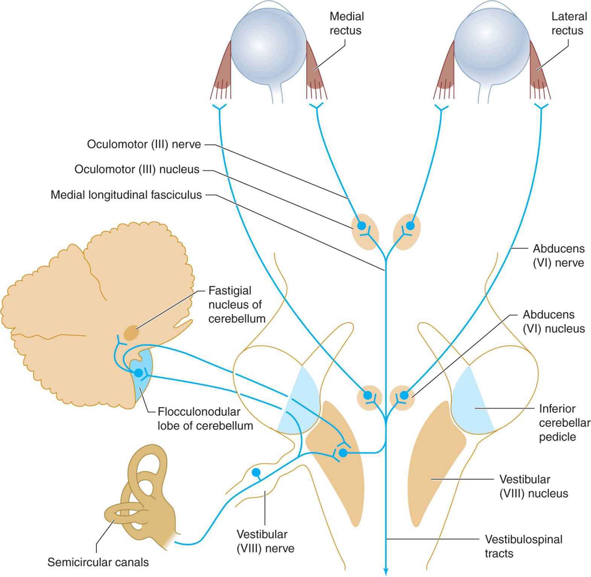
Figure 8-1. Peripheral and central vestibular pathways. The vestibular nerve terminates in the vestibular nucleus in the brainstem and midline cerebellar structures that project to the vestibular nucleus. From here, bilateral pathways in the medial longitudinal fasciculus ascend to the abducens (VI) and oculomotor (III) nuclei and descend to the spinal cord (vestibulospinal tracts).
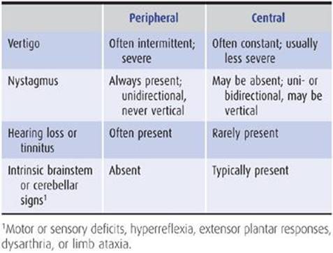
Table 8-1 Characteristics of peripheral and central vertigo.
PERIPHERAL VERTIGO
Peripheral vestibular lesions affect the labyrinth of the inner ear or the vestibular division of the vestibulocochlear (VIII) nerve. Vertigo from peripheral lesions tends to be intermittent, lasts for briefer periods, and produces more distress than vertigo of central origin (see next section). Nystagmus (rhythmic oscillation of the eyeballs) is always present with peripheral vertigo; it is usually unidirectional and never vertical. Peripheral lesions commonly produce additional symptoms of inner ear or vestibulocochlear (VIII) nerve dysfunction, such as hearing loss and tinnitus (the illusion of hearing a nonexistent sound, such as ringing in the ears).
CENTRAL VERTIGO
Vertigo of central nervous system origin usually results from lesions that affect the brainstem vestibular nuclei or their connections; rarely, vertigo can be produced by a cerebral cortical lesion, such as when it occurs as a symptom of complex partial seizures (Chapter 12).
Central vertigo may occur with or without nystagmus; if nystagmus is present, it can be vertical, unidirectional, or multidirectional and may differ in character in the two eyes. (Vertical nystagmus is oscillation in a vertical plane; nystagmus produced by upgaze or downgaze is not necessarily in the vertical plane.) Vertigo from central lesions may be accompanied by intrinsic brainstem or cerebellar signs, such as motor or sensory deficits, hyperreflexia, extensor plantar responses, dysarthria, or limb ataxia.
ATAXIA
Ataxia is incoordination or clumsiness of movement that is not the result of muscular weakness. It can be caused by vestibular, cerebellar, or sensory (proprioceptive) disorders. Ataxia can affect eye movement, speech (producing dysarthria), individual limbs, the trunk, stance, or gait (Table 8-2).
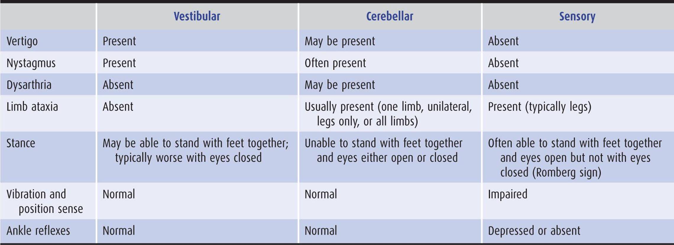
Table 8-2 Characteristics of vestibular, cerebellar, and sensory ataxia.
VESTIBULAR ATAXIA
The same central and peripheral lesions that cause peripheral or central vertigo (see preceding section) can also produce vestibular ataxia. Nystagmus is frequently present and is typically unilateral and most pronounced on gaze away from the side of vestibular involvement. Dysarthria is not a component of vestibular ataxia. Vestibular ataxia is gravity-dependent: Incoordination of the limbs becomes apparent only when the patient attempts to stand or walk.
CEREBELLAR ATAXIA
Cerebellar ataxia is produced by lesions of the cerebellum or its afferent or efferent connections in the cerebellar peduncles, red nucleus, pons, or spinal cord (Figure 8-2). Because of the crossed connection between the frontal cerebral cortex and the cerebellum, unilateral frontal disease can also occasionally mimic a disorder of the contralateral cerebellar hemisphere. The clinical manifestations of cerebellar ataxia consist of irregularities in the rate, rhythm, amplitude, and force of voluntary movements.
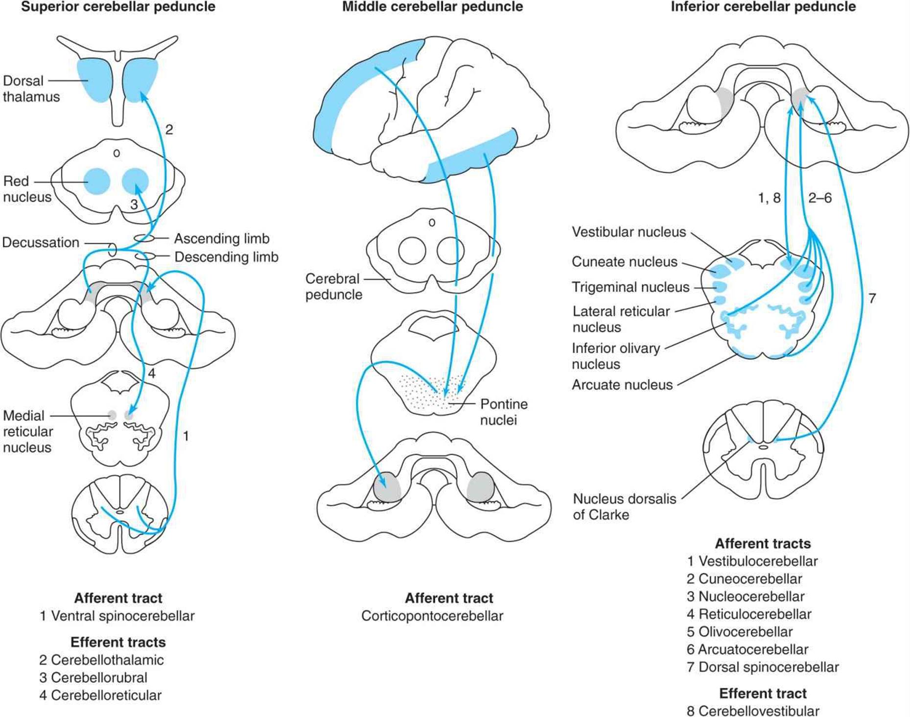
Figure 8-2. Cerebellar connections in the superior, middle, and inferior cerebellar peduncles. The peduncles are indicated by gray shading and the areas to and from which they project by blue shading.
Hypotonia
Cerebellar ataxia is commonly associated with hypotonia, which results in defective posture maintenance. Limbs are easily displaced by a relatively small force and, when shaken by the examiner, exhibit an increased range of excursion. The range of arm swing during walking may be similarly increased. Tendon reflexes take on a pendular quality, so that several oscillations of the limb may occur after the reflex is elicited, although neither the force nor the rate of the reflex is increased. When muscles are contracted against resistance that is then removed, the antagonist muscle fails to check the movement and compensatory muscular relaxation does not ensue promptly. This results in rebound movement of the limb.
Incoordination
In addition to hypotonia, cerebellar ataxia is associated with incoordination of voluntary movements. Simple movements are delayed in onset, and their rates of acceleration and deceleration are decreased. The rate, rhythm, amplitude, and force of movements fluctuate, producing a jerky appearance. Because these irregularities are most pronounced during initiation and termination of movement, their most obvious clinical manifestations include terminal dysmetria, or “overshoot” when the limb is directed at a target, and terminal intention tremor as the limb approaches the target. More complex movements tend to become decomposed into a succession of individual movements rather than a single smooth motor act (asynergia). Movements that involve rapid changes in direction or greater physiologic complexity, such as walking, are most severely affected.
Eye Movement Abnormalities
Because of the cerebellum’s prominent role in the control of eye movements, ocular abnormalities are a frequent consequence of cerebellar disease. These include nystagmus and related ocular oscillations, gaze paresis, and defective saccadic and pursuit movements.
Anatomic Basis of Clinical Signs
Various anatomic regions of the cerebellum (Figure 8-3) are functionally distinct, corresponding to the somatotopic organization of their motor, sensory, visual, and auditory connections (Figure 8-4).


Figure 8-3. Anatomic divisions of the cerebellum in midsagittal view (A), or unfolded (arrows) and viewed from behind (B).

Figure 8-4. Functional organization of the cerebellum. The view is similar to that in Figure 8-3B but is of a monkey rather than a human cerebellum. The three cerebellar homunculi represent areas to which proprioceptive and tactile stimuli project, and the stripes represent areas to which auditory and visual stimuli project.
1. Midline lesions—The middle zone of the cerebellum—the vermis and flocculonodular lobe and their associated subcortical (fastigial) nuclei—is involved in the control of axial functions, including eye movements, head and trunk posture, stance, and gait. Midline cerebellar disease therefore results in a clinical syndrome characterized by nystagmus and other disorders of ocular motility, dysarthria, oscillation of the head and trunk (titubation), instability of stance, and gait ataxia (Table 8-3). Selective involvement of the superior cerebellar vermis, as commonly occurs in alcoholic cerebellar degeneration, produces exclusively or primarily ataxia of gait, as would be predicted from the somatotopic map of the cerebellum (Figure 8-4).
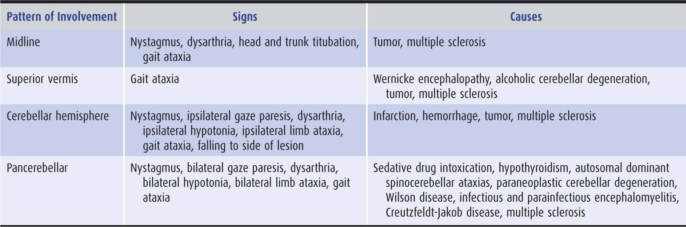
Table 8-3 Clinical patterns of cerebellar ataxia.
2. Hemispheric lesions—The lateral zones of the cerebellum (cerebellar hemispheres) help to coordinate movements and maintain tone in the ipsilateral limbs. The hemispheres also have a role in regulating ipsilateral gaze. Disorders affecting one cerebellar hemisphere cause ipsilateral hemiataxia and hypotonia of the limbs as well as nystagmus and transient ipsilateral gaze paresis (inability to look voluntarily toward the affected side). Involvement of the medial (paravermian) portion of either cerebellar hemisphere can also produce dysarthria.
3. Diffuse disease—Many cerebellar disorders—typically toxic, metabolic, and degenerative conditions—affect the cerebellum diffusely. The clinical picture in such states combines the features of midline and bilateral hemisphere disease.
SENSORY ATAXIA
Sensory ataxia results from disorders that affect the prop-rioceptive pathways in peripheral sensory nerves, sensory roots, posterior columns of the spinal cord, or medial lemnisci. Thalamic and parietal lobe lesions are rare causes of contralateral sensory hemiataxia.
Sensations of joint position and movement (kinesthesis) originate in pacinian corpuscles and unencapsulated nerve endings in joint capsules, ligaments, muscle, and periosteum. Such sensations are transmitted via heavily myelinated A fibers of primary afferent neurons, which enter the dorsal horn of the spinal cord and ascend uncrossed in the posterior columns (Figure 8-5). Proprioceptive information from the legs is conveyed in the medially located fasciculus gracilis, and information from the arms is conveyed in the more laterally situated fasciculus cuneatus. These tracts synapse on second-order sensory neurons in the nucleus gracilis and nucleus cuneatus in the lower medulla. The second-order neurons decussate as internal arcuate fibers and ascend in the contralateral medial lemniscus. They terminate in the ventral posterior nucleus of the thalamus, from which third-order sensory neurons project to the parietal cortex.
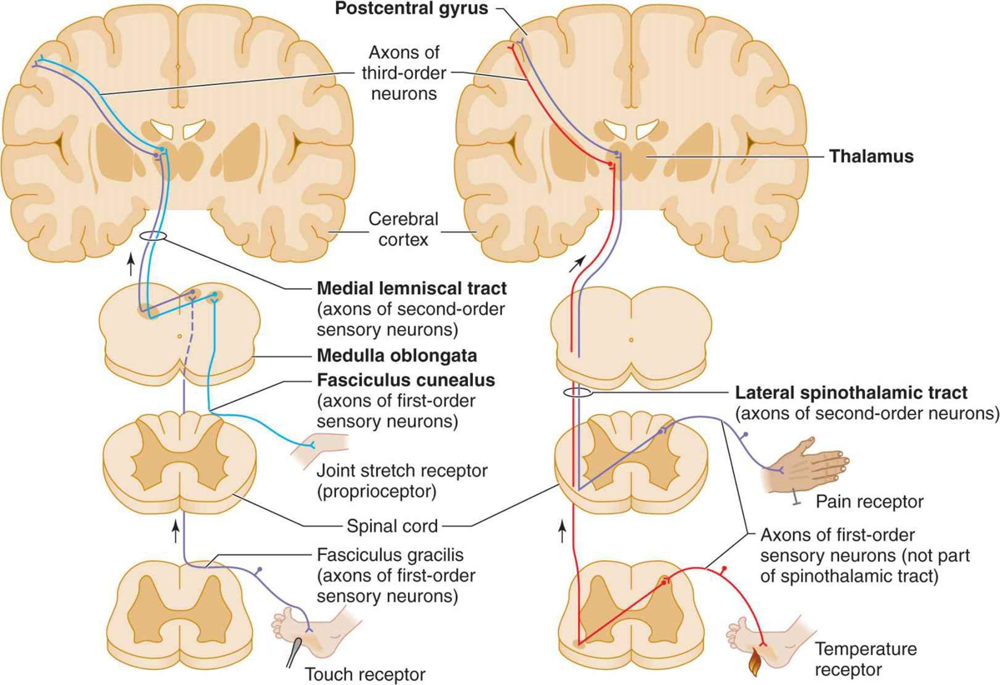
Figure 8-5. Pathways mediating proprioception (left, light blue) and other somatic sensory modalities (left and right). (Reprinted with permission from Fox SI. Human Physiology. 10th ed. Boston, MA: McGraw-Hill; 2008.)
Sensory ataxia from polyneuropathy or posterior column lesions typically affects the gait and legs in symmetric fashion; the arms are involved to a lesser extent or are spared entirely. Examination reveals impaired sensation of joint position in the affected limbs, and vibration sense is also commonly disturbed. Vertigo, nystagmus, and dysarthria are characteristically absent.
HISTORY
SYMPTOMS & SIGNS
Vertigo
True vertigo must be distinguished from a light-headed or presyncopal sensation. Vertigo is typically described as spinning, rotating, or moving, but when the description is vague, the patient should be asked specifically if the symptom is associated with a sense of movement.
The circumstances under which symptoms occur may also be diagnostically helpful. Vertigo is often brought on by changes in head position. The occurrence of symptoms upon arising after prolonged recumbency is a common feature of orthostatic hypotension, which may be immediately relieved by sitting or lying down. Orthostatic hypotension and other cerebral hypoperfusion states can lead to loss of consciousness, which is rarely associated with true vertigo.
Symptoms associated with vertigo may help to localize the site of the causal lesion. Hearing loss or tinnitus strongly suggests a disorder of the peripheral vestibular apparatus (labyrinth or vestibulocochlear [VIII] nerve). Dysarthria, dysphagia, diplopia, or focal weakness or sensory loss affecting the face or limbs points to a likely central (brainstem) lesion.
Ataxia
Ataxia associated with vertigo suggests a vestibular disorder, whereas ataxia with numbness or tingling in the legs is common in patients with sensory ataxia. Because proprioceptive deficits may, to some extent, be compensated for by other sensory cues, patients with sensory ataxia may report that their balance is improved by watching their feet when they walk or by using a cane or the arm of a companion for support. They thus find that they are much more unsteady in the dark and may experience particular difficulty in descending stairs.
ONSET & TIME COURSE
The mode of onset and time course may help to identify the cause of a disorder of equilibrium.
Sudden
Sudden onset of disequilibrium occurs with infarcts and hemorrhages in the brainstem or cerebellum (eg, lateral medullary syndrome, cerebellar hemorrhage, or infarction).
Episodic
Episodic disequilibrium of acute onset suggests transient ischemic attacks in the basilar artery distribution, benign positional vertigo, or Ménière disease. Disequilibrium from transient ischemic attacks is usually accompanied by cranial nerve deficits, neurologic signs in the limbs, or both. Ménière disease is usually associated with progressive hearing loss and tinnitus as well as vertigo.
Chronic & Progressive
Chronic, progressive disequilibrium evolving over weeks to months is most suggestive of a toxic or nutritional disorder (eg, vitamin B12 or vitamin E deficiency, nitrous oxide exposure). Evolution over months to years is characteristic of an inherited spinocerebellar degeneration.
MEDICAL HISTORY
The medical history should be scrutinized for evidence of diseases that affect the sensory pathways (vitamin B12 deficiency, syphilis) or cerebellum (hypothyroidism, paraneoplastic syndromes, tumors) and drugs that produce disequilibrium by impairing vestibular or cerebellar function (ethanol, sedative drugs, phenytoin, aminoglycoside antibiotics, quinine, salicylates).
FAMILY HISTORY
A hereditary degenerative disorder may be the cause of chronic, progressive cerebellar ataxia. Such disorders include spinocerebellar degenerations, Friedreich ataxia, ataxia-telangiectasia, and Wilson disease.
GENERAL PHYSICAL EXAMINATION
Various features of the general physical examination may provide clues to the underlying disorder.
1. Orthostatic hypotension is associated with certain sensory disorders that produce ataxia (eg, tabes dorsalis, polyneuropathies) and with some cases of spinocerebellar degeneration.
2. Skin may show oculocutaneous telangiectasia (ataxia-telangiectasia), or it may be dry, with brittle hair (hypothyroidism) or have a lemon-yellow coloration (vitamin B12 deficiency).
3. Pigmented corneal (Kayser-Fleischer) rings are seen in Wilson disease (Chapter 11).
4. Skeletal abnormalities may be present. Kyphoscoliosis is typical in Friedreich ataxia, hypertrophic or hyperextensible joints are common in tabes dorsalis, and pes cavus is a feature of certain hereditary neuropathies. Abnormalities at the craniocervical junctions may be associated with Arnold-Chiari malformations or other congenital anomalies that involve the posterior fossa.
NEUROLOGIC EXAMINATION
MENTAL STATUS EXAMINATION
Acute confusional state with ataxia characterizes ethanol or sedative drug intoxication and Wernicke encephalopathy. Dementia with cerebellar ataxia is seen in Wilson disease, Creutzfeldt-Jakob disease, hypothyroidism, paraneoplastic syndromes, and some spinocerebellar degenerations. Dementia with sensory ataxia suggests syphilitic taboparesis or vitamin B12 deficiency. Amnesia and cerebellar ataxia are associated with chronic alcoholism (Korsakoff amnestic syndrome, Chapter 5).
STANCE & GAIT
Observation of stance and gait is helpful in distinguishing among vestibular, cerebellar, and sensory ataxia. In any ataxic patient, the stance and gait are wide-based and unsteady, often associated with reeling or lurching movements.
Stance
The ataxic patient asked to stand with the feet together may show great reluctance or an inability to do so. With persistent urging, the patient may gradually move the feet closer together but will leave some space between them.
1. Patients with sensory ataxia and some with vestibular ataxia are, nevertheless, ultimately able to stand with the feet together, compensating for the loss of one source of sensory input (proprioceptive or labyrinthine) with another (visual). This compensation is demonstrated when the patient closes the eyes, eliminating visual cues. With sensory or vestibular disorders, unsteadiness increases and may result in falling (Romberg sign). With a vestibular lesion, the tendency is to fall toward the side of the lesion.
2. Patients with cerebellar ataxia are unable to compensate for their deficit by using visual input and are unstable on their feet whether the eyes are open or closed.
Gait
1. The gait in cerebellar ataxia is wide-based, often with a staggering quality that might suggest drunkenness. Oscillation of the head or trunk (titubation) may be present. If a unilateral cerebellar hemisphere lesion is responsible, there is a tendency to deviate toward the side of the lesion when the patient attempts to walk in a straight line or circle or marches in place with eyes closed. Tandem (heel-to-toe) gait, which requires walking with an exaggerated narrow base, is always impaired.
2. In sensory ataxia, the gait is also wide-based and tandem gait is poor. In addition, walking is typically characterized by lifting the feet high off the ground and slapping them down heavily (steppage gait) because of impaired proprioception. Stability may be dramatically improved by letting the patient use a cane or lightly rest a hand on the examiner’s arm for support. If the patient is made to walk in the dark or with eyes closed, gait is much more impaired.
3. Gait ataxia may also be a manifestation of conversion disorder (conversion disorder with motor symptom or deficit) or malingering. Determining this can be particularly difficult, as isolated gait ataxia without ataxia of individual limbs can also be produced by diseases that affect the superior cerebellar vermis. The most helpful observation in identifying factitious gait ataxia is that such patients often exhibit wildly reeling or lurching movements from which they are able to recover without falling. In fact, recovery of balance from such awkward positions requires excellent equilibrium.
CRANIAL NERVES
Abnormalities of extraocular (III, IV, and VI) and vestibulocochlear (VIII) nerve function are typically present with vestibular disease and often present with lesions of the cerebellum.
Ocular Alignment
The eyes are examined in the primary position of gaze (looking directly forward) to detect misalignment in the horizontal or vertical plane.
Eye Movements
1. The patient is asked to turn the eyes in each of the cardinal directions of gaze (left, up and left, down and left, right, up and right, down and right; Chapter 1) to determine whether ocular nerve palsy or gaze paresis (impaired ability to move the two eyes coordinately in any of the cardinal directions of gaze) is present.
2. Nystagmus—an abnormal involuntary oscillation of the eyes—is characterized in terms of the positions of gaze in which it occurs (gaze-evoked nystagmus), its amplitude, and the direction of its fast phase. Pendular nystagmus, usually the result of visual impairment that begins in infancy, has the same velocity in both directions of eye movement; jerk nystagmus is characterized by both fast (vestibular-induced) and slow (cortical) phases. The direction of jerk nystagmus is defined by the direction of the fast component.
3. Fast voluntary eye movements (saccades) are elicited by having the patient rapidly shift gaze from one target to another placed in a different part of the visual field. Slow voluntary eye movements (pursuits) are assessed by having the patient track a slowly moving target such as the examiner’s finger.
4. Peripheral vestibular disorders produce unidirectional horizontal jerk nystagmus that is maximal on gaze away from the involved side.
5. Central vestibular disorders can cause unidirectional or bidirectional horizontal nystagmus, vertical nystagmus, or gaze paresis.
6. Cerebellar lesions are associated with a wide range of ocular abnormalities, including gaze pareses, defective saccades or pursuits, nystagmus in any or all directions, and ocular dysmetria (overshoot of visual targets during saccadic eye movements).
Hearing
Preliminary examination of the vestibulocochlear (VIII) nerve should include otoscopic inspection of the auditory canals and tympanic membranes, assessment of auditory acuity in each ear, and Weber and Rinne tests (Table 8-4) performed with a 256-Hz tuning fork.

Table 8-4 Assessment of hearing loss.
1. In the Weber test, unilateral sensorineural hearing loss (from lesions of the cochlea or vestibulocochlear nerve) causes the patient to perceive the sound produced by a vibrating tuning fork placed at the vertex of the skull as coming from the normal ear. With a conductive (external or middle ear) disorder, sound is localized to the abnormal ear.
2. The Rinne test may also distinguish between sensorineural and conductive defects in the affected ear. Air conduction (tested by holding the vibrating tuning fork next to the external auditory canal) normally produces a louder sound than does bone conduction (tested by placing the base of the tuning fork over the mastoid bone). This pattern also occurs with sensorineural hearing loss due to vestibulocochlear nerve lesions but is reversed in the case of conductive hearing loss.
Positional Tests
When patients indicate that vertigo occurs with a change in position, the Nylen-Bárány or Dix-Hallpike maneuver (Figure 8-6) is used to try to reproduce the precipitating circumstance. The head is rapidly lowered 30 degrees below horizontal. This process is repeated with the head and eyes turned first to the right and then to the left. The eyes are observed for nystagmus, and the patient is asked to note the onset, severity, and cessation of vertigo.
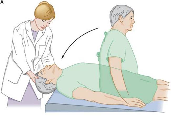

Figure 8-6. Test for positional vertigo and nystagmus. The patient is seated on a table with the head and eyes directed forward (A) and is then quickly lowered to a supine position with the head over the table edge, 45 degrees below horizontal. The patient’s eyes are then observed for nystagmus, and the patient is asked to report any vertigo. The test is repeated with the patient’s head and eyes turned 45 degrees to the right (B), and again with the head and eyes turned 45 degrees to the left (not shown).
Positional nystagmus and vertigo are usually associated with peripheral vestibular lesions and are most often a feature of benign positional vertigo. This is typically characterized by severe distress, a latency of several seconds between assumption of the position and the onset of vertigo and nystagmus, a tendency for the response to remit spontaneously (fatigue) as the position is maintained, and attenuation of the response (habituation) as the offending position is repeatedly assumed (Table 8-5). Positional vertigo can also occur with central vestibular disease.
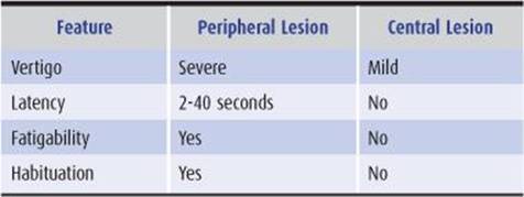
Table 8-5 Characteristics of positional nystagmus.
Caloric Testing
Disorders of the vestibuloocular pathways can be detected by caloric testing. Caloric testing should be preceded by careful otoscopic examination and should not be undertaken if the tympanic membrane is perforated. The patient is placed supine with the head elevated 30 degrees to bring the superficially situated lateral semicircular canal into the upright position. Each ear canal is irrigated in turn with cold (33°C) or warm (44°C) water for 40 seconds, with at least 5 minutes between tests. Warm water tends to produce less discomfort than cold.
1. In the normal, awake patient, cold-water caloric stimulation produces nystagmus with the slow phase toward and the fast phase away from the irrigated ear. Warm water irrigation produces the opposite response.
2. In patients with unilateral labyrinthine, vestibulocochlear (VIII) nerve, or vestibular nuclear dysfunction, irrigation of the affected side fails to cause nystagmus or elicits nystagmus that is later in onset or briefer in duration than on the normal side.
Other Cranial Nerves
Papilledema associated with disequilibrium suggests an intracranial mass lesion, usually in the posterior fossa, that is causing increased intracranial pressure. Optic neuropathy may be present in multiple sclerosis, neurosyphilis, or vitamin B12 deficiency. A depressed corneal reflex or facial palsy ipsilateral to the lesion (and the ataxia) can accompany cerebellopontine angle tumor. Weakness of the tongue or palate, hoarseness, or dysphagia results from lower brainstem disease.
MOTOR SYSTEM
Examination of motor function in the patient with a disorder of equilibrium should disclose the pattern and severity of ataxia and any associated pyramidal, extrapyramidal, or lower motor neuron involvement that might suggest a cause. The clinical features that help distinguish cerebellar disease from disease involving these other motor systems are summarized in Table 8-6.

Table 8-6 Clinical features distinguishing cerebellar from other motor systems disorders.
Muscle Tone
1. Hypotonia is characteristic of cerebellar disorders; with unilateral cerebellar hemispheric lesions, the ipsilateral limbs are hypotonic.
2. Extrapyramidal hypertonia (rigidity) may occur in disorders that affect both the cerebellum and basal ganglia (eg, Wilson disease, acquired hepatocerebral degeneration, Creutzfeldt-Jakob disease, and some autosomal dominant spinocerebellar ataxias).
3. Ataxia with spasticity may be seen in conditions that affect both the cerebellum and upper motor neuron pathways (eg, multiple sclerosis, posterior fossa tumors, congenital anomalies, vertebrobasilar ischemia or infarction, some autosomal dominant spinocerebellar ataxias, Friedreich ataxia, neurosyphilis, Creutzfeldt-Jakob disease, and vitamin B12 deficiency).
Coordination
1. Truncal stability is assessed by examination of gait (discussed earlier) and by observing the patient as he or she sits unsupported. In addition to gait ataxia, patients with midline or pancerebellar disease may show oscillation of the head or trunk (titubation) and truncal ataxia while seated; in the latter case, they tend to fall when they try to remain sitting upright.
2. Movement of the patient’s arm is observed as his or her finger tracks back and forth between his or her own nose or chin and the examiner’s finger. With mild cerebellar ataxia, an intention tremorcharacteristically appears near the beginning and end of each such movement, and the patient may overshoot the target.
3. When the patient is asked to raise the arms rapidly to a given height—or when the arms, extended and outstretched in front of the patient, are displaced by a sudden force—there may be overshooting (rebound). Impaired ability to check the force of muscular contractions can also be demonstrated by having the patient forcefully flex the arm at the elbow against resistance—and then suddenly removing the resistance. If the limb is ataxic, continued contraction without resistance may cause the hand to strike the patient, whose head should be turned to the side to avoid being hit in the face.
4. Ataxia of the legs is demonstrated by the supine patient’s inability to run the heel of the foot smoothly up and down the opposite shin.
5. Ataxia of any limb is reflected by irregularity in the rate, rhythm, amplitude, and force of rapid successive tapping movements.
Weakness
1. Pure vestibular, cerebellar, or sensory disorders do not cause weakness. However, some diseases affect these systems in combination with motor pathways.
2. Distal weakness can be caused by disorders that also produce sensory ataxia, such as polyneuropathies and Friedreich ataxia.
3. Paraparesis may be superimposed on ataxia in vitamin B12 deficiency, multiple sclerosis, foramen magnum lesions, or spinal cord tumors.
4. Ataxic quadriparesis, hemiataxia with contralateral hemiparesis, or ataxic hemiparesis suggests a brainstem lesion.
Abnormal Involuntary Movements
1. Titubation and intention tremor, discussed earlier, result from cerebellar disorders. Other abnormal movements can be seen in patients with diseases that affect both the cerebellum and other brain structures.
2. Asterixis may occur in hepatic encephalopathy, acquired hepatocerebral degeneration, or other metabolic encephalopathies.
3. Myoclonus occurs in the same conditions as asterixis and is a prominent manifestation of Creutzfeldt-Jakob disease.
4. Chorea may be associated with cerebellar signs in Wilson disease, acquired hepatocerebral degeneration, or ataxia-telangiectasia.
SENSORY SYSTEM
Joint Position Sense
In patients with sensory ataxia, joint position sense is always impaired in the legs and may be defective in the arms as well. Testing is by asking the patient to detect passive movement of the joints, beginning distally and moving proximally, to establish the upper level of deficit in each limb. With normal joint position sense, it should be possible to detect almost any displacement. Abnormalities of position sense also can be demonstrated by positioning one limb and having the patient, with eyes closed, place the opposite limb in the same position.
Vibration Sense
Vibration sense is frequently impaired in patients with sensory ataxia. The patient is asked to detect the vibration of a 128-Hz tuning fork placed over a bony prominence. Again, successively more proximal sites are tested to determine the upper level of the deficit in each limb. The patient’s threshold for appreciating the vibration is compared with the examiner’s own ability to detect it in the hand that holds the tuning fork.
REFLEXES
1. Tendon reflexes are typically hypoactive, with a pendular quality, in cerebellar disorders; unilateral cerebellar lesions produce ipsilateral hyporeflexia.
2. Hyporeflexia of the legs is a prominent manifestation of Friedreich ataxia, tabes dorsalis, and polyneuropathies that cause sensory ataxia.
3. Hyperactive reflexes and extensor plantar responses may accompany ataxia caused by multiple sclerosis, vitamin B12 deficiency, focal brainstem lesions, and certain autosomal dominant spinocerebellar ataxias.
INVESTIGATIVE STUDIES
BLOOD TESTS
Blood studies may disclose low vitamin B12 levels and hematologic abnormalities (macrocytic anemia, leukopenia with hypersegmented neutrophils, thrombocytopenia with giant platelets) associated with vitamin B12 deficiency, decreased levels of thyroid hormones in hypothyroidism, elevated hepatic enzymes and low ceruloplasmin and copper concentrations in Wilson disease, immunoglobulin deficiency and elevated α-fetoprotein in ataxia-telangiectasia, antibodies to Purkinje cell antigens in paraneoplastic cerebellar degeneration, or mutations associated with autosomal dominant spinocerebellar ataxias.
CEREBROSPINAL FLUID
The cerebrospinal fluid (CSF) shows elevated protein with cerebellopontine angle tumors (eg, acoustic neuroma), brainstem or spinal cord tumors, hypothyroidism, and some polyneuropathies. Increased protein with pleocytosis is commonly found with infectious or parainfectious encephalitis, paraneoplastic cerebellar degeneration, and neurosyphilis. CSF levels of 14–3–3 protein are increased in Creutzfeldt-Jakob disease. Although elevated pressure and bloody CSF characterize cerebellar hemorrhage, lumbar puncture is contraindicated if cerebellar hemorrhage is suspected. CSF VDRL is reactive in tabes dorsalis, and oligoclonal immunoglobulin G (IgG) bands may be present in multiple sclerosis or other inflammatory disorders.
BRAIN IMAGING
The computed tomography (CT) scan is useful for demonstrating posterior fossa tumors or malformations, cerebellar infarction or hemorrhage, and cerebellar atrophy associated with degenerative disorders. Magnetic resonance imaging (MRI) provides better visualization of posterior fossa lesions, including cerebellopontine angle tumors, and is superior to CT scanning for detecting the lesions of multiple sclerosis.
EVOKED POTENTIALS
Evoked potential testing, especially of optic pathways (visual evoked potentials), may be helpful in evaluating patients with suspected multiple sclerosis. Brainstem auditory evoked potentials can localize vestibular disease to the peripheral vestibular pathways and may help to identify cerebellopontine angle tumors.
CHEST X-RAY & ECHOCARDIOGRAPHY
The chest x-ray or echocardiogram may provide evidence of cardiomyopathy associated with Friedreich ataxia. The chest x-ray may also show a lung tumor in paraneoplastic cerebellar degeneration.
AUDIOMETRY
This is useful when vestibular disorders are associated with auditory impairment; such testing can distinguish conductive, labyrinthine, vestibulocochlear (VIII) nerve, and brainstem disease.
Tests of pure tone hearing are abnormal when sounds are transmitted through air with conductive hearing loss and when transmitted through either air or bone with labyrinthine or vestibulocochlear (VIII) nerve disorders.
Speech discrimination is markedly impaired with vestibulocochlear (VIII) nerve lesions and is impaired less with disorders of the labyrinth. Speech discrimination is normal in conductive or brainstem involvement.
ELECTRONYSTAGMOGRAPHY
This test can be used to detect and characterize nystagmus, including that elicited by caloric stimulation.
PERIPHERAL VESTIBULAR DISORDERS
A list of peripheral vestibular disorders and features helpful in the differential diagnosis is presented in Table 8-7.
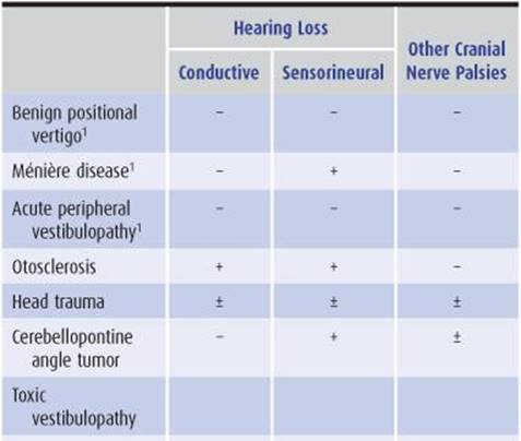
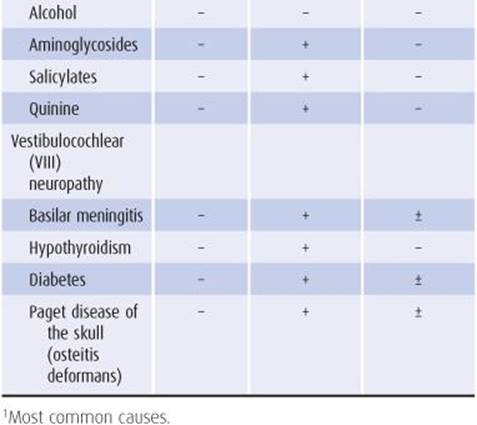
Table 8-7 Differential diagnosis of peripheral vestibular disorders.
BENIGN POSITIONAL VERTIGO
Positional vertigo occurs upon assuming a particular head position. It is usually associated with peripheral vestibular lesions, but also may be due to central (brainstem or cerebellar) disease.
PATHOGENESIS
Benign positional vertigo is the most common cause of vertigo of peripheral origin, accounting for approximately 30% of cases. The most frequently identified cause is head trauma, but in most instances, no cause can be determined. The pathophysiologic basis of benign positional vertigo is thought to be canalolithiasis—stimulation of the semicircular canal by debris floating in the endolymph.
CLINICAL FINDINGS
The syndrome is characterized by brief (seconds to minutes) episodes of severe vertigo that may be accompanied by nausea and vomiting. Symptoms may occur with any change in head position but are usually most severe in the lateral decubitus position with the affected ear down. Episodic vertigo typically continues for several weeks and then resolves spontaneously; in some cases, it is recurrent. Hearing loss is not a feature.
Peripheral and central causes of positional vertigo usually can be distinguished on physical examination by means of the Nylen-Bárány or Dix-Hallpike maneuver (Figure 8-6). Positional nystagmus always accompanies vertigo in the benign disorder and is typically unidirectional, rotatory, and delayed in onset by several seconds after assumption of the precipitating head position. If the position is maintained, nystagmus and vertigo resolve within seconds to minutes. If the maneuver is repeated successively, the response is attenuated. In contrast, positional vertigo of central origin tends to be less severe, and positional nystagmus may be absent. There is no latency, fatigue, or habituation in central positional vertigo.
TREATMENT
The mainstay of treatment in most cases of benign positional vertigo of peripheral origin is the use of repositioning (Epley) maneuvers that employ the force of gravity to move endolymphatic debris out of the semicircular canal and into the vestibule, where it can be reabsorbed. In one such maneuver (Figure 8-7), the head is turned 45 degrees in the direction of the affected ear (determined clinically, as described earlier), and the patient reclines to a supine position, with the head (still turned 45 degrees) hanging down over the end of the examining table. The head, still hanging down, is then turned 90 degrees in the opposite direction, to 45 degrees toward the opposite ear. Next, the patient rolls to a lateral decubitus position with the affected ear up, and the head still turned 45 degrees toward the unaffected ear and hanging down. Finally, the patient turns to a prone position and sits up.

Figure 8-7. Repositioning treatment for benign positional vertigo resulting from canalolithiasis. In the example shown, repositioning maneuvers are used to move endolymphatic debris out of the posterior semicircular canal (PSC) of the right ear and into the utricle (UT), the larger of two membranous sacs in the vestibule of the labyrinth, where this debris can be reabsorbed. The numbers (1-6) refer to both the position of the patient and the corresponding location of debris within the labyrinth. The patient is seated, and the head is turned 45 degrees to the right (1). The head is lowered rapidly to below the horizontal (2); the examiner shifts position (3); and the head is rotated rapidly 90 degrees in the opposite direction, so it now points 45 degrees to the left, where it remains for 30 seconds (4). The patient then rolls onto the left side without turning the head in relation to the body and maintains this position for another 30 seconds (5) before sitting up (6). This maneuver may need to be repeated until nystagmus is abolished. The patient must then avoid the supine position for at least 2 days. (Courtesy of Baloh RW. Reproduced with permission from Samuels MA, et al. Office Practice of Neurology. New York, NY: Churchill Livingstone; 1995.)
Vestibulo-suppressant drugs (Table 8-8) may also be useful in the acute period, and vestibular rehabilitation, which promotes compensation for vestibular dysfunction through the recruitment of other sensory modalities, may be helpful as well.
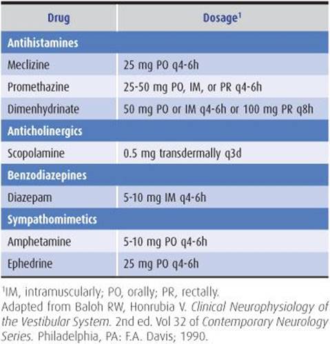
Table 8-8 Drugs used in the treatment of vertigo.
MÉNIÈRE DISEASE
Ménière disease is characterized by repeated episodes of vertigo lasting from minutes to days, accompanied by tinnitus and progressive sensorineural hearing loss.
PATHOGENESIS
Most cases are sporadic, but familial occurrence also has been described, and may show anticipation (earlier onset in successive generations). Rare cases appear to be related to mutations in the COCH(coagulation factor C homology) gene, which codes for cochlin, a main component of inner ear extracellular matrix. Onset of sporadic Ménière disease is usually between the ages of 20 and 50 years, and men are affected more often than women. The cause is thought to be an increase in the volume of labyrinthine endolymph (endolymphatic hydrops), but the pathogenetic mechanism is unknown.
CLINICAL FINDINGS
At the time of the first acute attack, patients already may have noted the insidious onset of tinnitus, hearing loss, and a sensation of fullness in the ear. Acute attacks are characterized by vertigo, nausea, and vomiting and recur at intervals ranging from weeks to years. Hearing deteriorates in a stepwise fashion, with bilateral involvement reported in 10% to 70% of patients. As hearing loss increases, vertigo tends to become less severe.
Physical examination during an acute episode shows spontaneous horizontal or rotatory nystagmus (or both) that may change direction. Although spontaneous nystagmus is characteristically absent between attacks, caloric testing usually reveals impaired vestibular function. The hearing deficit is not always sufficiently advanced to be detectable at the bedside. Audiometry shows low-frequency pure-tone hearing loss, however, that fluctuates in severity, as well as impaired speech discrimination and increased sensitivity to loud sounds.
As has been noted, episodes of vertigo tend to resolve as hearing loss progresses.
TREATMENT
Symptomatic management of acute exacerbations involves administration of antihistamines, benzodiazepines, or other drugs listed in Table 8-8. Between episodes, patients may be treated with a low-salt diet and diuretics, such as hydro-chlorothiazide (50 mg orally daily) plus triamterene (25 mg orally daily) or, where it is available, with the histamine H3 receptor antagonist betahistine (8-24 mg orally three times daily). In persistent, disabling cases resistant to orally administered drugs, transtympanic instillation of gentamicin or dexamethasone may be beneficial.
ACUTE PERIPHERAL VESTIBULOPATHY
This term is used to describe a spontaneous attack of vertigo of inapparent cause that resolves spontaneously and is not accompanied by hearing loss or evidence of central nervous system dysfunction. It includes disorders diagnosed as acute labyrinthitis or vestibular neuronitis. A recent antecedent febrile illness can sometimes be identified.
The disorder is characterized by vertigo, nausea, and vomiting of acute onset, typically lasting up to 2 weeks. Symptoms may recur, and some degree of vestibular dysfunction may be permanent. During an attack, the patient—who appears acutely ill—typically lies on one side with the affected ear upward and is reluctant to move his or her head. Nystagmus with the fast phase away from the affected ear is always present. The vestibular response to caloric testing is defective in one or both ears with about equal frequency. Auditory acuity is normal.
Acute peripheral vestibulopathy must be distinguished from central disorders that produce acute vertigo, such as stroke in the posterior cerebral circulation. Central disease is suggested by vertical nystagmus, altered consciousness, motor or sensory deficit, or dysarthria. Treatment is with a 10- to 14-day course of prednisone, 20 mg orally twice daily, the drugs listed in Table 8-8, or both.
OTOSCLEROSIS
Otosclerosis is caused by immobility of the stapes, the ear ossicle that transmits vibration of the tympanic membrane to the inner ear. Its most distinctive feature is conductive hearing loss, but sensorineural hearing loss and vertigo are also common; tinnitus is infrequent. Auditory symptoms usually begin before 30 years of age, and familial occurrence is common.
Vestibular dysfunction is most often characterized by recurrent episodic vertigo—with or without positional vertigo—and a sense of positional imbalance. More continuous symptoms may also occur, and the frequency and severity of attacks may increase with time. Vestibular abnormalities on examination include spontaneous or positional nystagmus of the peripheral type and attenuated caloric responses, which are usually unilateral. Hearing loss is always demonstrable by audiometry. It is usually of mixed conductive-sensorineural character and is bilateral in approximately two-thirds of patients.
In patients with episodic vertigo, progressive hearing loss, and tinnitus, otosclerosis must be distinguished from Ménière disease. Otosclerosis is suggested by a positive family history, a tendency toward onset at an earlier age, the presence of conductive hearing loss, or bilateral symmetric auditory impairment. Imaging studies also may be diagnostically useful.
Treatment with a combination of sodium fluoride, calcium gluconate, and vitamin D may be effective. If not, surgical stapedectomy should be considered.
HEAD TRAUMA
Head trauma is the most common identifiable cause of benign positional vertigo. Injury to the labyrinth is usually responsible for posttraumatic vertigo; however, fractures of the petrosal bone may lacerate the vestibulocochlear (VIII) nerve, producing vertigo and hearing loss. Hemotympanum or CSF otorrhea suggests such a fracture.
CEREBELLOPONTINE ANGLE TUMOR
PATHOGENESIS
The most common tumor in the cerebellopontine angle—a triangular region in the posterior fossa bordered by the cerebellum, lateral pons, and petrous ridge (Figure 8-8)—is the histologically benign acoustic neuroma. Also termed neurilemoma, neurinoma, or schwannoma, it typically arises from the neurilemmal sheath of the vestibular portion of the vestibulocochlear (VIII) nerve in the internal auditory canal. Less common tumors at this site include meningiomas and primary cholesteatomas (epidermoid cysts).
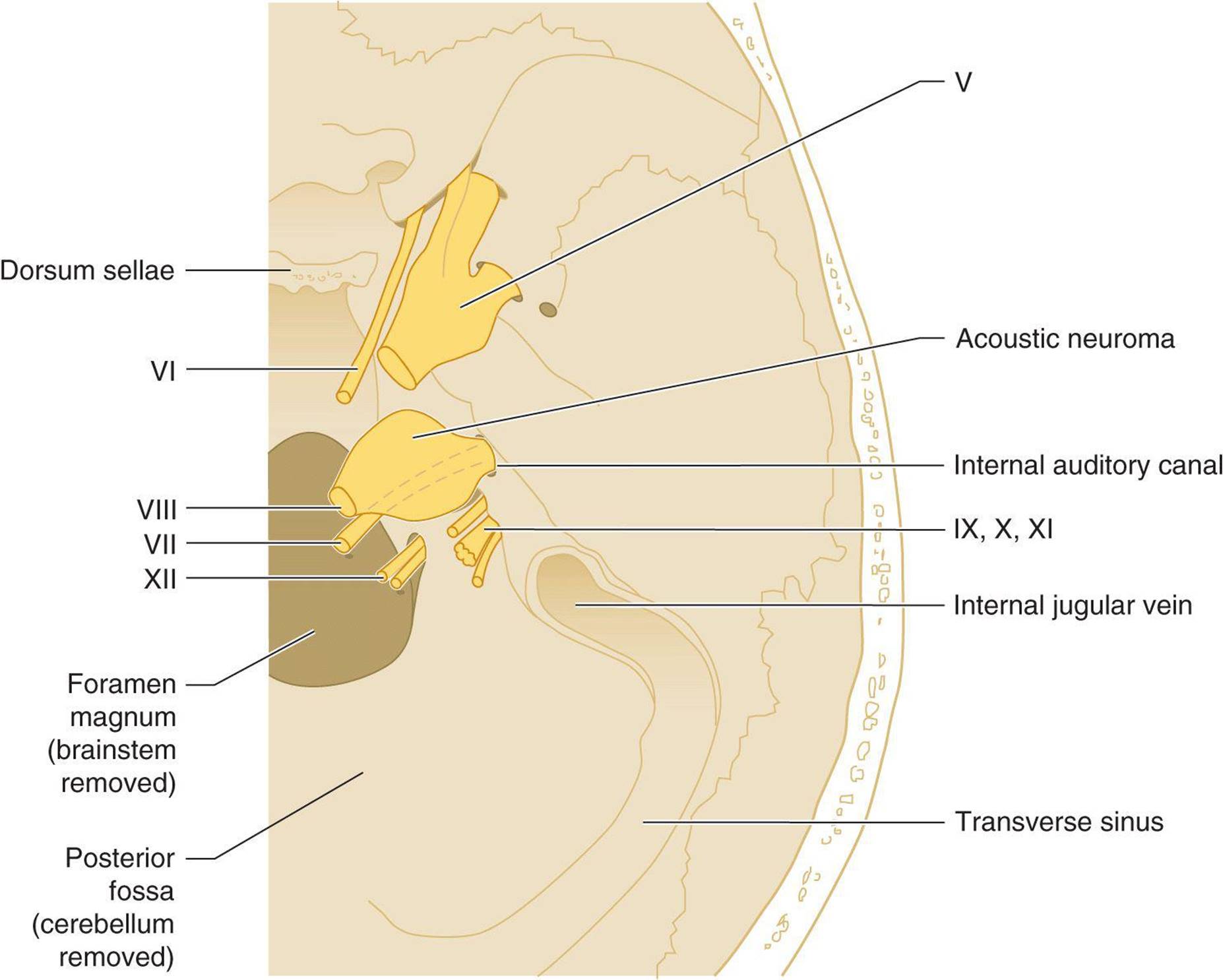
Figure 8-8. Cerebellopontine angle tumor, viewed from above, with the brain removed to permit the cranial nerves and base of the skull to be seen. The tumor, a neuroma arising from the vestibulocochlear (VIII) nerve, may compress adjacent structures, including the trigeminal (V) and facial (VII) nerves, the brainstem, and the cerebellum.
Acoustic neuromas occur most often as isolated lesions in patients 30 to 60 years old, but they may also be a manifestation of neurofibromatosis. Neurofibromatosis 1 (von Recklinghausen disease) is a common autosomal dominant disorder related to mutations in the gene for neurofibromin 1 (NF1), a tumor suppressor gene. In addition to unilateral acoustic neuromas, neurofibromatosis 1 is associated with café-au-lait spots on the skin, cutaneous neurofibromas, axillary or inguinal freckles, optic gliomas, iris hamartomas, and dysplastic bony lesions. Neurofibromatosis 2 is a rare autosomal dominant disorder caused by mutations in the gene for neurofibromin 2 (NF2), an inhibitor of contact-dependent cell proliferation. Its hallmark is bilateral acoustic neuromas, which may be accompanied by other central or peripheral nervous system tumors, including neurofibromas, meningiomas, gliomas, and schwannomas.
PATHOPHYSIOLOGY
Symptoms are produced by compression or displacement of the cranial nerves, brainstem, and cerebellum and by obstruction of CSF flow. Because of their anatomic relationship to the vestibulocochlear (VIII) nerve, the trigeminal (V) and facial (VII) nerves are often affected.
CLINICAL FINDINGS
Symptoms & Signs
Insidious hearing loss is usually the initial symptom. Less often, patients present with headache, vertigo, gait ataxia, facial pain, tinnitus, a sensation of fullness in the ear, or facial weakness. Although vertigo ultimately develops in 20% to 30% of patients, nonspecific unsteadiness is a more common complaint. Symptoms may be stable or progress over months or years.
Unilateral sensorineural hearing loss is the most common finding on examination. Other frequent abnormalities are ipsilateral facial palsy, depressed or absent corneal reflex, and sensory loss over the face. Ataxia, spontaneous nystagmus, other lower cranial nerve palsies, and signs of increased intracranial pressure are less common. Unilateral vestibular dysfunction can usually be demonstrated with caloric testing.
Laboratory Findings
Audiometry shows a sensorineural deficit with high-frequency pure-tone hearing loss, poor speech discrimination, and marked tone decay. CSF protein is elevated in approximately 70% of patients, usually in the range of 50 to 200 mg/dL. The most useful diagnostic radiologic study is MRI of the cerebellopontine angle. Acoustic neuromas may cause abnormal brainstem auditory evoked potentials.
DIFFERENTIAL DIAGNOSIS
Acoustic neuroma must be distinguished from other cerebellopontine angle tumors. Meningioma should be considered in patients whose initial symptoms indicate more than isolated vestibulocochlear (VIII) nerve disease. Cholesteatoma is suggested by conductive hearing loss, early facial weakness, or facial twitching, with normal CSF protein. Metastatic carcinoma may also present as a lesion in the cerebellopontine angle.
TREATMENT
Treatment is complete surgical excision. Patients with neurofibromatosis 2 may benefit from bevacizumab, a monoclonal antibody against the vascular endothelial growth factor, VEGFA. In untreated cases, severe complications can result from brainstem compression or hydrocephalus.
TOXIC VESTIBULOPATHIES
ALCOHOL
Alcohol causes acute positional vertigo because of its differential distribution between the cupula and endolymph of the inner ear. Alcohol-induced positional vertigo typically occurs within 2 hours after ingesting ethanol in amounts sufficient to produce blood levels >40 mg/dL. Alcohol initially diffuses into the cupula, later also diffuses into the endolymph, and then leaves the cupula before it leaves the endolymph. This produces two symptomatic phases of vertigo, which together last up to approximately 12 hours, separated by an asymptomatic interval of 1 to 2 hours. The clinical syndrome is characterized by vertigo and nystagmus in the lateral recumbent position and is accentuated when the eyes are closed. Other signs of alcohol intoxication, such as spontaneous nystagmus, dysarthria, and gait ataxia, are caused primarily by cerebellar rather than vestibular dysfunction.
AMINOGLYCOSIDES
Aminoglycoside antibiotics are widely recognized ototoxins that can produce both vestibular and auditory symptoms. Streptomycin, gentamicin, and tobramycin are the agents most likely to cause vestibular toxicity, and amikacin, kanamycin, and tobramycin are associated with hearing loss. Aminoglycosides concentrate in the perilymph and endolymph and exert their ototoxic effects by destroying sensory hair cells. The risk of toxicity is related to drug dosage, plasma concentration, duration of therapy, conditions—such as renal failure—that impair drug clearance, preexisting vestibular or cochlear dysfunction, and concomitant administration of other ototoxic agents.
Symptoms of vertigo, nausea, vomiting, and gait ataxia may begin acutely; physical findings include spontaneous nystagmus and the Romberg sign. The acute phase typically lasts 1 to 2 weeks and is followed by gradual improvement. Prolonged or repeated aminoglycoside therapy may be associated with a chronic, progressive vestibular dysfunction.
SALICYLATES
Salicylates, when used chronically and in high doses, can cause vertigo, tinnitus, and sensorineural hearing loss—all usually reversible when the drug is discontinued. Symptoms result from cochlear and vestibular end-organ damage. Salicylism is characterized by headache, tinnitus, hearing loss, vertigo, nausea, vomiting, thirst, hyperventilation, and sometimes a confusional state. Severe intoxication may be associated with fever, skin rash, hemorrhage, dehydration, seizures, psychosis, or coma. Laboratory findings include a high plasma salicylate level (≥0.35 mg/mL) and combined metabolic acidosis and respiratory alkalosis. Treatments include gastric lavage, activated charcoal, forced diuresis, peritoneal dialysis or hemodialysis, and hemoperfusion.
QUININE & QUINIDINE
Quinine and quinidine can produce cinchonism, which resembles salicylate intoxication in many respects. Manifestations include tinnitus, impaired hearing, vertigo, visual deficits (including disordered color vision), nausea, vomiting, abdominal pain, hot flushed skin, and sweating. Fever, encephalopathy, coma, and death can occur. Symptoms usually result from overdosage, but can also be due to idiosyncratic reactions to therapeutic doses.
CISPLATIN
Cisplatin is an antineoplastic drug used to treat solid tumors of the ovary, testis, uterine cervix, lung, head and neck, bladder, and other tissues. It causes ototoxicity, which is commonly bilateral and irreversible, in a high percentage of patients. Tinnitus, hearing loss, and vestibular dysfunction may all occur.
VESTIBULOCOCHLEAR NEUROPATHY
Involvement of the vestibulocochlear (VIII) nerve by systemic disease is an uncommon cause of vertigo. Basilar meningitis from bacterial, syphilitic, or tuberculous infection or sarcoidosis can lead to compression of the vestibulocochlear and other cranial nerves, but hearing loss is a more common consequence than vertigo. Metabolic disorders associated with vestibulocochlear neuropathy include hypothyroidism, diabetes, and Paget disease.
CEREBELLAR & CENTRAL VESTIBULAR DISORDERS
Many disorders can produce acute or chronic cerebellar dysfunction (Table 8-9). Several of these conditions may also be associated with central vestibular disorders, particularly Wernicke encephalopathy, vertebrobasilar ischemia or infarction, multiple sclerosis, and posterior fossa tumors. For this reason, cerebellar and central vestibular disorders are considered together here.
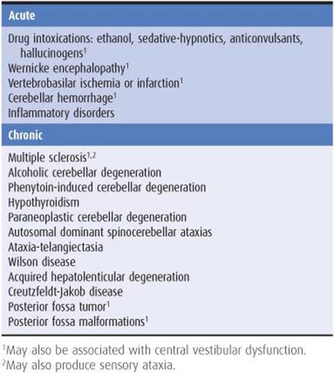
Table 8-9 Differential diagnosis of cerebellar ataxia.
ACUTE DISORDERS
DRUG INTOXICATION
Pancerebellar dysfunction—manifested by nystagmus, dysarthria, and limb and gait ataxia—is a prominent feature of drug intoxication. Drugs that can produce this syndrome include ethanol, sedative-hypnotics, anticonvulsants, and hallucinogens. The severity of symptoms is dose-related; therapeutic doses of sedatives or anticonvulsants commonly produce nystagmus, but other cerebellar signs imply toxicity.
Drug-induced cerebellar ataxia often is associated with confusion; ethanol and sedative drugs tend to cause somnolence, whereas hallucinogens are often associated with agitated delirium. In most cases, only general supportive care is necessary. The distinctive features of intoxication with each of these groups of drugs are discussed in detail in Chapter 4.
WERNICKE ENCEPHALOPATHY
Pathogenesis
Wernicke encephalopathy (see also Chapter 4) is an acute disorder comprising the clinical triad of ataxia, ophthalmoplegia, and confusion. It is caused by thiamine (vitamin B1 deficiency and is most common in chronic alcoholics, but may occur as a consequence of malnutrition from any cause.
Clinical Findings
Cerebellar and vestibular involvement both contribute to ataxia, which affects gait primarily or exclusively; the legs are ataxic in only approximately one-fifth of patients, and the arms in one-tenth. Dysarthria is rare. Other findings include an amnestic syndrome or global confusional state, horizontal or combined horizontal and vertical nystagmus, bilateral lateral rectus palsies, and absent ankle reflexes. Caloric testing shows bilateral or unilateral vestibular dysfunction. Conjugate gaze palsies, pupillary abnormalities, and hypothermia can also occur.
Diagnosis & Treatment
The diagnosis should be suspected in any patient who is alcoholic or otherwise at risk for malnutrition and is established by the clinical response to thiamine (100 mg intravenously). Ocular palsies tend to improve within hours and ataxia, nystagmus, and acute confusion within a few days. Horizontal nystagmus may persist. Ataxia is fully reversible in only approximately 40% of patients, in whom full recovery typically takes weeks to months.
VERTEBROBASILAR ISCHEMIA & INFARCTION
Transient ischemic attacks and strokes in the vertebrobasilar system (see also Chapter 13) are often associated with ataxia or vertigo.
Internal Auditory Artery Occlusion
Occlusion of the internal auditory artery (Figure 8-9), which supplies the vestibulocochlear (VIII) nerve, causes vertigo of central vestibular origin and unilateral hearing loss. This vessel may originate from the basilar or anterior inferior cerebellar artery. Vertigo is accompanied by nystagmus, with the fast phase directed away from the involved side. Hearing loss is unilateral and sensorineural in type.
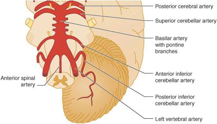
Figure 8-9. Principal arteries of the posterior fossa. (Reproduced with permission from Waxman SG. Clinical Neuroanatomy. 26th ed. New York, NY: Mc-Graw-Hill; 2010.)
Lateral Medullary Infarction
Lateral medullary infarction, which is usually caused by proximal vertebral artery occlusion, produces Wallenberg syndrome (Figure 8-10). Clinical manifestations vary, depending on the extent of infarction.

Figure 8-10. Lateral medullary infarction (Wallenberg syndrome) showing the area of infarction (shaded) and anatomic structures affected.
1. Vertigo, nausea, vomiting, and nystagmus result from involvement of the vestibular nuclei.
2. Hoarseness and dysphagia are caused by involvement of the dorsal motor nucleus of the vagus (X) nerve, nucleus solitarius, and nucleus ambiguous.
3. Ipsilateral Horner syndrome, limb ataxia, and loss of all sensation over the face and of light touch and position sense in the limbs are due to involvement of the descending sympathetic tract, inferior cerebellar peduncle, and spinal nucleus and tract of the trigeminal (V) nerve.
4. Contralateral impaired pin and temperature sense in the limbs results from involvement of the spinothalamic tract.
Cerebellar Infarction
The cerebellum is supplied by three arteries: the superior cerebellar, anterior inferior cerebellar, and posterior inferior cerebellar (Figure 8-11). The territory supplied by each of these vessels is highly variable, both from one individual to another and between the two sides of the cerebellum in a given patient. The superior, middle, and inferior cerebellar peduncles are typically supplied by the superior, anterior inferior, and posterior inferior cerebellar arteries, respectively.

Figure 8-11. Arterial supply of the cerebellum, viewed from below.
Occlusion of any cerebellar artery can cause cerebellar infarction; the clinical syndromes produced are usually distinguishable only by associated brainstem findings, if at all. Signs of cerebellar infarction include ipsilateral limb ataxia, lateropulsion (falling toward or, less commonly, away from the side of the lesion), and hypotonia. Other symptoms and signs that may be present, depending on the extent to which the cerebellum and brainstem are involved, are headache, nausea, vomiting, vertigo, nystagmus, dysarthria, ocular or gaze palsies, facial weakness or sensory loss, and contralateral hemiparesis or hemisensory deficit. Brainstem infarction or compression by cerebellar edema can result in coma and death.
The diagnosis of cerebellar infarction is made by CT scan or MRI, which allows differentiation between infarction and hemorrhage and should be obtained promptly. Brainstem compression is an indication for surgical decompression and resection of infarcted tissue, which can be lifesaving.
Paramedian Midbrain Infarction
Paramedian midbrain infarction caused by occlusion of the paramedian penetrating branches of the basilar artery affects the oculomotor (III) nerve root fibers and red nucleus (Figure 8-12). The result (Benedikt syndrome) is ipsilateral oculomotor (III) nerve involvement (producing medial rectus palsy with a fixed dilated pupil) and contralateral limb ataxia (typically affecting only the arm). Cerebellar signs result from involvement of the red nucleus, which receives a crossed projection from the cerebellum in the ascending limb of the superior cerebellar peduncle.

Figure 8-12. Paramedian midbrain infarction (Benedikt syndrome). The area of infarction is indicated by shading.
CEREBELLAR HEMORRHAGE
Pathogenesis
Cerebellar hemorrhage (see also Chapter 13) is usually due to hypertensive vascular disease; less common causes include anticoagulation, arteriovenous malformation, blood dyscrasia, tumor, and trauma. Hypertensive cerebellar hemorrhages are usually located in the deep white matter of the cerebellum and commonly extend into the fourth ventricle.
Clinical Findings
Hypertensive cerebellar hemorrhage causes the sudden onset of headache, which may be accompanied by nausea, vomiting, and vertigo, followed by gait ataxia and impaired consciousness, usually evolving over hours.
At presentation, patients can be fully alert, confused, or comatose. The blood pressure is typically elevated, and nuchal rigidity may be present. The pupils are often small and sluggishly reactive. Ipsilateral gaze palsy (with gaze preference away from the side of hemorrhage) and ipsilateral peripheral facial palsy are common. The gaze preference cannot be overcome by cold-water caloric stimulation. Nystagmus may be present, and the ipsilateral corneal reflex may be depressed. The patient, if alert, exhibits ataxia of stance and gait; limb ataxia is less common. In the late stage of brainstem compression, there is spasticity in the legs and extensor plantar responses are present.
Diagnosis & Treatment
The diagnosis of cerebellar hemorrhage can be missed or delayed, and death result, if gait is not tested in every patient with hypertension and either acute headache or depressed consciousness.
The CSF is frequently bloody, but lumbar puncture should be avoided if cerebellar hemorrhage is suspected, because it may lead to a herniation syndrome. The diagnostic procedure of choice is a CT scan. Treatment consists of surgical evacuation of the hematoma, a procedure that can be lifesaving.
INFLAMMATORY DISORDERS
Viral Infection
Cerebellar ataxia caused by viral infection is one of the principal manifestations of St. Louis encephalitis and is also seen in HIV-associated dementia and meningoencephalitis due to varicella, mumps, poliomyelitis, infectious mononucleosis, and lymphocytic choriomeningitis.
Bacterial Infection
Bacterial infection is an uncommon cause of cerebellar ataxia, but 10% to 20% of brain abscesses are located in the cerebellum, and ataxia may be a feature of meningitis due to Listeria monocytogenes in adults or Haemophilus influenzae in children. Legionella pneumophila pneumonia can also produce a cerebellar syndrome without clinical evidence of meningitis.
Acute Cerebellar Ataxia of Childhood
Acute cerebellar ataxia of childhood is characterized by severe gait ataxia that follows an acute viral infection or vaccination and usually resolves completely within months.
Acute Disseminated Encephalomyelitis
Idiopathic, immune-mediated demyelination in the cerebellar white matter may produce ataxia that is often associated with impaired consciousness, seizures, focal neurologic signs, or myelopathy (Chapter 9).
Fisher Variant of Guillain-Barré Syndrome
Ataxia, ophthalmoplegia, and areflexia constitute this variant of Guillain-Barré syndrome, which presents more typically with ascending paralysis (Chapter 9). Symptoms develop over a few days. Ataxia primarily affects the gait and trunk, with lesser involvement of the individual limbs; dysarthria is uncommon. CSF protein may be elevated. Respiratory insufficiency is rare, and the usual course is a gradual and often complete recovery over weeks to months.
CHRONIC DISORDERS
MULTIPLE SCLEROSIS
Pathogenesis
Multiple sclerosis (see also Chapter 9) is characterized clinically by remitting and relapsing neurologic dysfunction at multiple sites in the central nervous system, which may include vestibular, cerebellar, and sensory pathways. Symptoms and signs are associated with demyelination and axonal loss, which primarily affect white matter.
Clinical Findings
Nystagmus is one of the most common findings. Dysarthria also occurs frequently and vertigo less often. Gait ataxia is a presenting complaint in 10% to 15% of patients and is usually due to cerebellar involvement. Cerebellar signs are present in approximately one-third of patients on initial examination and ultimately develop in twice that number. Limb ataxia is common, usually bilateral, and tends to affect either both legs or all four limbs.
Diagnosis
The diagnosis relies heavily on a history of multiple episodes of neurologic dysfunction separated in both time and space. Evidence of subclinical lesions may be found in abnormalities such as optic neuritis, internuclear ophthalmoplegia, or pyramidal signs or from laboratory investigations. CSF analysis may reveal oligoclonal bands, elevated IgG, increased protein, or a mild lymphocytic pleocytosis. Visual, auditory, or somatosensory evoked response recording (Chapter 2) can also document sub-clinical sites of involvement. The CT scan or MRI shows areas of demyelination.
Treatment
Treatment is discussed in Chapter 9.
ALCOHOLIC CEREBELLAR DEGENERATION
Pathogenesis
A characteristic cerebellar syndrome may develop in chronic alcoholics, probably as a result of nutritional deficiency. Degenerative changes in the cerebellum are largely restricted to the superior vermis (Figure 8-13), which is also the site of cerebellar involvement in Wernicke encephalopathy.
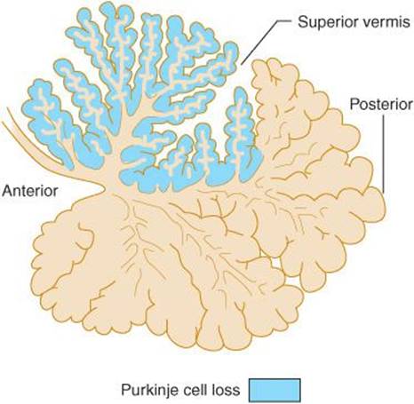
Figure 8-13. Distribution of disease in alcoholic cerebellar degeneration. Midsagittal view of the cerebellum showing loss of Purkinje cells, confined largely to the superior vermis.
Clinical Features
Alcoholic cerebellar degeneration is most common in men and usually has its onset between the ages of 40 and 60 years. Affected patients typically have a history of daily or binge drinking lasting 10 or more years with associated dietary inadequacy. Most have experienced other medical complications of alcoholism, such as liver disease, delirium tremens, Wernicke encephalopathy, or polyneuropathy. The disorder usually has an insidious onset and progresses gradually, eventually reaching a plateau level of deficit. Progression over weeks to months is most common, but in occasional cases, ataxia appears abruptly.
Gait ataxia is a universal feature and is almost always the problem that brings the patient to attention. The legs are also ataxic on heel-knee-shin testing in approximately 80% of patients. Common associated findings include distal sensory deficits in the feet and absent ankle reflexes, which result from polyneuropathy. Ataxia of the arms, nystagmus, dysarthria, hypotonia, and truncal instability are seen less frequently. CT scan or MRI may show cerebellar atrophy (Figure 8-14), but this is a nonspecific finding.

Figure 8-14. CT scan in alcoholic cerebellar degeneration, showing marked atrophy of the cerebellar vermis with relative sparing of the cerebellar hemispheres. (Courtesy of A. Gean.)
Treatment
No specific treatment is available, but patients should receive thiamine because of the apparent role of thiamine deficiency in the pathogenesis of Wernicke encephalopathy, a closely related syndrome. Abstinence from alcohol, combined with adequate nutrition, may help prevent progression.
TOXIN-INDUCED CEREBELLAR DEGENERATION
Purkinje cells and granule cells of the cerebellum are selectively vulnerable to a variety of toxins. These compounds may cause cerebellar degeneration associated with nystagmus, dysarthria, and ataxia affecting the limbs, trunk, and gait. In addition to alcohol (discussed in the preceding section), drugs and other toxins that can produce this syndrome include the anticonvulsant, phenytoin; the mood stabilizer, lithium; the antiarrhythmic, amiodarone; the anticancer drugs, fluorouracil and cytarabine; the abused inhalant, toluene; and the heavy metals, lead, mercury, and thallium. Treatment is discontinuation of the offending agents and, for fluorouracil, administration of thiamine (vitamin B1). However, toxin-induced cerebellar syndromes may be irreversible.
HYPOTHYROIDISM
Hypothyroidism can cause a subacute or chronically progressive cerebellar syndrome, which is most common in middle-aged or elderly women. Symptoms evolve over months to years. Systemic symptoms of myxedema usually precede the cerebellar disorder, but patients occasionally present first with ataxia.
Gait ataxia is universally present and is the most prominent finding. Limb ataxia is also common and may be asymmetric. Dysarthria and nystagmus occur less frequently. Other neurologic disorders related to hypothyroidism may coexist with cerebellar involvement, including sensorineural hearing loss, carpal tunnel syndrome, neuropathy, or myopathy.
Laboratory studies show decreased blood levels of thyroid hormones, elevated thyroid-stimulating hormone, and often increased CSF protein.
Replacement therapy with levothyroxine 25 to 50 μg, increased gradually to 100 to 200 μg/d orally, usually produces definite but incomplete improvement.
PARANEOPLASTIC CEREBELLAR DEGENERATION
Pathogenesis
Cerebellar degeneration can occur as a remote (paraneo-plastic) effect of systemic cancer. Lung cancer (especially small-cell), ovarian cancer, Hodgkin disease, and breast cancer are the most commonly associated neoplasms.
Paraneoplastic degeneration affects the cerebellar vermis and hemispheres diffusely. The pathogenic mechanism in many cases appears to involve onconeural antibodies, which cross-react with tumor cell antigens and cerebellar Purkinje cells.
Clinical Findings
Cerebellar symptoms may appear before or after the diagnosis of systemic cancer and typically develop over months. Although the disorder usually progresses steadily, it may stabilize; remission has been described with treatment of the underlying neoplasm.
Gait and limb ataxia are characteristically prominent, and dysarthria occurs in most cases. The limbs may be affected asymmetrically. Nystagmus is rare. Paraneoplastic involvement of other regions of the nervous system may produce associated dysphagia, dementia, memory disturbance, pyramidal signs, or neuropathy.
Diagnosis
Onconeural antibodies, such as anti-Yo (ovarian and breast cancer), anti-Hu (small-cell lung cancer), or anti-Ri (breast cancer), can sometimes be detected in the blood (Table 8-10). The CSF may show a mild lymphocytic pleocytosis or elevated protein.

Table 8-10 Onconeural antibodies associated with paraneoplastic cerebellar degeneration and related paraneoplastic syndromes.
The diagnosis of paraneoplastic cerebellar degeneration is most difficult when neurologic symptoms precede discovery of underlying cancer. The frequent occurrence of dysarthria and dysphagia helps to distinguish this condition from the cerebellar syndromes produced by chronic alcoholism or hypothyroidism. Ataxia of the arms also suggests that alcohol is an unlikely cause. Wernicke encephalopathy should always be considered because of the susceptibility of patients with cancer to malnutrition.
Treatment
Treatment is directed at the underlying tumor, supplemented in some cases with immunotherapy (eg, intravenous immunoglobulin 400-1,000 mg/d to a total dose of 2-3 g; methylprednisolone up to 1 g/d intravenously; prednisone 1 mg/kd/d orally; plasma exchange; cyclophosphamide 2 mg/kg/d orally; or rituximab 375 mg/m2 intravenously monthly for 4 months).
AUTOSOMAL DOMINANT SPINOCEREBELLAR ATAXIA
Autosomal dominant spinocerebellar ataxia (SCA) encompasses a group of genetically and clinically heterogenous disorders (Table 8-11).

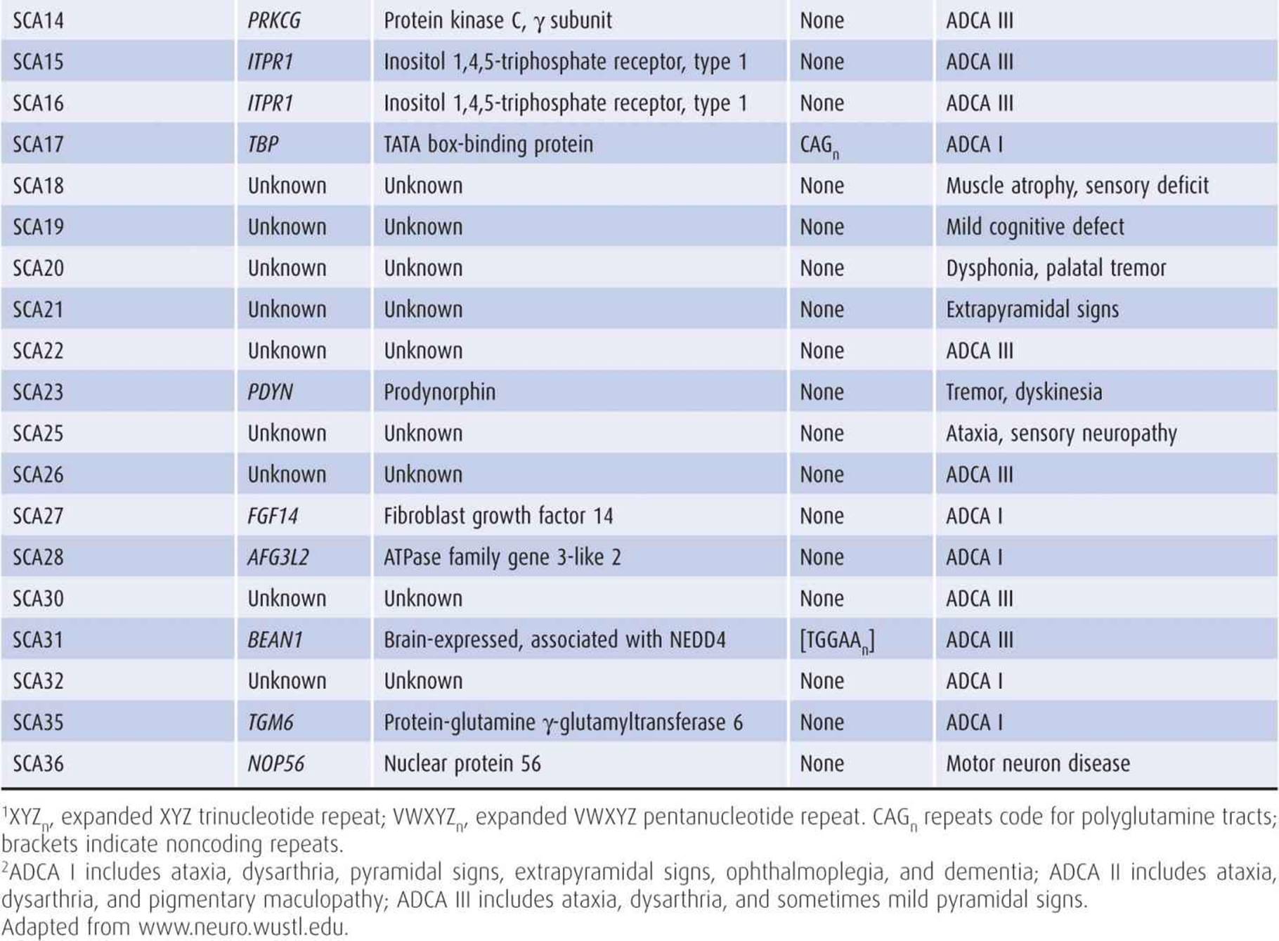
Table 8-11 Genetic and clinical features of autosomal dominant spinocerebellar ataxias (SCA).
Genetics
At least three types of mutations can produce autosomal dominant SCA: expansion of CAG trinucleotide repeats coding for polyglutamine (polyQ) tracts, expansion of tri- or pentanucleotide repeats in noncoding regions, and point mutations (Figure 8-15). Of these, the polyQ disorders are the most common and best characterized and affect a wide range of protein classes, including ion channels, receptors, enzymes, and cytoskeletal proteins.
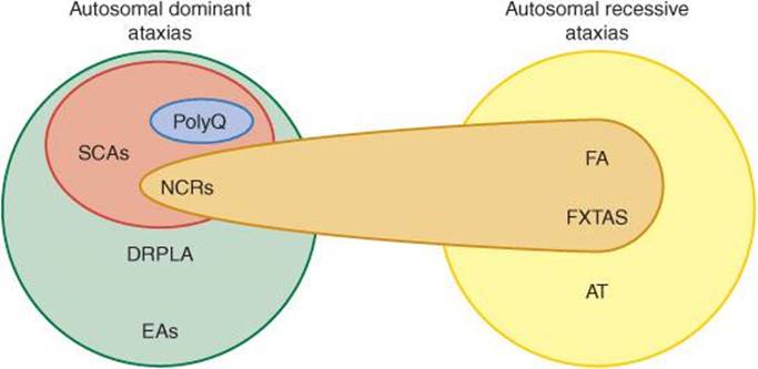
Figure 8-15. Genetic mechanisms in hereditary ataxias. Autosomal dominant ataxias include the spinocerebellar ataxias (SCAs), dentatorubral pallidoluysian atrophy (DRPLA), and the episodic ataxias (EAs). Autosomal recessive ataxias include Friedreich ataxia (FA), fragile X–associated tremor-ataxia syndrome (FXTAS), and ataxia-telangiectasia (AT). Two distinctive modes of autosomal dominant inheritance are observed in some, but not all, SCAs. In one of these (blue in figure), there is pathologic expansion of a CAG trinucleotide repeat in the coding region of an affected gene, which is translated into an abnormally long polyglutamine (PolyQ) tract within the protein (eg, SCA1, 2, 3, 6, 7, 8, 12, or 17). In the other (brown in figure), there is pathologic expansion of a tri- or pentanucleotide repeat in noncoding regions (NCRs) of the protein, which, although not translated, interferes with protein function (eg, SCA8, 10, or 31). Two autosomal recessive ataxias, FA and FXTAS, also involve trinucleotide repeats in noncoding regions.
A striking feature of polyQ disorders is that the underlying trinucleotide expansion is unstable and tends to enlarge further with time. This accounts for the phenomenon of anticipation, in which the age at onset decreases, the disease severity increases, or both, in successive generations.
In addition to SCAs, polyQ disorders include spinal bulbar muscular atrophy (Kennedy disease, Chapter 9) and Huntington disease (Chapter 11).
Pathogenesis
PolyQ expansions confer a toxic gain of function on the target protein. The abnormally long polyQ tract predisposes the protein to conformational changes, misfolding, and proteolytic cleavage (Figure 8-16). As a consequence, protein fragments are generated that are prone to aggregate and, in some cases, translocate from the cytoplasm to the nucleus. Neuronal dysfunction and death are thought to result from some combination of direct toxicity of abnormal proteins or their cytoplasmic or nuclear aggregates; impaired proteosomal function, axonal transport, or nuclear function; and protein-protein interactions.
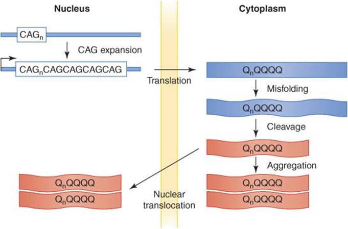
Figure 8-16. Proposed mechanisms of polyQ protein processing and toxicity. In polyQ diseases including several autosomal dominant spinocerebellar ataxias, a gene containing a CAG trinucleotide repeat (CAGn) undergoes mutation by expansion of the repeat. The resulting abnormal protein (QnQQQQ) contains an abnormally long polyQ tract, which induces conformational changes that promote misfolding. The misfolded protein is subject to proteolytic cleavage, which generates abnormal and possibly toxic fragments, which may also have an increased tendency to be translocated from the cytoplasm to the nucleus, to aggregate, or both. As a result of these events, neuronal function is impaired, and neurons may eventually die. How neurotoxicity and neuronal death ultimately occur is unknown, but there may be multiple mechanisms, and these may differ across polyQ diseases. Possible contributing factors include direct toxicity of misfolded and cleaved protein monomers or oligomers, or of cytoplasmic or nuclear aggregates (red in figure); impaired proteosomal degradation, axonal transport, or nuclear function; and interactions between polyQ proteins and other cellular proteins.
Clinical Findings
The autosomal dominant SCAs show considerable clinical variability, even within a given family. In general, they are associated with an adult-onset, slowly progressive cerebellar syndrome in which gait ataxia is an early and prominent feature. Other manifestations are dysarthria, diplopia, and limb ataxia. Extracerebellar findings are common, including cognitive, pyramidal, extrapyramidal, motor neuron, peripheral nerve, or macular involvement.
The most common SCAs are 1, 2, 3, 6, and 7. SCA1 produces gait ataxia, limb ataxia, and dysarthria, with brainstem involvement but little cognitive abnormality. SCA2 is notable for the association of ataxia and dysarthria with slow saccadic eye movements and polyneuropathy. SCA3 (Machado-Joseph disease) is especially common in patients of Portuguese ancestry; ataxia is accompanied by eyelid retraction, reduced blinking, external ophthalmoplegia, dysarthria, dysphagia, and sometimes parkinsonism or peripheral neuropathy. SCA6 is comparatively less severe, progresses more slowly, and is more limited to cerebellar involvement than other SCAs. SCA7 is distinguished by retinal degeneration leading to blindness, in addition to ataxia.
Atrophy of the cerebellum and sometimes also of the brainstem may be apparent on CT or MRI scans (Figure 8-17). However, definitive diagnosis is by demonstrating one of the known SCA gene defects on genetic testing. There is no specific treatment for the spinocerebellar ataxias, but occupational and physical therapy and devices to assist ambulation may be helpful, and genetic counseling may be indicated.

Figure 8-17. CT scan in spinocerebellar atrophy, showing an atrophic cerebellum and brainstem. (Courtesy of A. Gean.)
OTHER AUTOSOMAL DOMINANT CEREBELLAR ATAXIAS
Dentatorubral Pallidoluysian Atrophy
Dentatorubral pallidoluysian atrophy (DRPLA) is a dominantly inherited disorder that results from a polyglutamine expansion in the ATN1 gene coding for the protein atrophin 1. DRPLA causes ataxia, chorea, dementia, seizures, and myoclonus. Because of the prominent extrapyramidal features, this disorder is discussed in Chapter 11.
Episodic Ataxias
Episodic ataxias are autosomal dominant disorders characterized by transient attacks of cerebellar ataxia that may be precipitated by physical or emotional stress. Episodic ataxia 1 (EA1) results from mutations in the KCN1A gene, which codes for the Kv1.1 voltage-gated potassium channel. Attacks last from seconds to minutes and may occur many times per day; myokymia—a quivering, involuntary movement of muscle—commonly occurs between episodes. EA2 is caused by mutations in the CACNA1A gene, which codes for the α1A subunit of the P/Q-type voltage-gated calcium channel; this gene is also affected in SCA6 and familial hemiplegic migraine (Chapter 6). Attacks are more prolonged than in EA2, typically lasting for hours, and nystagmus and slowly progressive ataxia persist between acute episodes. Acetazolamide (500 mg orally four times daily) can often prevent or relieve acute symptoms in EA2. EA5 likewise affects voltage-gated calcium channels, but in this case the mutation is in CACNB4, which encodes the β subunit. EA6 is due to mutations in SLC1A3, which encodes the EAAT1 glial glutamate transporter. Glutamate uptake is reduced, leading to enhanced excitatory input onto cerebellar Purkinje cells. EA1 and EA6 are thought to impair channel function through dominant negative effects, whereas EA2 involves haploin-sufficiency; the mechanism in EA5 is uncertain.
ATAXIA-TELANGIECTASIA
Pathogenesis
Ataxia-telangiectasia (also known as Louis-Bar syndrome) is an inherited autosomal recessive disorder with onset in infancy. It results from loss-of-function mutations in the ataxia-telangiectasia mutated (ATM) gene, which codes for a serine/threonine protein kinase related to phosphatidylinositol 3-kinase. Deletions, insertions, and substitutions all have been described. A defect in the repair of DNA double strand breaks is thought to be involved in pathogenesis.
Clinical Findings
Ataxia-telangiectasia is characterized by progressive cerebellar ataxia, oculocutaneous telangiectasia, sinopulmo-nary infections, and lymphoid tumors. All patients suffer from progressive pancerebellar degeneration characterized by nystagmus, dysarthria, and gait, limb, and trunk ataxia. Choreoathetosis, loss of vibration and position sense in the legs, areflexia, and disorders of voluntary eye movement are almost universal findings. Mental deficiency is commonly observed in the second decade.
Oculocutaneous telangiectasia usually appears in the teen years. The bulbar conjunctivae are typically affected first, followed by sun-exposed areas of the skin, including the ears, nose, face, and antecubital and popliteal fossae. The vascular lesions, which rarely bleed, spare the central nervous system.
Immunologic impairment usually becomes evident later in childhood, with recurrent sinopulmonary infections in more than 80% of patients. Malignancies occur in approximately one-third of patients and include non-Hodgkin lymphoma, leukemia, and Hodgkin disease.
Other common clinical findings are progeric changes of the skin and hair, hypogonadism, and insulin-resistant diabetes mellitus. The characteristic laboratory abnormalities include decreased circulating levels of IgG2, IgA, and IgE and elevation of α-fetoprotein and carcinoembryonic antigen levels.
Because the vascular and immunologic manifestations of ataxia-telangiectasia occur later than the neurologic symptoms, the condition may be confused with Friedreich ataxia, which also manifests in childhood (see later). Ataxia-telangiectasia can be distinguished by its earlier onset (before age 4 years), associated choreoathetosis, and the absence of kyphoscoliosis.
There is no specific treatment for ataxia-telangiectasia, but antibiotics are useful in the management of infections. X-rays should be avoided because of the hypersensitivity to ionizing radiation present in this disorder.
WILSON DISEASE
Cerebellar symptoms may occur in Wilson disease, a disorder of copper metabolism characterized by copper deposition in a variety of tissues. Wilson disease is an autosomal recessive disorder that results from mutations in the ATP7B gene, which codes for the β polypeptide of a copper-transporting ATPase. Because extrapyramidal features are usually the most prominent neurologic manifestations, Wilson disease is discussed in more detail in Chapter 11.
CREUTZFELDT-JAKOB DISEASE
Creutzfeldt-Jakob disease is described in Chapter 5 as a prion disease that causes dementia. Cerebellar signs are present in approximately 60% of patients, and patients present with ataxia in approximately 10% of cases. Cerebellar involvement is diffuse, but the vermis is often most severely affected. In contrast to most other cerebellar disorders, depletion of granule cells is frequently more striking than Purkinje cell loss.
Patients with cerebellar manifestations of Creutzfeldt-Jakob disease typically complain first of gait ataxia. Dementia is usually evident at this time, and cognitive dysfunction always develops eventually. Nystagmus, dysarthria, truncal ataxia, and limb ataxia are all present initially in approximately one-half of patients with the ataxic form of Creutzfeldt-Jakob disease. The course is characterized by progressive dementia, myoclonus, and extrapyramidal and pyramidal dysfunction. Death typically occurs within 1 year after onset.
POSTERIOR FOSSA TUMORS
Tumors of the posterior fossa cause cerebellar symptoms when they arise in the cerebellum or compress it from without. The most common cerebellar tumors of childhood are astrocytomas and medulloblastomas, whereas metastases from primary sites outside the nervous system predominate in adults (Table 8-12).
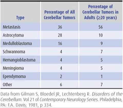
Table 8-12 Tumors of the cerebellum.
Clinical Findings
Patients with cerebellar tumors present with headache from increased intracranial pressure or with ataxia. Nausea, vomiting, vertigo, cranial nerve palsies, and hydrocephalus are common. The nature of the clinical findings varies with the location of the tumor. Most metastases are located in the cerebellar hemispheres, causing asymmetric cerebellar signs. Medulloblastomas and ependymomas, on the other hand, tend to arise in the midline, with early involvement of the vermis and hydrocephalus.
Diagnosis & Treatment
As in the case of most brain tumors, the CT scan or MRI is extremely useful for diagnosis, but biopsy may be required for histologic characterization. Methods of treatment include surgical resection, irradiation, and chemotherapy. Corticosteroids are useful in controlling tumor-associated edema.
1. Metastases—from the lung and breast and less often from other sites—are the most common tumors of the cerebellum, especially in adults. The site of the primary tumor may or may not be evident at the time the patient presents with central nervous system involvement. If the site is not evident, careful examination of the breasts and skin, chest x-ray, urinalysis, and tests for the presence of occult blood in the stool may lead to a diagnosis. The prognosis for patients with cerebellar metastases is usually worse than for patients with supratentorial lesions. Patients with carcinoma of the breast tend to survive longer than those with primary lung tumors.
2. Cerebellar astrocytomas usually occur between the ages of 2 and 20 years, but older patients can also be affected. These tumors are often histologically benign and cystic in appearance. Symptoms of increased intracranial pressure, including headache and vomiting, typically precede the onset of cerebellar dysfunction by several months. If complete surgical resection is possible, cerebellar astrocytoma is potentially curable.
3. Medulloblastoma is common in children but rare in adults. These tumors appear to originate from either cerebellar granule neuron progenitor cells with activating mutations in the Hedgehog signaling pathway or dorsal brainstem progenitor cells with activating mutations in the Wnt pathway. In contrast to astrocytomas, medulloblastomas tend to be highly malignant. They often spread through the subarachnoid space and ventricles and may metastasize outside the nervous system. Whereas most childhood medulloblastomas are located in the midline, adult-onset tumors usually arise laterally. Headache, vomiting, ataxia, and visual deterioration are common presenting symptoms. Hemiataxia is a frequent finding in adults because of the hemispheric location of most tumors. Gait ataxia, papilledema, nystagmus, facial palsy, and neck stiffness are also common. Without treatment, medulloblastoma causes death within a few months after presentation. Treatment with partial surgical resection, decompression, and craniospinal irradiation may prolong survival for years. Developing the tumors in adulthood and being female are favorable prognostic factors.
4. Acoustic neuromas have been discussed previously as a cause of vestibular nerve dysfunction. Growth of these or other less common tumors of the cerebellopontine angle may result in compression of the ipsilateral cerebellar hemisphere, causing hemiataxia in addition to the earlier symptoms of vertigo and hearing loss. These tumors are histologically benign and often fully resectable.
5. Hemangioblastoma is a rare benign tumor that usually affects adults. It can be an isolated abnormality or a feature of von Hippel-Lindau disease, which results from a dominant mutation in the VHL tumor suppressor gene. Associated features of von Hippel-Lindau disease include retinal hemangioblastoma; cysts of the kidney, pancreas, or other viscera; and polycythemia. Patients typically present with headache, and common examination findings include papilledema, nystagmus, and ataxia. Treatment is by surgical resection.
6. Meningiomas of the posterior fossa constitute 9% of all meningiomas. They are benign tumors, derived from arachnoidal cap cells of the meninges, and involve the cerebellum indirectly by compression. The locations of posterior fossa meningiomas (in decreasing order of frequency) include the posterior surface of the petrous bone, the tentorium cerebelli, the clivus, the cerebellar convexities, and the foramen magnum. Meningiomas grow slowly and usually present with headache, although tumors of the cerebellopontine angle or clivus may come to attention when they give rise to cranial nerve or brainstem symptoms. Where possible, complete surgical resection is curative.
7. Ependymomas most commonly arise from the walls or choroid plexus of the fourth ventricle. Like medulloblastomas, they are malignant tumors that seed through the ventricular system and usually occur in children. Because of their location they produce early hydro-cephalus; cerebellar signs caused by compression are late or minor manifestations. Surgical resection, cranio-spinal irradiation, and shunting procedures to relieve hydrocephalus may prolong survival, but widespread dissemination of the tumors and postoperative recurrences are common.
POSTERIOR FOSSA MALFORMATIONS
Developmental anomalies affecting the cerebellum and brainstem may present with vestibular or cerebellar symptoms in adulthood. This occurs most commonly with type I (adult) Arnold-Chiari malformation, which consists of downward displacement of the cerebellar tonsils through the foramen magnum. The clinical manifestations of this malformation are related to cerebellar involvement, obstructive hydrocephalus, brainstem compression, and syringomyelia (a cyst, or syrinx, in the spinal cord). Type II Arnold-Chiari malformation is associated with menin-gomyelocele (protrusion of the spinal cord, nerve roots, and meninges through a fusion defect in the vertebral column) and has its onset in childhood.
Cerebellar ataxia in the type I malformation usually affects the gait and is bilateral; in some cases it is asymmetric. Hydrocephalus leads to headache and vomiting. Compression of the brainstem by herniated cerebellar tissue may be associated with vertigo, nystagmus, and lower cranial nerve palsies. Syringomyelia typically produces a capelike distribution of defective pain and temperature sensation.
Arnold-Chiari malformation can be diagnosed by CT or MRI studies that demonstrate cerebellar tonsillar herniation. Patients with headache, neck pain, hydrocephalus, or other symptoms related to compression of the cerebellum or brainstem may benefit from surgical decompression of the foramen magnum. Neuropathic pain may respond to antidepressants or anticonvulsants (see Chapter 12).
SENSORY ATAXIAS
Sensory ataxia is usually the result of impaired proprioceptive sensation due to lesions of the peripheral sensory nerves (sensory neuropathy), dorsal root ganglia (sensory neuronopathy), or posterior columns of the spinal cord (myelopathy) (Table 8-13). Clinical findings include defective joint position and vibration sense in the legs and sometimes the arms, unstable stance with Romberg sign, and a gait of slapping or steppage quality.
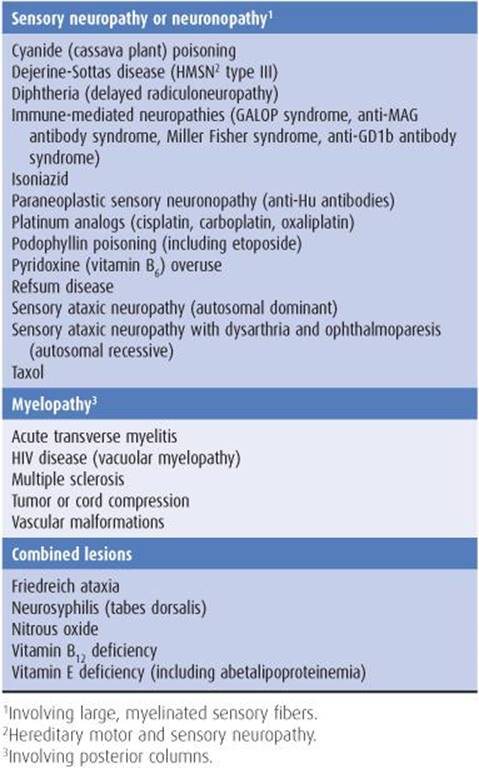
Table 8-13 Causes of sensory ataxia.
SENSORY NEUROPATHY OR NEURONOPATHY
Polyneuropathies that affect large myelinated sensory fibers and sensory neuronopathies (which target dorsal root ganglia) are the peripheral disorders most likely to present with ataxia. Prominent examples include the Hu antibody-positive sensory neuronopathy associated with small-cell lung cancer, sensory neuronopathy from consumption of high doses of pyridoxine, and the Fisher variant of the Guillain-Barré syndrome. These are discussed in more detail in Chapter 10.
MYELOPATHY
Myelopathies that affect the posterior columns can also cause ataxia. A common cause of this syndrome is multiple sclerosis, discussed earlier as a cause of cerebellar ataxia and in Chapter 9 as a cause of myelopathy.
COMBINED LESIONS
Several diseases can affect both peripheral and central sensory pathways (Figure 8-18). Examples include neurosyphilis (tabes dorsalis) and combined systems disease from vitamin B12 deficiency, which are discussed elsewhere as causes of dementia (Chapter 5) or sensory disturbance (Chapter 10). Another example is Friedreich ataxia, discussed next.

Figure 8-18. Principal sites of spinal cord disease (shading) in disorders producing sensory ataxia.
FRIEDREICH ATAXIA
Friedreich ataxia is an autosomal recessive disorder with onset in childhood and the most common cause of hereditary ataxia. It results from an expanded GAA trinucleotide repeat in a noncoding region of the FXN gene, which causes loss-of-function of a mitochondrial protein, frataxin. The key pathologic findings are degeneration of the dorsal root ganglia, large myelinated axons of peripheral sensory nerves, corticospinal tracts, and dentate nuclei of the cerebellum, with secondary involvement of the spinocerebellar tracts, posterior columns, and dorsal nucleus of Clarke.
Clinical Findings
The average age at onset is approximately 13 years, with longer GAA repeats correlating with earlier onset. The initial symptom is usually progressive gait ataxia, followed by limb ataxia, dysarthria, and sensory gait ataxia. Neurologic examination classically shows knee and ankle areflexia, impaired joint position and vibration sense in the legs, and leg (and sometimes arm) weakness, as well as extensor plantar responses. Pes cavus (high-arched feet with clawing of the toes caused by weakness and wasting of the intrinsic foot muscles) is a widely recognized sign, but may also occur in other neurologic disorders (eg, Charcot-Marie-Tooth disease).
Severe progressive kyphoscoliosis may lead to chronic restrictive lung disease. Cardiomyopathy may result in congestive heart failure, arrhythmia, and death. Other abnormalities include visual impairment from optic atrophyand diabetes mellitus.
Friedreich ataxia can usually be differentiated from other cerebellar and spinocerebellar degenerations by its early onset and the presence of prominent sensory impairment, areflexia, skeletal abnormalities, and cardiomyopathy.
Treatment & Prognosis
There is no current treatment for the neurologic manifestations of Friedreich ataxia. The antioxidant, idebenone, may be beneficial for the associated cardiomyopathy, and is under study for neurologic symptoms. Orthopedic procedures or devices are useful to improve kyphoscoliosis and gait disorder. The average duration of symptomatic illness is approximately 25 years, with death occurring at a mean age of approximately 40 years. Cardiomyopathy and infection are the usual causes of death.
REFERENCES
General
Bovo R, Ciorba A, Martini A. Vertigo and autoimmunity. Eur Arch Otorhinolaryngol. 2010;267:13-19.
Brunberg JA, Expert Panel on Neurologic Imaging. Ataxia. Am J Neuroradiol. 2008;29:1420-1422.
Kaski D, Seemungal BM. The bedside assessment of vertigo. Clin Med. 2010;10:402-405.
Kerber KA. Vertigo and dizziness in the emergency department. Emerg Med Clin Nortn Am. 2009;27:39-50.
Vestibular Disorders
Asthagiri AR, Parry DM, Butman JA, et al. Neurofibromatosis type 2. Lancet. 2009;373:1974-1986.
Dieterich M, Brandt T. Functional brain imaging of peripheral and central vestibular disorders. Brain. 2008;131:2538-2552.
Gonzalez CM, González FM, Trinidad A, et al. Medical management of Ménière’s disease: a 10-year case series and review of literature. Eur Arch Otorhinolaryngol. 2010;267:1371-1376.
Helminski JO, Zee DS, Janssen I, Hain TC. Effectiveness of particle repositioning maneuvers in the treatment of benign paroxysmal positional vertigo: a systematic review. Phys Ther. 2010;90:663-678.
Horak FB. Postural compensation for vestibular loss. Ann NY Acad Sci. 2009;1164:76-81.
Plotkin SR, Stemmer-Rachamimov AO, Barker FG 2nd, et al. Hearing improvement after bevacizumab in patients with neurofibromatosis type 2. N Engl J Med. 2009;361:358-367.
Pong WW, Gutmann DH. The ecology of brain tumors: lessons learned from neurofibromatosis-1. Oncogene. 2011;30: 1135-1146.
Schreiber BE, Agrup C, Haskard DO, Luxon LM. Sudden sensorineural hearing loss. Lancet. 2010;375:1203-1211.
Wippold FJ II, Turski PA. Vertigo and hearing loss. Am J Neuroradiol. 2009;30:1623-1625.
Cerebellar Disorders
Bauer PO, Nukina N. The pathogenic mechanisms of polyglutamine diseases and current therapeutic strategies. J Neurochem. 2009;110:1737-1765.
Biton S, Barzilai A, Shiloh Y. The neurological phenotype of ataxiatelangiectasia: solving a persistent puzzle. DNA Repair (Amst). 2008;7:1028-1038.
Brown K, Mastrianni JK. The prion diseases. J Geriat Psychiatr Neurol. 2010;23:277-298.
Dalmau J, Rosenfeld MR. Paraneoplastic syndromes of the CNS. Lancet Neurol. 2008;7:327-340.
Fernández AA, Guerrero AI, Martínez MI, et al. Malformations of the craniocervical junction (Chiari type I and syringomyelia: classification, diagnosis and treatment). BMC Musculoskelet Disord. 2009;10(suppl 1):S1.
Galvin R, Bråthen G, Ivashynka A, et al. EFNS guidelines for diagnosis, therapy and prevention of Wernicke encephalopathy. Eur J Neurol. 2010;17:1408-1418.
Greenlee JE. Treatment of paraneoplastic neurologic disorders. Curr Treat Options Neurol. 2010;12:212-230.
Jen JC, Graves TD, Hess EJ, et al. Primary episodic ataxias: diagnosis, pathogenesis and treatment. Brain. 2007;130:2484-2493.
Klockgether T. Sporadic ataxia with adult onset: classification and diagnostic criteria. Lancet Neurol. 2010;9:94-104.
Lavin MF. Ataxia-telangiectasia: from a rare disorder to a paradigm for cell signalling and cancer. Nat Rev Mol Cell Biol. 2008;9:759-769.
Millen KJ, Gleeson JG. Cerebellar development and disease. Curr Opin Neurobiol. 2008;18:12-19.
Mulholland PJ. Susceptibility of the cerebellum to thiamine deficiency. Cerebellum. 2006;5:55-63.
Ng JMY, Curran T. The Hedgehog’s tale: developing strategies for targeting cancer. Nat Rev Cancer. 2011;11:493-501.
Paulson HL. The spinocerebellar ataxias. J Neuroophthalmol. 2009;29:227-237.
Pelosof LC, Gerber DE. Paraneoplastic syndromes: an approach to diagnosis and treatment. Mayo Clin Proc. 2010;85:838-854.
Shakkottai VG, Paulson HL. Physiologic alterations in ataxia. Channeling changes into novel therapies. Arch Neurol. 2009;66:1196-1201.
Takahashi T, Katada S, Onodera O. Polyglutamine diseases: Where does toxicity come from? What is toxicity? Where are we going? J Mol Cell Biol. 2010;2:180-191.
Ye BS, Kim YD, Nam HS, Lee HS, Nam CM, Heo JH. Clinical manifestations of cerebellar infarction according to specific lobular involvement. Cerebellum. 2010;9:571-579.
Sensory Disorders
Delmas P, Hao J, Rodat-Despoix L. Molecular mechanisms of mechanotransduction in mammalian sensory neurons. Nat Rev Neurosci. 2011;12:139-153.
Koeppen AH. Friedreich’s ataxia: pathology, pathogenesis, and molecular genetics. J Neurol Sci. 2011;303:1-12.
Langan RC, Zawistoski KJ. Update on vitamin B12 deficiency. Am Fam Physician. 2011;83:1425-1430.
Marmolino D. Friedreich’s ataxia: past, present and future. Brain Res Rev. 2011;67:311-330.
Pestronk A. Neuromuscular Disease Center Web site. St. Louis, MO: Washington University; 2011. http://neuromuscular.wustl.edu/.
Stadelmann C, Wegner C, Brück W. Inflammation, demyeli-nation, and degeneration—recent insights from MS pathology. Biochem Biophs Acta. 2011;1812:275-282.
Teive HA, Haratz S, Zavala J, Munhoz RP, Scola RH, Werneck LC. Lhermitte’s sign and vitamin B12 deficiency: case report. Sao Paulo Med J. 2009;127:171-173.