
The International Association for the Study of Pain (IASP) suggests the terms complex regional pain syndrome type I (CRPS type I) for reflex sympathetic dystrophy and CRPS type II for causalgia. The sole differentiating criterion between CRPS type I and type II is the presence of a known nerve injury in CRPS type II. CRPS type I usually follows an initiating noxious event, is not limited to the distribution of a single nerve, and is apparently disproportionate to the inciting event. At some point, there is associated edema, changes in skin blood flow, abnormal sudomotor activity in the region of the pain, allodynia, or hyperalgesia. CRPS type I is associated with a variety of precipitating factors (Table 51.1); trauma is the most common precipitating event (Fig. 51.1A and B).
I. PATHOPHYSIOLOGY
Various hypotheses include peripheral mechanisms, central mechanisms, or psychogenic factors.
A. Peripheral mechanisms do not address the spread of pain beyond a dermatomal territory and pain occurring in patients without nerve injury. They are of four types.
1. After an inciting event, a subset of C-polymodal nociceptors develop sensitivity to sympathetic stimulation and thus may be stimulated by noradrenalin. An alternative explanation is that noradrenalin may act indirectly through the release of prostaglandins that stimulate the nociceptors.
2. Sympathetic efferents cause abnormal activation of peripheral nociceptors.
3. Aberrant nerve sprouts generated at the site of injury develop into neuromas.
4. Artificial synapses form at the site of nerve injury and allow “ephaptic” transmission between sympathetic efferent and sensory afferent fibers.
B. Central mechanisms are of two types.
1. Self-sustaining loops of abnormal interneuronal firing in the dorsal horn, after being propagated by a peripheral irritative focus, give rise to ascending projections of pain and descending sympathetic hyperactivity.
2. Long-term sensitization or “wind-up” of wide-dynamic-range neurons in the spinal cord resulting from ongoing nociceptive stimulation from the periphery. The sensitized wide-dynamic-range neurons then respond to activity in large diameter A-mechanoreceptors that are activated by light touch. The pain threshold is reduced and previously subthreshold stimuli are then perceived as painful.
C. Psychogenic factors. A major issue in the diagnosis and management of CRPS is the lack of properly controlled comparison studies of placebo with sympathetic blockade. Furthermore, a fair number of patients with neuropathic pain improve with injections of placebo. In some patients, symptoms may be conversion–somatization of an underlying psychiatric condition, because these patients seem to respond to cognitive psychotherapy.
II. DIAGNOSIS AND COURSE
A. Diagnosis.
1. IASP diagnostic criteria include the following:
a. Presence of an initiating noxious event or a cause of immobilization.
b. Persistent pain, allodynia, or hyperalgesia, with ongoing pain disproportionate to the inciting event.
c. Evidence at some time of edema, changes in skin blood flow, or abnormal sudomotor activity in the region of the pain.
d. Diagnosis excluded by the existence of conditions that would otherwise account for the degree of pain and dysfunction.
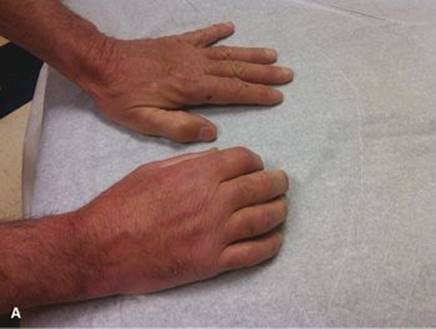
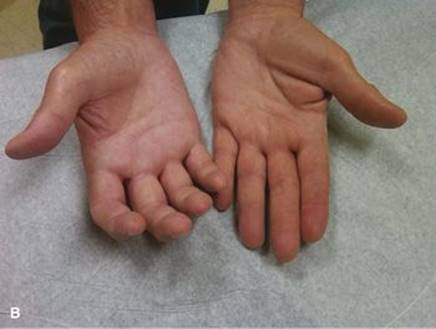
FIGURE 51.1 A: and B: A 59-year old man was drilling a hole in concrete when the drill kicked back and jerked his hand. Patient did not seek medical care for 2 weeks. When he did seek medical care, he found he had crushed two wrist bones, the trapezium, and triquetral. The patient was casted for 4 weeks and then developed severe pain and soreness of the right hand. On examination the dorsum of the right hand was purplish-reddish and very shiny, and there was more hair on the right hand than on the left. (Courtesy of Dr. Jose Biller.)
TABLE 51.1 Precipitating Factors in the Development of CRPS Type I
|
Soft-Tissue Injury |
Malignancy |
|
Fracture |
Arthritis |
|
Sprain |
Bursitis |
|
Joint dislocation |
Peripheral nerve injury |
|
Operative procedures |
Carpal tunnel release |
|
Immobilization with a cast or splint |
Venipuncture |
|
Arthroscopic surgery |
Myocardial infarction |
|
Brachial plexopathy |
Polymyalgia rheumatica |
|
Radiculopathy |
Myelopathy |
|
Stroke |
Dental extraction |
|
Spinal cord injury |
Prolonged bed rest |
|
Drugs (isoniazid, phenobarbital, ergotamine and cyclosporine) |
Criteria (b) through (d) must be satisfied.
2. Modified diagnostic criteria. A study validated the IASP diagnostic criteria but showed that the CRPS criteria have inadequate specificity and are likely to lead to overdiagnosis. The investigators proposed the following modified research diagnostic criteria for CRPS:
a. Continuing pain disproportionate to any inciting event.
b. At least one symptom in each of the four following categories:
(1) Sensory: hyperesthesia.
(2) Vasomotor: temperature asymmetry, skin color changes, skin color asymmetry, or a combination of these signs.
(3) Sudomotor edema: edema, sweating changes, sweating asymmetry, or combination of these symptoms.
(4) Motor/trophic: decreased range of motion, motor dysfunction (weakness, tremor, and dystonia), trophic changes (hair, nails, and skin), or a combination of these symptoms.
c. At least one sign in two or more of the following categories:
(1) Sensory: evidence of hyperalgesia to pin prick or allodynia to light touch.
(2) Vasomotor: evidence of temperature asymmetry, skin color changes, asymmetry, or a combination of these signs.
(3) Sudomotor/edema: evidence of edema, sweating changes, sweating asymmetry, or a combination of these symptoms.
(4) Motor/trophic: evidence of decreased range of motion, motor dysfunction (weakness, tremor, and dystonia), trophic changes (hair, nails, and skin), or a combination of these symptoms.
These research criteria may help prevent overdiagnosis of CRPS and may improve the ability to differentiate CRPS from other types of neuropathic pain.
3. Diagnosis of CRPS type I is generally made on the basis of history and clinical findings. Laboratory testing is utilized to exclude other diagnosis. Clinical features are summarized in Table 51.2. CRPS in children is often under recognized by physicians resulting in diagnostic delays. The lower extremities are more commonly affected than the upper extremities. Minor trauma remains the most frequent cause. The female preponderance is much greater among children than among adults; patients are typically pubertal adolescent girls.
A therapeutic response to sympathetic neural blockade should be carefully evaluated to identify responders to this diagnostic procedure. The pain-relieving effect should outlast the expected 6- to 12-hour effect of the local anesthetic.
4. Differential diagnosis. CRPS type II (causalgia), unrecognized local lesions (e.g., fracture, strain, and sprain), traumatic vasospasm, cellulitis, Raynaud’s disease, thromboangiitis obliterans, arterial/venous thrombosis, diabetic painful neuropathy, radicular syndromes, and gout.
5. Methods used to aid in difficult-to-diagnose cases of and various severities of CRPS type I include radiography and scintigraphy. Most laboratory tests for CRPS are based on detection of asymmetric vascular or sudomotor function between the affected and contralateral unaffected side.
a. Radiography. Plain radiographs may show patchy osteopenia in one half of all patients. Plain radiography remains useful in detecting or excluding other bony abnormalities.
b. Scintigraphy. A three-phase technetium bone scan (TPBS) is helpful in confirming the diagnosis and staging of CRPS type I. The sensitivity and specificity ranges from 54% to 100% and 85% to 98%, respectively. Given a variety of presentations of CRPS, a TPBS not only confirms the diagnosis, but it also helps exclude other diagnoses such as degenerative arthritis, benign or malignant bony lesions, or even metabolic bone diseases such as Paget’s disease, osteomyelitis, stress fracture, bone infarction, Reiter’s disease, and thoracic outlet syndrome, particularly if utilized in combination with SPECT/CT. A TPBS is used in three ways.
(1) Blood flow phase. Rapid-sequence images of the involved extremity are obtained after intravenous (IV) injection of a radionuclide tracer to evaluate the vascularity of a region.
(2) Blood pool phase. Images are obtained immediately after the blood flow phase to evaluate regional perfusion, including that of soft tissue.
(3) Bone scan phase. Static images are obtained 2 to 3 hours after initial injection to detect abnormal osteoblastic activity, reflected locally as increased periarticular uptake in the affected extremity.
c. Quantitative sudomotor axon reflex testing (QSART). Resting sweat output and stimulated sweat output as measured by QSART can help detect sudomotor asymmetry between the affected and contralateral unaffected side. Thermography and infrared thermometry are also used to measure skin temperature as an index of blood flow.
TABLE 51.2 Clinical Features of CRPS Type I
|
Feature |
Example |
|
Autonomic deregulation |
Temperature, vasomotor, and sudomotor instability |
|
Blood flow alterations |
Hyperhydrosis, hypohydrosis, edema, and discoloration |
|
Sensory abnormalities |
Hyperalgesia burning pain, hyperpathia, allodynia, dysesthesia and hemisensory impairment |
|
Motor dysfunction |
Weakness, tremor, and dystonia |
|
Trophic changes |
Skin thinning, hair loss, brittle nails, and changes in structure of both superficial and deep tissues |
|
Psychological disturbances |
Anxiety, depression, and suicidal ideation |
|
Radiologic changes |
Patchy osteoporosis, soft-tissue edema, and articular erosion |
B. Course. CRPS type I can progress through three stages (Fig. 51.2).
1. Stage 1 (acute). Pain is described as aching or burning, aggravated by physical contact or emotional upset, and typically restricted to a vascular or peripheral nerve or root territory. Some patients report paresthesias or burning distal pain. Hyperalgesia may be present, as may allodynia to light touch, thermal stimulation, deep pressure, or joint movement. Tissue swelling and local vascular, bony, and trophic changes occur in the affected part. Radiography may show diffuse bony changes. TPBS may show increased radionuclide uptake in all phases. Stage 1 usually occurs 1 to 3 months after injury.
2. Stage 2 (dystrophic) is characterized by spontaneous burning pain radiating proximally or distally from the site of injury and associated with pronounced hyperpathia, decreased hair growth, brittle nails, and indurated edematous tissue. Radiography may show patchy osteoporosis. TPBS shows normalization in the blood flow and blood pool phases. The bone scan phase remains intense. Stage 2 usually occurs 3 to 6 months after injury.
3. Stage 3 (atrophic). Pain tends to subside or diminish in intensity. The skin is cool, thin, and shiny. Irreversible trophic changes occur with subcutaneous atrophy and wasted fingertips. Radiographs may show severe patchy osteopenia. TPBS shows reduced blood flow and blood pool phases and bone scan phase normalization. Stage 3 usually occurs 6 months to years after injury.
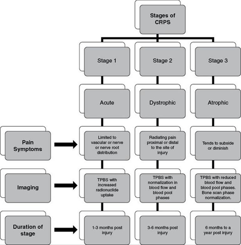
FIGURE 51.2 Stages and course of CRPS.
III. PREVENTION
Prevention is best accomplished through early recognition and treatment. Unnecessary use of braces, casts, splints, and immobilization is best avoided. If CRPS type I is suspected, heat rather than cold should be applied. Alcohol is better avoided.
IV. MANAGEMENT
Treatment goals early in the course of the disease are cessation of aberrant sympathetic hyperactivity or hypoactivity, desensitization of normal sensory pathways transmitting pain, and maintenance of normal musculoskeletal function. Overviews of the various treatment modalities available for patients with CRPS are presented in Figure 51.3. Step-wise approach for the management of upper- and lower-extremity CRPS type I are presented in algorithms in Figures 51.4 and 51.5. Management is individualized, but overall could be divided into noninvasive and invasive methods.
Noninvasive Methods
A. Pharmacologic therapy.
1. Abortive therapy.
a. Anti-inflammatory agents. Administration of nonsteroidal anti-inflammatory drugs (NSAIDs) usually is begun in the care of patients with early, mild symptoms, particularly when an inflammatory process (e.g., arthritis) is a component of the painful condition. Dosage should be maximized for optimal control of pain. NSAIDs are of minimal beneficial value, however, in the management of long-standing CRPS type I.
b. Systemic corticosteroids. In addition to having anti-inflammatory effects, corticosteroids may have inhibitory effects on spontaneous neural discharge. The effects are only temporary, and these agents do not inhibit the sympathetic reflex mechanism in CRPS type I.
(1) A standardized regimen of high initial dosage and subsequent tapering of dosage consists of prednisone (or an equivalent) at 15 mg four times a day, 10 mg four times a day, 10 mg three times a day, 10 mg twice a day, 15 mg every morning, 10 mg every morning, and 5 mg every morning for 4 days each.
(2) Injections of depot steroids directly into inflamed muscles and joints may greatly reduce pain in an affected extremity and serve adjunctively as a therapy for CRPS type I.
(3) Potential harmful effects of steroids include osteoporosis, hyperglycemia, adrenal suppression, glucose intolerance, and sodium and water retention.
c. Narcotic analgesics rarely are needed in the management of CRPS type I. Intermittent use of narcotic analgesics as a prophylactic measure may be beneficial before exercise therapy. In severe, dystrophic stages associated with long-standing pain not controlled with other medical therapy or nerve block procedures, narcotic analgesics may become a necessary adjunct to long-term care. In such cases, administration by means of an intrathecal morphine pump may be appropriate. The risks of tolerance and dependence that attend long-term narcotic use must be carefully weighed against the benefits.
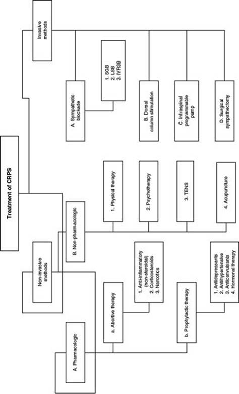
FIGURE 51.3 Overview of treatment options in patients with CRPS. SGB, stellate ganglion block; LSB, lumbar sympathetic block.
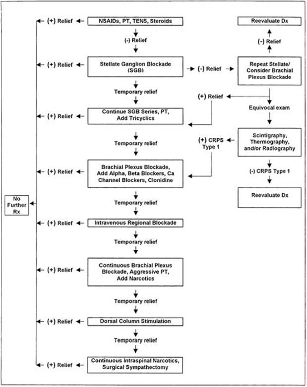
FIGURE 51.4 Upper-extremity CRPS algorithm. (+), present; (–), absent; Dx, diagnosis; Rx, treatment; Ca, calcium.
2. Prophylactic therapy.
a. Antidepressants are effective adjuvants and are commonly used in CRPS type I. The choice of drug depends on the patient’s medical profile and previous response to antidepressants.
(1) Amitriptyline is the prototype tricyclic antidepressant and is the most widely used. Two weeks of treatment may be needed before the analgesic effect is observed. Treatment should begin with small doses of 10 to 25 mg at bedtime. The dose should be increased 10 to 25 mg every 1 to 2 weeks while the patient is examined for side effects until a beneficial effect is achieved or a maximum of 150 mg is reached.
(a) Antidepressants can interfere with the reuptake mechanism of bretylium, an agent commonly used in an IV regional sympathetic blockade (IVRSB) technique. Antidepressant therapy should be discontinued for at least 2 weeks before initiation of an IV bretylium block.
(b) Complications of antidepressant use includes sedation, orthostatic hypotension, dry mouth, blurred vision, and urinary retention.

FIGURE 51.5 Lower-extremity CRPS algorithm.
b. Antihypertensives.
(1) Calcium channel blockers are used to manage in CRPS type I for their peripheral vasodilatory effects as well as their antagonistic effects with norepinephrine on arterial and venous smooth muscles.
(a) Nifedipine has the most vasodilatory effect of all the calcium channel blockers. A hypothermic extremity, as typically observed in CRPS type I, is an appropriate indication.
(b) Side effects of calcium channel blockers include an increase in pain, hypotension, myocardial depression, and cold intolerance as a result of peripheral vasodilatation.
(2) α2-Agonists have been studied in recent years to determine their analgesic effects on chronic pain states.
(a) Clonidine, a centrally acting α2-agonist, decreases sympathetic outflow and vasodilatation. Clonidine administered transdermally by means of application of a patch inhibits norepinephrine release from peripheral presynaptic adrenergic terminals. Clinically, it produces substantial reduction in hyperalgesia in response to mechanical stimuli confined primarily to the skin beneath the patch. Reports of systemic analgesic effects of clonidine are conflicting. Therefore, this agent may be most useful for patients with CRPS type I limited to only small areas. The recommended method of application is to place a patch over an area of allodynia, starting with 0.1 mg placed every 3 days and increasing the dosage in 0.1-mg increments every 12 days, to a dose of 0.3 mg. The skin should be checked for desensitization underneath the patch on replacement, and tolerance of side effects should be documented before the dosage is increased. The localized analgesia effect typically occurs within 36 to 48 hours and subsides within 1 week of discontinuance.
(b) Clonidine administered in a 300-mg dose within the epidural space produces effective analgesia for patients with refractory CRPS type I, but long-term relief has not been fully addressed.
(c) Significant side effects of clonidine include hypotension, bradycardia, and sedation.
(3) α1-Antagonists have been shown to be of some help to patients with CRPS type I.
(a) Terazosin, an oral α1-antagonist, appears to be effective for some patients and can be used in a once daily dosing regimen.
(b) Terazosin can be started at 1 mg at bedtime and titrated slowly upward to 3 mg at bedtime.
(c) Use of α-blockers is limited by their prominent side effects of hypotension, reflex tachycardia, fatigue, and dizziness.
c. Anticonvulsants stabilize abnormal hyperexcitability in both peripheral and central neurons and are thus hypothesized to inhibit excessive discharge of regional sympathetic nerves. Phenytoin at a dosage of 100 mg four times a day or carbamazepine at 200 mg three times a day may be used for the management of dysesthesia.
d. Hormonal therapy—calcitonin, given parenterally or intranasally, is widely used for CRPS type I pain in the United Kingdom. It has analgesic activity and inhibits bone resorption activity and therefore seems like a valuable aid in the management of certain types of osteoporosis.
B. Nonpharmacologic therapy.
1. Physical therapy (PT) is a useful adjunct in the management of CRPS type I. It is used to control and minimize dystrophic changes in muscles and joints. Compliance increases if adequate pain relief can be achieved before the initiation of PT.
a. Range-of-motion and stretching exercises are used for the progressive stretching of restrictive tissue for maintenance of flexibility. Aggressive treatment in the early stages has been shown to improve prognosis. It is recommended to utilize sensory stimulation, particularly with self-massage in order to keep the involved part as mobile as possible from onset of the disease process.
b. Strengthening exercises entail progressive resistance to the musculoskeletal system to maintain strength and coordination of the muscles. These exercises include isotonic, isometric, isokinetic, and aerobic exercises. If a lower extremity is involved, therapy should focus on a gradual increase in the weight-bearing capability of the limb.
c. Deep friction massage is useful as a desensitization technique in the care of patients who can tolerate such manipulation.
d. PT has been reported by many to be the mainstay of treatment for children.
2. Psychotherapy. Patients with CRPS type I experience a range of behavioral changes, including depression, anxiety, suicidal ideation, and drug addiction. Patients in whom these traits develop at or before the time of injury are considered at higher risk of development and maintenance of symptoms of CRPS type I. Therefore all such patients should be considered for psychological consultation. Psychotherapeutic management of CRPS type I includes counseling and cognitive-behavioral techniques.
a. Counseling is a means whereby the patient is helped to cope with the pain, irrespective of how well it may be controlled with pharmacologic or physical treatments. The services of a psychiatrist, psychologist, or social worker are used when appropriate.
b. Cognitive-behavioral techniques include biofeedback, relaxation, and hypnosis.
3. Transcutaneous electrical nerve stimulation (TENS). According to the gate theory put forth by Melzack and Wall in 1965, painful transmission from the periphery carried by afferent C fibers causes a loss of interneuronal large-diameter A-fiber inhibition in the dorsal horns of the spinal cord. Both sets of fibers synapse on interneurons in the substantia gelatinosa of laminae II and III before secondary fibers transmit across the midline to ascending tracts and eventually synapse in the thalamus and higher cortical centers. TENS is thought to provide pain relief by closing this gating mechanism through preferential stimulation of large-diameter A fibers and inhibition of the smaller fibers within the substantia gelatinosa or by causing the release of endorphins, enkephalins, or both. Overall, TENS relieves symptoms of CRPS type I for most children, but only for approximately 25% of adults.
4. Alternative pain management. Acupuncture has been proposed as a possible management strategy.
Invasive Methods
A. Sympathetic blockade. The mechanism of continued pain relief from sympathetic blockade is not completely understood. Relief of symptoms of early CRPS type I through inhibition of sympathetic activity seems to be a paradoxic effect if one considers that this stage is typically characterized by sympathetic hypoactivity consisting of vasodilation, redness, warmth, and sweating. Possible explanations for this effect include blockade of afferent nerve fibers transmitting pain, blockade of efferent fibers eliminating sensitization of nociceptors, and inhibition of localized vasospasm.
Sympathetic blockade can be performed on the cervical sympathetic chain, primarily the stellate ganglion, for head, neck, and upper-extremity symptoms, and on the lumbar sympathetic chain for lower-extremity manifestations. For sympathetic blockade to be used as a therapeutic intervention, the following criteria should be met:
· The blockade should demonstrate a desired effect. Efficacy is assessed subjectively by the degree of pain relief and objectively by the degree of functional improvement of the extremity.
· The effect should last longer than the known duration of the local anesthetic.
· The patient must be willing to continue a series of sympathetic blockades for the therapy to be effective.
Once a sympathetic blockade has been shown effective, a series of frequent blocks is performed. Initially, the interval between procedures should roughly equal the time it takes for the pain to return and limit rehabilitation. This typically equates to intervals ranging from daily to no more than weekly. After the first three to five blockades, the time interval is extended to approximately 1.5 times the period of pain relief and improved function. Medical therapy, PT, and desensitization techniques are used in conjunction. As long as the patient’s condition continues to improve, this therapy is continued. Resolution of symptoms typically occurs with four to eight blocks. If pain remains after sympathetic blockade, the diagnosis of CRPS type I should be reconsidered.
The patient’s condition is considered refractory if pain relief is incomplete or if the painful condition returns after a series of sympathetic blockades has been performed.
1. SGB. It involves instillation of local anesthetic at the anterior tubercle of C6. These blockades are repeated one to three times a week, up to 10 in a series, until a long-term effect is achieved. If pain relief remains inadequate, a brachial plexus block or IV regional bretylium block is attempted. Cervical epidural block with a local anesthetic as well as a narcotic has been tried in a patient with CRPS from electrical injuries.
a. Indications. CRPS type I and CRPS type II of the upper extremity are the two most common indications for SGB.
b. Complications. Injections may result in Horner’s syndrome and temporary hoarseness. Bilateral SGBs is not recommended because of possible bilateral recurrent laryngeal nerve paralysis and loss of cardioaccelerator activity. Phrenic nerve blockade results in temporary paralysis of a hemidiaphragm. Injections into the vertebral artery may result in seizures or cerebral air embolism. Intradural injections can result in unconsciousness, respiratory paralysis, seizures, and sometimes cardiovascular collapse. Other complications include brachial plexus dysfunction, pneumothorax, and osteitis of the transverse process.
2. Lumbar sympathetic block. The abdominal portion of the sympathetic trunk that supplies the lower extremities is anterolateral to the vertebral bodies of L1–3 along the medial margin of the psoas major muscle. Blockade of the lumbar sympathetic nerves can be performed with a spinal, epidural, or peripheral nerve block, but relief after lumbar sympathetic blockade most clearly delineates the cause of the pain as sympathetically mediated.
a. Indications include sympathetically mediated pain, postherpetic neuralgia, phantom limb pain, and stump pain.
b. Complications of lumbar sympathetic blockade include back pain, somatic nerve block, intraspinal anesthesia, intravascular injection, kidney trauma, and bowel perforation.
3. IVRSB. Bretylium, like guanethidine, selectively inhibits peripheral sympathetic nerve transmission by entering the preganglionic nerve endings by means of active transport and displacing norepinephrine from its storage sites. Its concentration builds up at these sites and prevents reuptake of norepinephrine and release of any remaining norepinephrine in response to neuronal stimulation. The norepinephrine depletion results in impairment and eventual loss of sympathetic adrenergic nerve function. This blockade lasts for many hours, for days, and sometimes for weeks because of the strong binding and slow elimination of the drug. In sufficient concentration, guanethidine can cause permanent damage to the norepinephrine reuptake pump. An IV regional blockade technique with ketorolac at a dose of 60 mg in saline solution or 0.5% lidocaine to volumes of 40 ml in the upper extremity and 50 ml in the lower extremity has produced prolonged pain relief with no serious side effects in some patients. IV regional blocks utilizing ketorolac and lidocaine produced only short-term pain reduction in patients with CRPS involving the lower extremity after four serial injections.
a. Indications. IVRSB with bretylium or guanethidine remains an alternative for patients who do not respond to block procedures. This procedure is less invasive than blocks and can be performed by physicians unfamiliar with the use of regional anesthesia. It is also useful for patients for whom regional anesthesia is contraindicated.
b. Complications. Placement of an Esmarch bandage and tourniquet on an extremity may be intolerable to a patient with painful CRPS type I. Agent not taken up by the tissue on release of the tourniquet may enter the systemic circulation and cause hypertension and tachycardia. Orthostatic hypotension can result from the chemical sympathetic blockade. Bretylium in high concentrations has local anesthetic and neuromuscular blocking properties. These drugs should not be administered to patients taking monoamine oxidase inhibitors or to patients with known pheochromocytoma. Other side effects include dysrhythmia, dizziness, diarrhea, edema, and nausea.
4. IV anesthetic dose. In patients with refractory CRPS, an almost complete resolution could be obtained with a combination of ketamine and midazolam infusion at anesthetic doses. Ketamine has reported to be successful orally as well, but further studies are recommended.
B. Dorsal column stimulation (DCS). If no response to conventional pharmacologic treatment is noted within 12 to 16 weeks, interventional techniques like spinal cord stimulation is recommended for refractory CRPS type I. The mechanisms of DCS are explained by the gate-control theory of pain and an increase in production of endogenous endorphins.
1. Indications. DCS is indicated for patients with chronic intractable pain. Patient-selection criteria include failure of conservative therapies, absence of drug abuse or history of psychological disorders, absence of contraindication to spinal implantation, and a successful trial of DCS with temporary electrodes.
2. Complications include infection, arachnoiditis, a high long-term failure rate, and mechanical failure.
C. Intraspinal opioid programmable pump. Preganglionic sympathetic intermediolateral columns of the spinal cord are modulated by projection from multiple supraspinal nuclei, including the nucleus raphae magnus. Intrathecal administration of morphine may be effective in relieving refractory CRPS type I by increasing nucleus raphae magnus inhibition of sympathetic outflow, in addition to its inhibitory effects on nociceptive neurons. Moreover, continuous intrathecal administration of morphine with a programmable pump allows stable CSF concentrations. The result is fewer episodes of pain breakthrough or drug overdose. Intrathecal therapy may also reduce the overall dosage requirements and delay the development of tolerance.
1. Indications. The intraspinal opioid programmable pump is reserved for treatment for patients with CRPS type I who are unresponsive to all other forms of less invasive measures. A successful trial of intrathecal morphine placed percutaneously is required.
2. Complications. An important disadvantage of an intraspinal opioid infusion pump is the high cost of implanting and maintaining the device. In addition, physical tolerance and dependence complicate therapy for many patients. Other problems include infection, arachnoiditis, mechanical failure, unremitting breakthrough pain, and opioid tolerance and dependence.
D. Surgical sympathectomy. For upper-extremity CRPS type I, surgical sympathectomy involves extensive ablation of the thoracic sympathetic ganglia from T1 to T6 or T7 through a transthoracic approach. Surgical sympathectomy for lower-extremity CRPS type I involves ablation of the lumbar sympathetic chain from L1 to L4. The pain relief derived from surgery may be inadequate or transient owing either to incomplete sympathetic denervation or to subsequent nerve regeneration.
1. Indications. Surgical sympathectomy usually is reserved for patients with CRPS type I who have been able to obtain definite but only temporary relief from repeated sympathetic nerve blocks and remain incapacitated by the disease.
2. Complications. Potential morbidity, including wound complications, permanent Horner’s syndrome, and painful neuralgia, warrants exhaustive physical, pharmacologic, and psychological therapy before consideration of surgical sympathectomy.
V. REFERRALS
Patients with CRPS type I should be referred to a specialist in chronic pain treatment or a neurologist if
· signs and symptoms remain persistent despite a 6-week trial of conservative measures,
· pain is severe and intractable any time after the inciting event,
· a new-onset neurologic deficit is noted, or
· signs and symptoms have spread to other parts of the body.

Recommended Readings
Besson JM, Chaouch A. Peripheral and spinal mechanisms of nociception. Physiol Rev. 1987;67:67–186.
Bruehl S, Harden RN, Galer BS, et al. External validation of IASP diagnostic criteria for complex regional pain syndrome and proposed research diagnostic criteria. Pain. 1999;81:147–154.
Demangeat JL, Constantinesco A, Brunot B, et al. Three-phase bone scanning in reflex sympathetic dystrophy of the hand. J Nucl Med. 1988;29:26–32.
Eckmann MS, Ramamurthy S, Griffin JG. Intravenous regional ketorolac and lidocaine in the treatment of complex regional pain syndrome of the lower extremity: a randomized, double-blinded, crossover study. Clin J Pain. 2011;27(3):203–206.
Gobelet C, Waldburger M, Meier JL. The effect of adding calcitonin to physical treatment on reflex sympathetic dystrophy. Pain. 1992;48:171–175.
Kiefer RT, Rohr P, Ploppa A, et al. Complete recovery from intractable complex regional pain syndrome, CRPS-type I, following anesthetic ketamine and midazolam. Pain Prac. 2007;7(2):147–150.
Lee J, Nandi P. Early aggressive treatment improves prognosis in complex regional pain syndrome. Practitioner. 2011;255(1736):23–26.
Low AK, Ward K, Wines AP. Pediatric complex regional pain syndrome. Pediatr Orthop. 2007;27(5):567–572.
Lynch ME. Psychological aspect of reflex sympathetic dystrophy: a review of the adult and pediatric literature. Pain. 1992;49:337–347.
Mackey S, Feinberg S. Pharmacologic therapies for complex regional pain syndrome. Curr Pain Headache Rep. 2007;11(1):38–43.
Melzack R, Wall PD. Pain mechanisms: a new theory. Science. 1965;150:971–975.
Nitzsche EU. Nuclear medicine imaging for diagnosis of CRPS I. Handchir Mikrochir Plast Chir. 2011;43(1):20–24. Epub 2011 Jan 11.
Rauck RL, Eisenach JC, Jackson K, et al. Epidural clonidine treatment for reflex sympathetic dystrophy. Anesthesiology. 1993;79:1163–1169.
Rommel O, Gehling M, Dertwinkel R, et al. Hemisensory impairment in patients with complex regional pain syndrome. Pain. 1999;80(1–2):95–101.
Rowbotham MC. Pharmacologic management of complex regional pain syndrome. Clin J Pain. 2006;22(5):425–429.
Sandroni P, Low PA, Ferrer T, et al. Complex regional pain syndrome I (CRPS I): prospective study and laboratory evaluation. Clin J Pain. 1998;14:282–289.
Sprague M, Chang JC. Integrative approach focusing on acupuncture in the treatment of chronic complex regional pain syndrome. J Altern Complement Med. 2011;17(1):67–70. Epub 2011 Jan 5.
Stanton-Hicks M, Janig W, Hassenbusch S, et al. Reflex sympathetic dystrophy: changing concepts and taxonomy. Pain. 1995;63:127–133.
Stanton-Hicks M. Complex regional pain syndrome: manifestations and the role of neurostimulation in its management. J Pain Symptom Manage. 2006;31(4 suppl):S20–S24.
Villanueva-Perez VL, Cerda-Olmedo G, Samper JM, et al. Oral ketamine for the treatment of type I complex regional pain syndrome. Pain Pract. 2007;7(1):39–43.