
Alan Ashworth
INTRODUCTION
Cancer cells may harbor defects in DNA repair pathways leading to genomic instability. This can foster tumorigenesis but also provides a weakness that can be exploited therapeutically. Tumors with compromised ability to repair double-strand DNA breaks by homologous recombination, including those with defects in the BRCA1 and BRCA2 genes, are highly sensitive to blockade of the repair of DNA single-strand breaks, via the inhibition of the enzyme poly(ADP-ribose) (PARP). This provides the basis for a synthetic lethal approach to cancer therapy, which is showing considerable promise in the clinic.
CELLULAR DNA REPAIR PATHWAYS
DNA is continually damaged by environmental exposures and endogenous activities, such as DNA replication and cellular free-radical generation, which cause diverse lesions including base modifications, double-strand breaks (DSB), single-strand breaks (SSB), and intrastrand and interstrand cross-links.1 These aberrations are repaired by distinct repair pathways, which are coordinated to maintain the stability and integrity of the genome. This faithful repair of DNA damage is an essential prerequisite for the maintenance of genomic integrity and cellular and organismal viability. Where one DNA strand is affected and the intact complementary strand is available as a template, the base-excision repair (BER), nucleotide-excision repair, or mismatch repair pathways are used and these pathways are highly efficient at repairing damage. DSBs, more problematic than SSBs because the complementary strand is not available as a template, are repaired by the homologous recombination (HR) or nonhomologous end-joining (NHEJ) pathways.1
Endogenous base damage, including SSBs, is the most common DNA aberration and it has been estimated that the average cell may repair 10,000 such lesions every day. BER is an important pathway for the repair of SSBs and involves the sensing of the lesion followed by the recruitment of a number of other proteins. PARP-1 (poly[ADP]ribose polymerase) is a critical component of the major “short-patch” BER pathway. PARP is an enzyme, discovered over 40 years ago,2 that produces large branched chains of poly(ADP) ribose (PAR) from NAD+. In humans, there are 17 members of the PARP gene family but most of these are poorly characterized.3,4 The abundant nuclear protein PARP-1 senses and binds to DNA nicks and breaks, resulting in activation of catalytic activity causing poly(ADP)ribosylation of PARP-1 itself as well as other acceptor proteins including histones. This modification may signal the recruitment of other components of DNA repair pathways as well as modify their activity. The highly negatively charged PAR that is produced around the site of damage may also serve as an antirecombinogenic factor. In addition to the BER pathway PARP enzymes have been implicated in numerous cellular pathways.3,4
Two main DSB repair pathways are available within eukaryotic cells: NHEJ and HR.5,6 HR can be further subdivided into the gene conversion (GC) and single-strand annealing (SSA) subpathways.1 Both GC and SSA rely on sequence homology for repair whereas NHEJ uses no, or little, homology.2,3 NHEJ is the most important pathway for the repair of DSBs during G0, G1, and early S phases of the cell cycle, although it is likely active throughout the cell cycle.7,8 This form of DSB repair usually results in changes in DNA sequence at the break site and, occasionally, in the joining of previously unlinked DNA molecules, potentially resulting in gross chromosomal rearrangements such as translocations.9 GC uses a homologous sequence, preferably the sister chromatid, as a template to resynthesize the DNA surrounding the DSB, and therefore generally results in accurate repair of the break. Repair by GC is critically dependent on the recombinase function of RAD51 and is facilitated by a number of other proteins. SSA also involves the use of homologous sequences for the repair of DSBs, but unlike GC, SSA is RAD51-independent and involves the annealing of DNA strands formed after resection at the DSB. The detailed mechanism of SSA is still obscure but it frequently results in the loss of one of the homologous sequences and deletion of the intervening sequence.9 SSA is a potentially important pathway of mutagenesis because a significant fraction of mammalian genomes consist of repetitive elements. GC and SSA are cell-cycle regulated and are most active in S-G2 phases of the cell cycle.10
THE DEVELOPMENT OF PARP INHIBITORS
PARP inhibitors were originally developed as chemopotentiators, which are agents that enhance the effects of DNA damage—a common mechanism of action of drugs used to treat cancer. The rationale was that inhibition of the repair of chemotherapy-induced DNA damage might give greater efficacy. Early studies using relatively nonspecific PARP inhibitors such as 3-aminobenzamide, demonstrated potential synergy with alkylating agents.11 Subsequent studies with more potent PARP inhibitors demonstrated synergy with temozolomide, an observation that was taken into a clinical trial with AG014699,12 a PARP inhibitor developed by Pfizer. This agent is now being developed by Clovis. Although the major focus of this chapter is the use of PARP inhibitors in synthetic lethal therapeutic strategies, their use in chemopotentiation in combination with chemotherapy remains under active investigation, as described later.
BRCA1 AND BRCA2 MUTATIONS AND DNA REPAIR
Heterozygous germline mutations in the BRCA1 and BRCA2 genes confer a high risk of breast (up to 85% lifetime risk) and ovarian (10% to 40%) cancer in addition to a significantly increased risk of pancreatic, prostate, and male breast cancer.13 The genes have been classified as tumor suppressors, because the wild-type BRCA allele is frequently lost in tumors, a phenomenon that occurs by a variety of mechanisms. The BRCA1 and BRCA2 genes encode large proteins that likely function in multiple cellular pathways, including transcription, cell-cycle regulation, and the maintenance of genome integrity. However, the roles of BRCA1 and BRCA2 in DNA repair have been best documented.14
BRCA1- and BRCA2-deficient cells are highly sensitive to ionizing radiation and display chromosomal instability, which is likely to be a direct consequence of unrepaired DNA damage.14 The similar genomic instability in BRCA1- and BRCA2-deficient cells and the interaction of both BRCA1 and BRCA2 with RAD51 suggested a functional link between the three proteins in the RAD51-mediated DNA damage repair process. However, although BRCA2 is directly involved in RAD51-mediated repair, affecting the choice between GC and SSA, BRCA1 acts upstream of these pathways15; both GC and SSA are reduced in BRCA1-deficient cells, placing BRCA1 before the branch point of GC and SSA.15
BRCA1 has a role in signaling DNA damage and cell-cycle checkpoint regulation,14,15 whereas BRCA2 has a more direct role in DNA repair itself. BRCA2 is thought to promote genomic stability through a role in the error-free repair of DSBs by GC via association with RAD51. Aberrations in BRCA2-deficient cells arise at least in part by the use of the SSA pathway. NHEJ, however, is apparently unaffected in BRCA2-deficient cells.14,15 Loss of BRCA2, therefore, results in the repair of DSBs by preferential utilization of an error-prone mechanism, which potentially explains the apparent chromosome instability associated with BRCA2 deficiency.15
The physical interaction between BRCA2 and RAD51 is essential for error-free DSB repair. BRCA2 is required for the localization of RAD51 to sites of DNA damage, where RAD51 forms the nucleoprotein filament required for recombination. The foci of the RAD51 protein are apparent in the nucleus after certain forms of DNA damage and these likely represent sites of repair by HR; BRCA2-deficient cells do not form RAD51 foci in response to DNA damage.15 Two different domains within BRCA2 interact with RAD51, the eight BRC repeats in the central part of the protein and a distinct domain, TR2, at the C-terminus.16
PARP-1 INHIBITION AS A SYNTHETIC LETHAL THERAPEUTIC STRATEGY FOR THE TREATMENT OF BRCA-DEFICIENT CANCERS
Synthetic lethality is defined as the situation when a mutation in either of two genes individually has no effect, but combining the mutations leads to death.17 This effect was first described and studied in genetically tractable organisms such as Drosophila and yeast.17,18 This effect can arise because of a number of different gene–gene interactions. Examples include two genes in separate semiredundant or cooperating pathways, and two genes acting in the same pathway where loss of both critically affects flux through the pathway. The implication is that targeting one of these genes in a cancer where the other is defective should be selectively lethal to the tumor cells but not toxic to the normal cells. In principle, this should lead to a large therapeutic window.19 The original suggestion that the concept of synthetic lethality could be used in the selection or development of cancer therapeutics came from Hartwell et al.,18 and from experiments performed in yeast. Synthetic lethal screens have now been performed in a number of model organisms20 and in human cells,21 and these have revealed multiple potential gene–gene interactions, some of which could be exploited clinically. However, synthetic lethal therapies have not been clinically used until recently, when evidence has been provided for PARP-1 inhibition as a potential synthetic lethal approach for the treatment of BRCA-mutation–associated cancers.
PARP-1 inhibition causes failure of the repair of SSB lesions but does not affect DSB repair.22 However, a persistent DNA SSB encountered by a DNA replication fork will cause stalling of the fork and may result in either fork collapse or the formation of a DSB.23 Therefore, the loss of PARP-1 increases the formation of DNA lesions that might be repaired by GC. As a loss of function of either BRCA1 or BRCA2 impairs GC,14,15 a loss of PARP-1 function in a BRCA1- or BRCA2-defective background could result in the generation of replication-associated DNA lesions normally repaired by sister chromatid exchange. If so, this might lead to cell-cycle arrest and/or cell death. Therefore, PARP inhibitors could be selectively lethal to cells lacking functional BRCA1 or BRCA2 but might be minimally toxic to normal cells. This would indicate a synthetic lethal interaction between PARP and BRCA1 or BRCA2. Exemplifying this principle, potent inhibitors of PARP were applied to cells deficient in either BRCA1 or BRCA2. Cell survival assays showed that cell lines lacking wild-type BRCA1 or BRCA2 were extremely sensitive to these agents compared with heterozygous mutant or wild-type cells.24,25
To explain these observations, a model was proposed whereby persistent single-strand gaps in DNA caused by PARP inhibition when encountered by a replication fork might trigger fork arrest, collapse, and/or a DSB.26Alternatively, PARP-1 trapped on DNA by the inhibition of enzyme activity might also cause a fork collapse. Normally, these DSBs would be repaired by RAD51-dependent GC.14,15 However, in the absence of BRCA1 or BRCA2, the replication fork cannot be restarted and collapses, causing persistent chromatid breaks. When repaired by the alternative error-prone DSB repair mechanisms of SSA or NHEJ, large numbers of chromatid aberrations would be induced, leading to cell lethality.26 The idea that the defect in GC is being targeted in BRCA-deficient cells is supported by the demonstration that deficiency in other genes implicated in HR also confers sensitivity to PARP inhibitors.27 This further suggests that this approach may be more widely applicable in the treatment of sporadic cancers with impairments of the HR pathway or BRCAness28 (see the following).
INITIAL CLINICAL RESULTS TESTING SYNTHETIC LETHALITY OF PARP INHIBITORS AND BRCA MUTATION
Phase I studies29 established that olaparib (AstraZeneca, London, UK; formerly KU-0059436, KuDOS Pharmaceuticals, Cambridge, UK) could be administered safely as a single agent at a dose of 400 mg twice per day. Side effects were classified as mild and were unlike those typically experienced with cytotoxic chemotherapy. Significant and durable responses were observed in patients with germ-line BRCA1or BRCA2 mutations and breast ovary or prostate cancer. Of the 19 mutation carriers enrolled, 9 had an objective response defined by Response Evaluation Criteria in Sold Tumors (RECIST) criteria and 12 had stable disease for more than 4 months in duration. A similar magnitude of clinical responses was observed in an expanded cohort.30 These observations are impressive because the cohort had been heavily pretreated and most were resistant to a wide range of chemotherapies.29,30
Phase II studies were subsequently performed in advanced breast and ovarian cancers arising in BRCA1 and BRCA2 mutation carriers.31,32 The reported response rate was 41% in the breast study and 52% in the ovarian group; both groups had been heavily pretreated. Again, the drug was well tolerated. Another study of BRCA1/2 carriers with ovarian cancer compared olaparib with pegylated liposomal doxorubicin (PLD).33 There was no significant difference in the response rates, but there were some differences in the patient characteristics and an unexpectedly high rate of response to PLD.
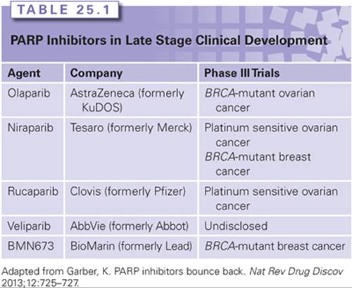
There are also reports of responses to PARP inhibitors in BRCA2 mutation carriers with prostate34 and pancreatic35 cancer. A number of other PARP inhibitors are in clinical development (Table 25.1), and some of these have shown efficacy in the treatment of cancers arising in BRCA1 or BRCA2 mutation carriers.36,37
THE USE OF PARP INHIBITORS IN SPORADIC CANCERS
Germline mutations in BRCA1 or BRCA2 are relatively common in hereditary breast and ovarian cancer. However, inactivation of BRCA genes by mutation in sporadic cancers is rare, at least in breast cancer, which may seem to limit the application of PARP inhibitors to a wider range of patients. However, many tumors display features in common with BRCA-deficient tumors, including similar defects in DNA repair due to either epigenetic mutation of BRCA1, such as promoter methylation, or mutation of other components of BRCA-associated pathways.28 This BRCAness may make these tumors also susceptible to PARP inhibition.28 For example, phosphatase and tensin homolog (PTEN)mutations, which occur with a frequency estimated at 50% to 80% in sporadic tumors,38 may cause PARP inhibitor sensitivity in preclinical models, possibly because PTEN-null cells display BRCAness phenotypes, such as the inability to efficiently repair certain forms of DNA damage.39
Traditional histopathologic methods and, more recently, gene expression profiling approaches have shown the phenotypic overlap between triple-negative breast cancers, basal-like breast cancers, and BRCA1familial breast cancers.40,41 In gene expression profiling studies, it has been observed that BRCA1 familial cancers strongly segregate with basal-like tumors and share features such as high-grade and pushing margins.28,40,41 Although the overlap is not absolute, it leads to the hypothesis that there may be a subset of sporadic breast cancers that exhibits features of BRCAness, including deficiencies in HR and that may be susceptible to treatment with drugs such as PARP inhibitors.26
There have been several studies of PARP inhibitors in sporadic ovarian cancer. A study by Lederman42 showed in a maintenance study following the response to platinum therapy a significant benefit in terms of progression-free survival (PFS) of olaparib compared to placebo. This was even more pronounced when the subgroup of BRCA mutation carriers were examined.43 In both cases, the overall survival (OS) advantage was less than the PFS, but in the case of the BRCA mutation group, this reached statistical significance. Gelmon44 also showed activity in sporadic ovarian cancer. In contrast, a study in sporadic triple-negative breast cancer failed to observe any benefit, although the study was small and the patients were heavily pretreated.44
Iniparib (initially reported as a PARP inhibitor) showed an overall survival benefit in a Phase II trial of triple-negative breast cancer in combination with gemcitabine and carboplatin compared with chemotherapy alone.45However, a subsequent Phase III study showed no improvement in PFS.45 The reasons for this are uncertain, but significant questions have been raised about whether iniparib is indeed a bona fide PARP inhibitor. Therefore, it is now generally conceded that studies of iniparib have no implications for PARP inhibitors as a drug class.46
Which population of patients lacking a BRCA1 or BRCA2 mutation might benefit from PARP inhibitors remains unclear. This is likely to require the development of a clinical test to identify prospectively tumors with intrinsic sensitivity. Presently, most efforts are directed at developing assays of DNA repair deficiency.47
MECHANISMS OF RESISTANCE TO PARP INHIBITORS
Resistance to targeted therapy frequently occurs, but it was unclear how resistance might arise to a synthetic lethal therapy.48 Potential mechanisms of resistance to PARP inhibitors have, however, been elucidated both directly in vitro, in mouse models, and in the clinic.48 An in vitro model for resistance was developed by producing cells from the highly PARP inhibitor–sensitive BRCA2-deficient cell line CAPAN1, which carries a c.6174delT BRCA2frameshift mutation. CAPAN1 cells cannot form damage-induced RAD51 foci, are defective for HR, and are extremely sensitive to treatment with PARP inhibitors.49 PARP inhibitor–resistant clones were highly resistant (over 1,000-fold) to the drug and were also cross-resistant to the DNA cross-linking agent cisplatin, but not to the microtubule-stabilizing drug docetaxel. PARP inhibitors and cisplatin both exert their effects on BRCA-deficient cells by increasing the frequency of misrepaired DSBs in the absence of effective HR. Therefore, this observation indicates that the resistance of PARP inhibitor–resistant clones to PARP inhibitors might be because of restored HR. This contention was supported by the acquisition in PARP inhibitor–resistant clone cells of the ability to form RAD51 foci after PARP inhibitor treatment or exposure to irradiation.
DNA sequencing of PARP inhibitor–resistant clones revealed the unexpected presence of novel BRCA2 alleles that resulted in the elimination of the c.6174delT mutation and restoration of an open reading frame.49 Therefore, in this case, resistance arises because of gain of function mutations in the synthetic lethal partner (BRCA2) rather than the direct drug target (PARP). Alternative mechanisms of PARP inhibitor resistance have also been described.48 A mouse model of BRCA1-associated mammary gland cancer demonstrated the efficacy of olaparib in vivo and was used to study mechanisms of resistance.50Resistance seemed to be caused by the upregulation of ABCB1a/b, which encode P-glycoprotein pumps; this effect could be reversed with the P-glycoprotein inhibitor tariquidar. In addition, other alterations in DNA repair pathways have been proposed to compensate for BRCA1 deficiency resulting in PARP inhibitor deficiency.48
Studies of the mechanisms of resistance to PARP inhibitors in patient material are still at an early stage. Initial studies addressed the mechanism of resistance to platinum salts in BRCA mutation carriers. Cisplatin and carboplatin are part of the standard of care for the treatment of ovarian cancer, including individuals with BRCA1 or BRCA2 mutations. Platinum salts are thought to exert their BRCA-selective effects by a similar mechanism to PARP inhibitors.15 Clinical observations suggest that BRCA mutation carriers with ovarian cancer usually respond better to these agents than patients without BRCAmutations51,52; however, resistance does eventually occur. To investigate this effect, BRCA1 and BRCA2 have been sequenced in tumor material from mutation carriers.49,53 These studies revealed mutations in BRCA1 or BRCA2 that restored the open reading frame and likely contributed to platinum resistance. These observations suggest that specific mutations in BRCA1 or BRCA2 and sensitivity to therapeutics in cell lines and patients can be suppressed by intragenic deletion. Presumably, these mutations occur randomly and are then selected for by differential drug sensitivity. Therefore, the best use of these agents is likely to be earlier in the disease process when the disease burden is smaller, which will reduce the probability of resistance based on stochastic genetic reversion. Recently, similar observations of revertant BRCA alleles were made in two patients who became resistant after an initial response to olaparib.54 Although preliminary, these results suggest that this mechanism is responsible for at least some of the clinical resistance observed. Doubtless, as with other targeted therapies, multiple resistance mechanisms will be implicated as further patients are studied.48
PROSPECTS
Currently, the treatments for cancers arising in carriers of BRCA1 or BRCA2 mutations are the same as those that occur sporadically matched for tumor pathology and age of onset. However, tumors in BRCA1or BRCA2 mutation carriers lack wild-type BRCA1 or BRCA2, but normal tissues retain a single wild-type copy of the relevant gene. This is a potentially targetable alteration that provides the basis for new mechanism-based approaches to the treatment of cancer. The biochemical difference in capacity to carry out HR between the tumor and normal tissues, in a BRCA1 or BRCA2 carrier, provides the rationale for this approach. Inhibiting the DNA repair protein PARP results in the generation of specific DNA lesions that require BRCA1 and BRCA2 specialized repair function(s) for their removal. Preclinical data indicate that tumors defective in wild-type BRCA1 or BRCA2 could be much more sensitive to PARP inhibition than unaffected heterozygous tissues, providing a potentially large therapeutic window. The safety and efficacy of this approach is currently being tested in clinical trials, which, if successful, may lead to registration for routine clinical use of one or more PARP inhibitors.37
Synthetic lethality by combinatorial targeting of DNA repair pathways may have usefulness as a therapeutic approach beyond familial cancers. The majority of solid tumors also exhibit genomic instability and aneuploidy. This suggests that pathways involved in the maintenance of genomic stability are dysfunctional in a significant proportion of neoplastic disorders.47 Understanding which specialized DNA damage response and repair pathways are abrogated in sporadic tumor subtypes may allow for the development of therapies that target the residual repair pathways on which the cancer, but not normal tissue, is now completely dependent. These potential therapies may significantly improve response rates while causing fewer treatment-related toxicities. However, these approaches may be associated with mechanism-associated resistance, and careful consideration of their optimal use will be required.
REFERENCES
1. Hoeijmakers JH. Genome maintenance mechanisms for preventing cancer. Nature 2001;411:366–374.
2. Chambon P, Weill JD, Mandel P. Nicotinamide mononucleotide activation of new DNA-dependent polyadenylic acid synthesizing nuclear enzyme. Biochem Biophys Res Commun 1963;11:39–43.
3. Amé JC, Spenlehauer C, de Murcia G. The PARP superfamily. Bioessays 2004;26:882–893.
4. Otto H, Reche PA, Bazan F, et al. In silico characterization of the family of PARP-like poly(ADP-ribosyl)transferases (pARTs). BMC Genomics 2005;6:139.
5. van Gent DC, Hoeijmakers JH, Kanaar R. Chromosomal stability and the DNA double-stranded break connection. Nat Rev Genet 2001;2:196–206.
6. Shin DS, Chahwan C, Huffman JL, et al. Structure and function of the double-strand break repair machinery. DNA Repair (Amst) 2004;3:863–873.
7. Takata M, Sasaki MS, Sonoda E, et al. Homologous recombination and non-homologous end-joining pathways of DNA double-strand break repair have overlapping roles in the maintenance of chromosomal integrity in vertebrate cells. EMBO J 1998;17:5497–5508.
8. Rothkamm K, Krüger I, Thompson LH, et al. Pathways of DNA double-strand break repair during the mammalian cell cycle. Mol Cell Biol 2003;23:5706–5715.
9. Stark JM, Pierce AJ, Oh J, et al. Genetic steps of mammalian homologous repair with distinct mutagenic consequences. Mol Cell Biol 2004;24:9305–9316.
10. Elliott B, Richardson C, Jasin M. Chromosomal translocation mechanisms at intronic alu elements in mammalian cells. Mol Cell 2005;17:885–894.
11. Durkacz BW, Omidiji O, Gray DA, et al. (ADP-ribose)n participates in DNA excision repair. Nature 1980;283(5747):593–596.
12. Tertoli L, Graziani G. Chemosensitisation by PARP inhibitors in cancer therapy. Pharmacol Res 2005;52:25–33.
13. Wooster R, Weber BL. Breast and ovarian cancer. N Engl J Med 2003;348:2339–2347.
14. Gudmundsdottir K, Ashworth A. The roles of BRCA1 and BRCA2 and associated proteins in the maintenance of genomic stability. Oncogene 2006;25:5864–5874.
15. Tutt AN, Lord CJ, McCabe N, et al. Exploiting the DNA repair defect in BRCA mutant cells in the design of new therapeutic strategies for cancer. Cold Spring Harb Symp Quant Biol 2005;70:139–148.
16. Lord CJ, Ashworth A. RAD51, BRCA2 and DNA repair: a partial resolution. Nat Struct Mol Biol 2007;14:461–462.
17. Dobzhansky T. Genetics of natural populations: Xiii. Recombination and variability in populations of Drosophila pseudoobscura. Genetics 1946;31:269–290.
18. Hartwell LH, Szankasi P, Roberts CJ, et al. Integrating genetic approaches into the discovery of anticancer drugs. Science 1997;278:1064–1068.
19. Kaelin WG Jr. The concept of synthetic lethality in the context of anticancer therapy. Nat Rev Cancer 2005;5:689–698.
20. Ooi SL, Pan X, Peyser BD, et al. Global synthetic-lethality analysis and yeast functional profiling. Trends Genet 2006;22:56–63.
21. Iorns E, Lord CJ, Turner N, et al. Utilizing RNA interference to enhance cancer drug discovery. Nat Rev Drug Discov 2007;6:556–568.
22. Noël G, Giocanti N, Fernet M, et al. Poly(ADP-ribose) polymerase (PARP-1) is not involved in DNA double-strand break recovery. BMC Cell Biol 2003;4:7.
23. Haber JE. DNA recombination: the replication connection. Trends Biochem Sci 1999;24:271–275.
24. Farmer H, McCabe N, Lord CJ, et al. Targeting the DNA repair defect in BRCA mutant cells as a therapeutic strategy. Nature 2005;434:917–921.
25. Bryant HE, Schultz N, Thomas HD, et al. Specific killing of BRCA2-deficient tumours with inhibitors of poly(ADP-ribose) polymerase. Nature 2005;434:913–917.
26. Ashworth A. A synthetic lethal therapeutic approach: PARP inhibitors for the treatment of cancers deficient in double-strand break repair. J Clin Oncol 2008;26:3785–3790.
27. McCabe N, Turner NC, Lord CJ, et al. Deficiency in the repair of DNA damage by homologous recombination and sensitivity to poly(ADP-ribose) polymerase inhibition. Cancer Res 2006;66:8109–8115.
28. Turner N, Tutt A, Ashworth A. Hallmarks of ‘BRCAness’ in sporadic cancers. Nat Rev Cancer 2004;4:814–819.
29. Fong PC, Boss DS, Yap TA, et al. Inhibition of poly(ADP-ribose) polymerase in tumors from BRCA mutation carriers. N Engl J Med 2009;361:123–134.
30. Fong PC, Yap TA, Boss DS, et al. Poly(ADP)-ribose polymerase (PARP) inhibition: frequent durable responses in BRCA carrier ovarian cancer correlating with platinum-free interval. J Clin Oncol 2010;28:2512–2519.
31. Audeh MW, Carmichael J, Penson RT, et al. Oral poly(ADP-ribose) polymerase inhibitor olaparib in patients with BRCA1 or BRCA2 mutations and recurrent ovarian cancer: a proof-of-concept trial. Lancet 2010;376:245–251.
32. Tutt A, Robson M, Garber JE, et al. Oral poly(ADP-ribose) polymerase inhibitor olaparib in patients with BRCA1 or BRCA2 mutations and advanced breast cancer: a proof-of-concept trial. Lancet 2010;376:235–244.
33. Kaye SB, Lubinski J, Matulonis U, et al. Phase II, open-label, randomized, multicenter study comparing the efficacy and safety of olaparib, a poly (ADP-ribose) polymerase inhibitor, and pegylated liposomal doxorubicin in patients with BRCA1 or BRCA2 mutations and recurrent ovarian cancer. J Clin Oncol2012;30:372–379.
34. Sandhu SK, Omlin A, Hylands L et al. Poly (ADP-ribose) polymerase (PARP) inhibitors for the treatment of advanced germline BRCA2 mutant prostate cancer. Ann Oncol 2013;24:1416–1418.
35. Fogelman DR, Wolff RA, Kopetz S, et al. Evidence for the efficacy of Iniparib, a PARP–1 inhibitor, in BRCA2-associated pancreatic cancer. Anticancer Res 2011;31:1417–1420.
36. Maxwell KN, Domchek SM. Cancer treatment according to BRCA1 and BRCA2 mutations Nat Rev Clin Oncol 2012;9:520–528.
37. Garber K. PARP inhibitors bounce back. Nat Rev Drug Discov 2013;12:725–727.
38. Salmena L, Carracedo A, Pandolfi PP. Tenets of PTEN tumor suppression. Cell 2008;133:403–414.
39. Mendes-Pereira AM, Martin SA, Brough R, et al. Synthetic lethal targeting of PTEN mutant cells with PARP inhibitors. EMBO Mol Med 2009;1:315–322.
40. Foulkes WD, Stefansson IM, Chappuis PO, et al. Germline BRCA1 mutations and a basal epithelial phenotype in breast cancer. J Natl Cancer Inst 2003;95:1482–1485.
41. Turner NC, Reis-Filho JS. Basal-like breast cancer and the BRCA1 phenotype. Oncogene 2006;25:5846–5853.
42. Ledermann J, Harter P, Gourley C, et al. Olaparib maintenance therapy in platinum-sensitive relapsed ovarian cancer. N Engl J Med 2012;366:1382–1392.
43. Ledermann JA, Harter P, Gourley C. Olaparib maintenance therapy in patients with platinum-sensitive relapsed serous ovarian cancer (SOC) and a BRCA mutation (BRCAm). J Clin Oncol 2013;31 (suppl; abstr 5505).
44. Gelmon KA, Tischkowitz M, Mackay H, et al. Olaparib in patients with recurrent high-grade serous or poorly differentiated ovarian carcinoma or triple-negative breast cancer: a phase 2, multicentre, open-label, non-randomised study. Lancet Oncol 2011;12:852–861.
45. O’Shaughnessy J, Osborne C, Pippen J, et al. Iniparib plus chemotherapy in metastatic triple-negative breast cancer. N Engl J Med 2011;3:205–214.
46. Mateo J, Ong M, Tan DS, et al. Appraising iniparib, the PARP inhibitor that never was—what must we learn? Nat Rev Clin Oncol 2013;10:688–696.
47. Lord CJ, Ashworth A. The DNA damage response and cancer therapy. Nature 2012;481:287–294.
48. Lord CJ, Ashworth A. Mechanisms of resistance to therapies targeting BRCA-mutant cancers. Nat Med 2013;19:1381–1388.
49. Edwards S, Brough R, Lord CJ, et al. Resistance to therapy caused by intragenic deletion in BRCA2. Nature 2008;451(7182):1111–1115.
50. Rottenberg S, Jaspers JE, Kersbergen A, et al. High sensitivity of BRCA1-deficient mammary tumors to the PARP inhibitor AZD2281 alone and in combination with platinum drugs. Proc Natl Acad Sci U S A 2008;105:17079–17084.
51. Cass I, Baldwin RL, Varkey T, et al. Improved survival in women with BRCA-associated ovarian carcinoma. Cancer 2003;97:2187–2195.
52. Pal T, Permuth-Wey J, Kapoor R, et al. Improved survival in BRCA2 carriers with ovarian cancer. Fam Cancer 2007;6:113–119.
53. Sakai W, Swisher EM, Karlan BY, et al. Secondary mutations as a mechanism of cisplatin resistance in BRCA2-mutated cancers. Nature 2008;451:1116–1120.
54. Barber LJ, Sandhu S, Chen L, et al. Secondary mutations in BRCA2 associated with clinical resistance to a PARP inhibitor. J Pathol 2013;229:422–429.