
Approach to the Problem
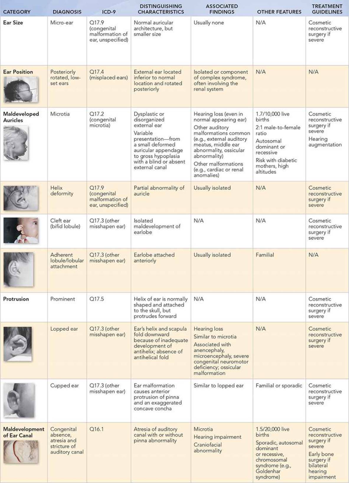
Normally, there is a wide variation of shapes, sizes, and positions of ears in children. Most auricular growth (85%) is completed by 3 years of age, cartilaginous formation by 5 to 6 years, and ear width and its distance from the scalp by 10 years. Abnormalities in ear shape or position may occur as isolated findings or as part of a complex of congenital anomalies. Congenital malformations of the external ear, which occur in 1:10,000 to 1:20,000 live births, include problems with ear size (e.g., micro-ear), position (e.g., posteriorly rotated, low-set ears), maldevelopment (e.g., anotia, microtia, cleft earlobe, lobular attachment), and protrusion (e.g., prominent, lopped, cupped).
Malformed or underdeveloped auricles are frequently seen with genetic problems such as Beckwith–Wiedemann syndrome (creased lobes), CHARGE association (lopped or cupped ears), facio-auriculo-vertebral spectrum (Goldenhar syndrome; microtia), Levy–Hollister (cupped ears), or Trisomy 21 (small ears). Low-set ears commonly occur in syndromes such as Noonan, Smith–Lemli–Opitz, Treacher Collins, and Trisomy 18. Several studies have found an association between renal anomalies and various ear abnormalities. Although the underlying etiology is unclear, the anomalies usually are not found as isolated findings, but rather as components of more complex congenital syndromes, such as Beckwith–Wiedemann or Trisomy 18.
When inspecting the external ear, it is important to evaluate its position, size, shape, and symmetry compared with the other ear. The protrusion angle of the ear should not exceed 15 degrees in children. Fifteen percent of the auricle (the superior attachment of the pinna) should be above the horizontal line (an imaginary line drawn from the inner canthus through the outer canthus). The angle between the vertical axis (the line perpendicular to the horizontal line) and the longitudinal axis of the ear (superior aspect of the outer helix to the inferior border of the earlobe) is normally between 10 and 30 degrees. In addition, the length of an ear can be roughly estimated by measuring the distance between the arch of the eyebrow and the base of the ala nasi.
Key Points in the History
• Because hearing impairment is associated with microtia, lopped, or cupped ears, and with meatal atresia, it is essential to ask about hearing and language development.
• Underlying renal anomalies should be considered when children with ear abnormalities have a history of deafness or a maternal history of gestational diabetes.
• Children with microtia often have hearing loss on the side of their normal-appearing auricle.
• Familial inheritance patterns are seen with abnormal earlobe attachments and cupped ears; therefore, asking about a family history of ear abnormalities may be helpful.
Key Points in the Physical Examination
• Children with posteriorly rotated, low-set ears should be inspected carefully for other congenital abnormalities.
• When a child has protruding ears, normal auricular architecture distinguishes prominent ears from lopped or cupped ears.
• Micro-ears, unlike maldeveloped auricles such as microtia, are small but normally formed.
• Marked skull molding can make normal auricles appear protruded.
• Abnormal facial features, including small chin, midfacial or nose hypoplasia, and highly arched eyebrows, can give the false impression of low-set, posteriorly rotated ears.
• Normally developed helices distinguish intrauterine compression abnormalities from the array of helix deformities. Also, intrauterine positioning effects do not generally result in symmetric abnormalities.
• Evaluation for renal anomalies should be considered when a patient with an auricular abnormality has other dysmorphic features, including facial asymmetry, choanal atresia, micrognathia, colobomas of the eye, branchial cysts, cardiac abnormalities, imperforate anus, and/or distal limb abnormalities.
|
PHOTOGRAPHS OF SELECTED DIAGNOSES |

Figure 17-1 Normal anatomy of external ear. (Used with permission from Fletcher MA, ed. Physical Diagnosis in Neonatology. Philadelphia, PA: Lippincott–Raven Publishers; 1998:285.)

Figure 17-2 Posteriorly rotated, low-set ears. (Used with permission from Fletcher MA, ed. Physical Diagnosis in Neonatology. Philadelphia, PA: Lippincott–Raven Publishers; 1998:287.)

Figure 17-3 Microtia. (Used with permission from Cotton RT, Myer CM III, eds. Practical Pediatric Otolaryngology. Philadelphia, PA: Lippincott–Raven Publishers; 1999:345.)

Figure 17-4 Helix deformity. (Used with permission from Fletcher MA, ed. Physical Diagnosis in Neonatology. Philadelphia, PA: Lippincott–Raven Publishers; 1998:288.)
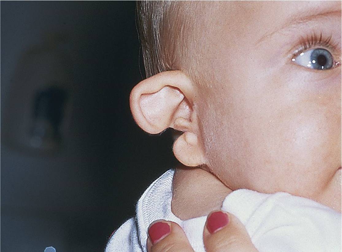
Figure 17-5 Cleft ear. (Courtesy of Steven D. Handler, MD, MBE.)

Figure 17-6 Adherent lobule/lobular attachment. (Used with permission from Fletcher MA, ed. Physical Diagnosis in Neonatology. Philadelphia, PA: Lippincott–Raven Publishers; 1998:289.)
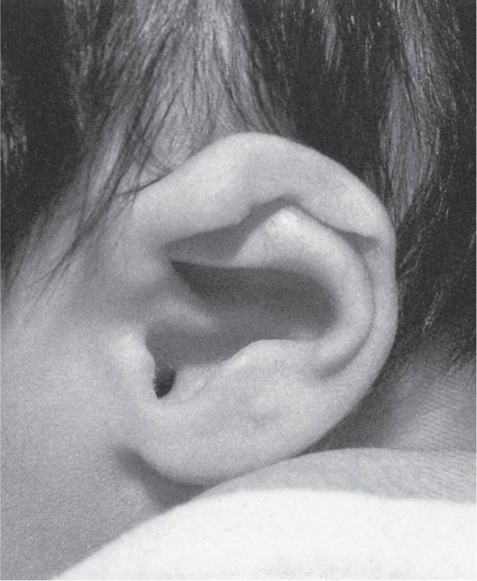
Figure 17-7 Lopped ear. (Used with permission from Fletcher MA, ed. Physical Diagnosis in Neonatology. Philadelphia, PA: Lippincott–Raven Publishers; 1998:297.)
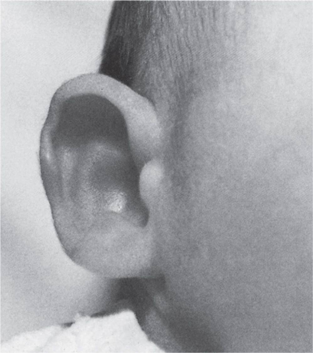
Figure 17-8 Cupped ear. (Used with permission from Fletcher MA, ed. Physical Diagnosis in Neonatology. Philadelphia, PA: Lippincott–Raven Publishers; 1998:298.)

Figure 17-9 Meatal atresia. (Courtesy of Steven D. Handler, MD, MBE.)
DIFFERENTIAL DIAGNOSIS
Other Diagnoses to Consider
• Intrauterine compression, such as folded helix
• Marked skull molding (appears “protruded”)
• Craniofacial disproportion forms such as severe microcephaly (ear appears large)
• Appearance of “low-set” or “posteriorly rotated” ears when abnormal facial features are present, such as a small chin, midface hypoplasia, or highly arched eyebrows
• Epidermal nevus on ear
• Arteriovenous malformation
When to Consider Further Evaluation or Treatment
• Malformed or underdeveloped auricles, as well as low-set ears, are often associated with more complex congenital syndromes or part of genetic disorders. Maldeveloped ears should be evaluated by otolaryngology and/or plastic surgery, particularly when considering cosmetic, reconstructive surgery.
• Closely monitor hearing, with periodic hearing evaluation, and language development in children with microtia, lopped, and cupped ears, as well as meatal atresia.
• Renal anomalies, often diagnosed by renal ultrasound, should be considered if a child with an ear abnormality has a history of deafness, a maternal history of gestational diabetes, or other dysmorphic features.
SUGGESTED READINGS
Bellucci RJ. Congenital aural malformation: Diagnosis and treatment. Otolaryngol Clin North Am. 1981;14:95–124.
Bordley JE, Brookhouser PE, Tucker GK, eds. Ear, Nose and Throat Disorders in Children. New York, NY: Raven Press; 1986.
Cotton RT, Myer CM III, eds. Practical Pediatric Otolaryngology. Philadelphia, PA: Lippincott–Raven Publishers; 1999.
Fletcher MA, ed. Physical Diagnosis in Neonatology. Philadelphia, PA: Lippincott–Raven Publishers; 1998.
Jones KL. Smith’s Recognizable Patterns of Human Malformation. 7th ed. Philadelphia, PA: Saunders; 2013.
Wang RY, Earl DL, Ruder RO, et al. Syndromic ear anomalies and renal ultrasounds. Pediatrics. 2001;108(2):E32.