
Approach to the Problem
An understanding of the natural history; a careful physical examination including range of motion, neurovascular examination, visual inspection, and palpation; and serial measurements allow proper diagnosis for most pedal (foot) deformities. Most importantly, the timing of presentation is critical. Congenital pedal deformities include metatarsus adductus, metatarsus varus, calcaneovalgus foot, rocker-bottom foot (congenital vertical talus), and clubfoot (talipes equinovarus). Pes planus (flatfeet) may be of either the soft or the rigid variety, and both are typically noticed as the child begins to ambulate. Flat feet are common in children because arch development occurs primarily before 4 years of age, and because the development has a wide variation in the rate or onset in any given child. The treatment of these defects is overwhelmingly conservative, and surgery is reserved for older children with severe deformities that will not improve over time.
Key Points in the History
• Intoeing and outtoeing may result from more proximal, tibial, femoral, or hip defects, such as long bone torsion, bowlegs, or knock-knees.
• While the majority of clubfoot and pes planus deformities are sporadic, these deformities have also been linked to inherited defects.
• The timing of ambulation in children with metatarsus adductus and pes planus is not generally delayed.
• Metatarsus adductus is the most common congenital foot deformity. It occurs more frequently in women, is more common on the left side than on the right side, and typically improves with time.
• Metatarsus adductus, clubfoot, and flexible pes planus are infrequently associated with pain, whereas rigid pes planus may be associated with significant discomfort.
• Trauma, occult infection, a foreign body, tarsal coalition, bone tumors, or osteochondrosis of the tarsal navicular bone may cause a stiff and painful flat foot.
• The presence of systemic symptoms, such as fever, may be suggestive of more serious foot disorders, such as infection or malignancy.
• When metatarsus adductus is also associated with hindfoot inversion, plantar flexion, a hypoplastic ipsilateral calf, or a slightly shortened tibia, a diagnosis of clubfoot should be considered.
Key Points in the Physical Examination
• With a clubfoot defect, there is extreme plantar flexion of the ankle (equinus). The heel is in varum (medial deviation), and the sole is kidney shaped (adducted and supinated) when viewed from the bottom. Also, callous and hyperpigmentation may be present on the dorsolateral clubfoot. The foot will have a deep medial skin furrow and, in bilateral cases, the soles of the feet will face each other. The Achilles tendon is tight, and there is limited dorsiflexion.
• In evaluation of metatarsus adductus, the forefoot has a convex, lateral border, and a crease over the medial midfoot is visible. The metatarsals are deviated medially. Also, the forefoot easily corrects to midline with gentle pressure.
• A calcaneovalgus foot appears flat, the heel is angled away from midline, and the ankle is in dorsiflexion so that the dorsal foot rests against the tibia. The ankle typically has limited dorsiflexion.
• Congenital vertical talus appears similar to calcaneovalgus, but is a rigid deformity.
• Flexible pes planus (flatfoot) is identified by appearance of the pedal arch when the child stands on his or her toes. Upon standing, however, the arch disappears.
• Rigid pes planus (flatfoot) is indicated by the lack of pedal arching while standing normally and standing on one’s toes.
• A compensatory flatfoot may occur as the result of a tight heel cord.
• Substantial pain upon gentle palpation, erythema, or localized firmness may be suggestive of more significant underlying pathology, such as cellulitis, osteomyelitis, joint infection, or malignancy.
|
PHOTOGRAPHS OF SELECTED DIAGNOSES |

Figure 49-1 Clubfeet. Bilateral clubfeet in an infant with notable metatarsus adductus. (Courtesy of Gerardo Cabrera-Meza, MD.)

Figure 49-2 Clubfeet. Dorsal view of clubfeet in an infant with plantar flexion and foot inversion. (Courtesy of Gerardo Cabrera-Meza, MD.)
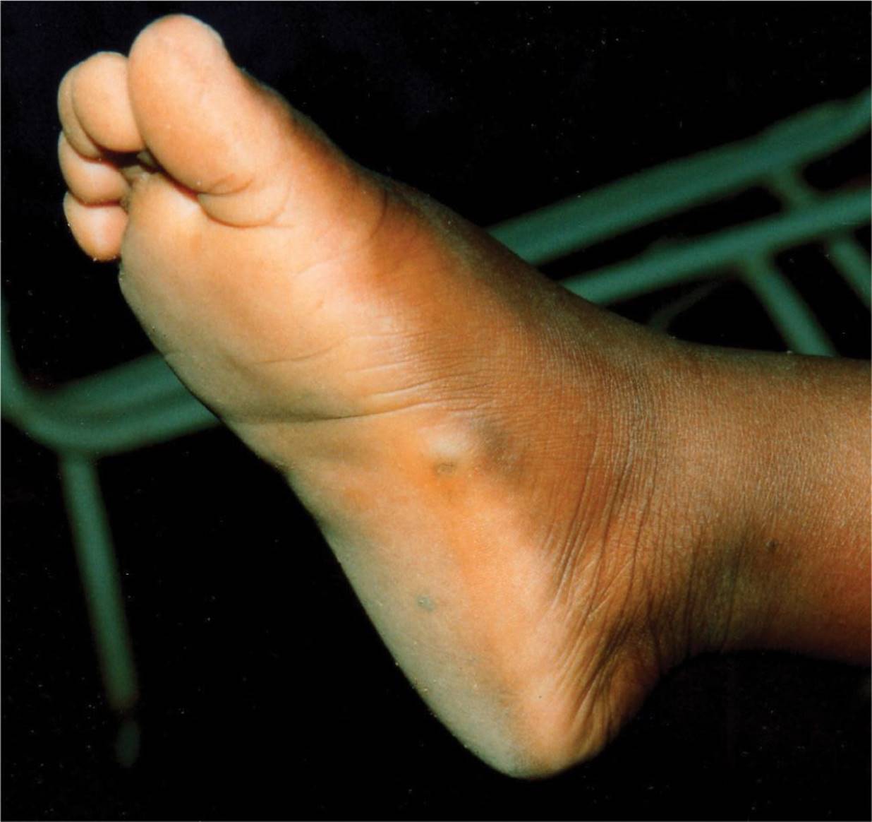
Figure 49-3 Pes planus. Arch absent in non weight-bearing position in this child with rigid pes planus. (Courtesy of Tom Thacher, MD.)

Figure 49-4 Pes planus. Mild pronation noted in this child with pes planus. (Courtesy of Sujata R. Tipnis, MD.)
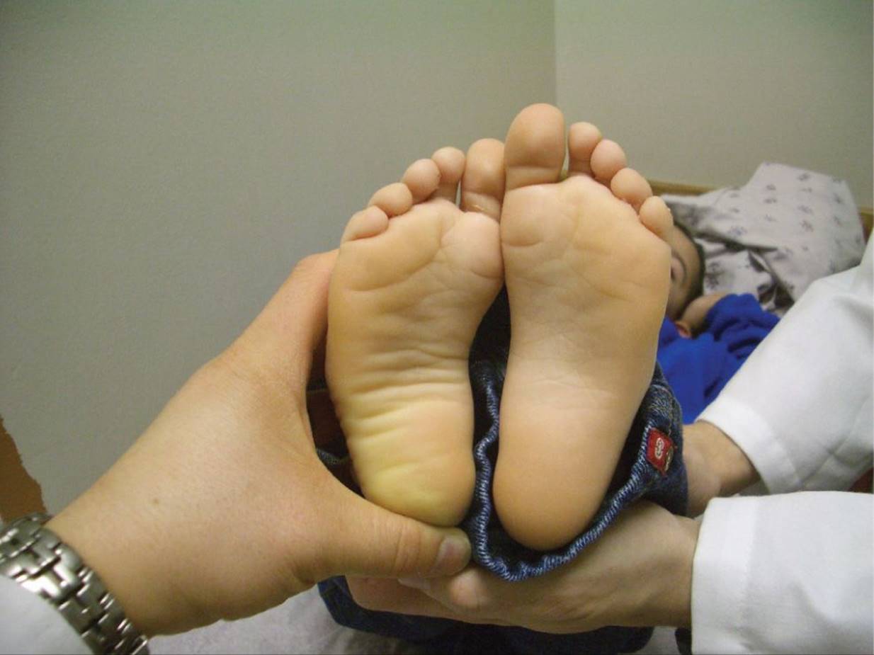
Figure 49-5 Metatarsus adductus. The convex (“C”) shape of the child’s right foot suggests metatarsus adductus. (Courtesy of Paul S. Matz, MD.)

Figure 49-6 Rocker-bottom feet. Gentle curvature to the bottom of this infant’s feet is typical of rocker-bottom feet and may be associated with Patau syndrome or Edward syndrome. (Courtesy of Gerardo Cabrera-Meza, MD.)
DIFFERENTIAL DIAGNOSIS
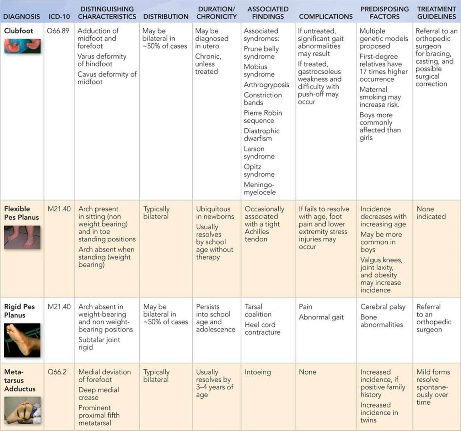
Other Diagnoses to Consider
• Trauma
• Infection
• Cavus foot (high-arched foot)
• Trauma (sprains, strains, or fractures)
• Infection
• Cavus foot (high-arched foot)
• Neoplasm
When to Consider Further Evaluation or Treatment
• Clubfoot deformity should be referred to an orthopedic surgeon for bracing, casting, and possible surgical correction.
• Flexible pes planus and metatarsus adductus should be simply observed as treatment is generally not indicated.
• A stiff or painful flat foot should be referred to an orthopedic surgeon.
• A tight heel cord should be further evaluated by an orthopedic surgeon and/or by a neurologist if hypertonicity is present.
SUGGESTED READINGS
Dietz FR. Intoeing—fact, fiction and opinion. Am Fam Physician. 1994;50(6):1249–1259.
Horn BD, Davidson RS. Current treatment of clubfoot in infancy and childhood. Foot Ankle Clin. 2010;15(2):235–243.
Kasser JR. The foot. In: Morrissy RT, Weinstein SL, eds. Lovell and Winter’s Pediatric Orthopaedics. 6th ed. Philadelphia, PA: Lippincott Williams & Wilkins; 2006:1258–1328.
McCrea JD. Pediatric Orthopedics of the Lower Extremity: An Instructional Handbook. Mount Kisco, NY: Futura Publishing Company, Inc.; 1985.
Roye BD, Hyman J, Roye DP Jr. Congenital idiopathic talipes equinovarus. Pediatr Rev. 2004;25(4):124–130.
Sass P, Hassan G. Lower extremity abnormalities in children. Am Fam Physician. 2003;68(3):461–468.
Scherl SA. Common lower extremity problems in children. Pediatr Rev. 2004;25(2):52–62.
Yagerman SE, Cross MB, Positano R, et al. Evaluation and treatment of symptomatic pes planus. Curr Opin Pediatr. 2011;23(1):60–67.