
Approach to the Problem
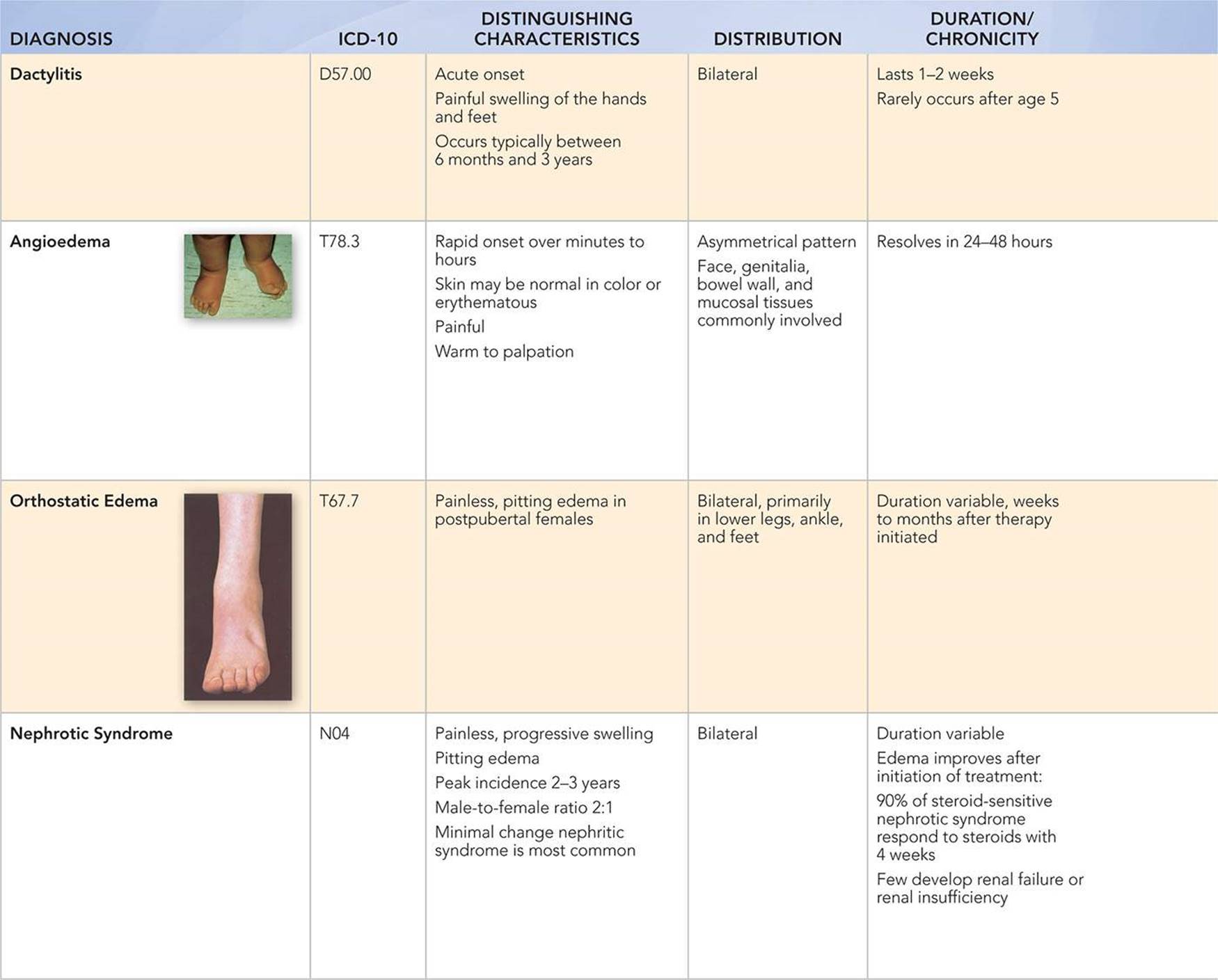
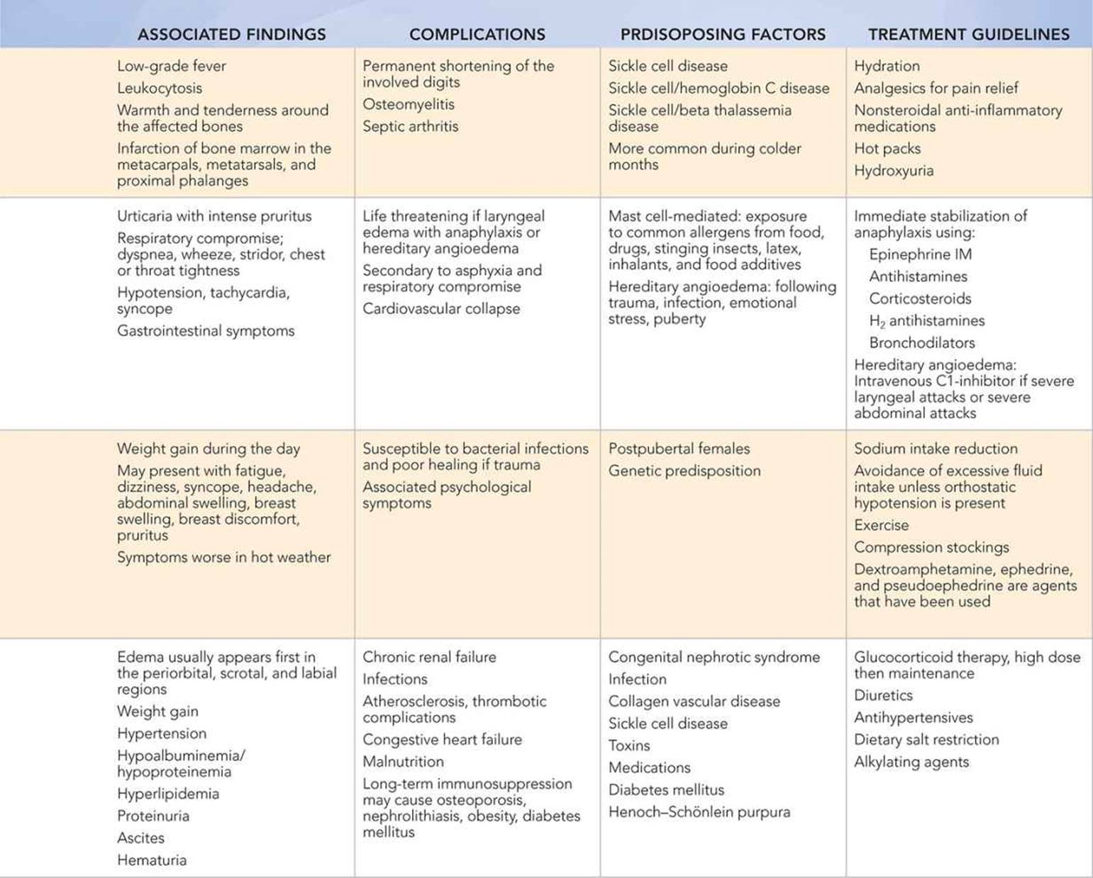
Foot swelling or edema is a common manifestation of many disease states. It may be part of a localized or generalized process. Unilateral foot swelling may develop from a variety of causes, including trauma (e.g., sprains, fractures), infection (e.g., cellulitis, osteomyelitis, abscess), angioedema (i.e., allergic reaction vs hereditary), vasculitis (e.g., Henoch–Schönlein purpura, Kawasaki disease), insect bites, snake bites with envenomation, lymphedema (i.e., hereditary, acquired), soft-tissue tumors (e.g., benign—fibromas, malignant—rhabdomyosarcoma, other sarcomas), or bone tumors (e.g., benign—exostosis and unicameral bone cyst, malignant—osteosarcoma and Ewing sarcoma). Pain may be a significant complaint with all of these etiologies except for angioedema, lymphedema, and benign tumors. Historical factors such as chronicity of symptoms (recurrent vs acute onset), environmental exposures, systemic symptoms, a family history of angioedema or lymphedema, underlying renal/cardiac/hepatic disease, medications, and a thorough physical examination will help determine the etiology of foot swelling. Bilateral painless foot swelling is more likely due to an underlying systemic condition such as nephrotic syndrome, heart failure, cardiomyopathy, cirrhosis, malnutrition, hypoproteinemia, renal failure, or pregnancy.
Key Points in the History
• The typical ankle sprain is an inversion injury that occurs in the plantar-flexed position. The lateral stabilizing ligaments are most commonly affected in this type of injury.
• Ankle sprains in children are less common than fractures because the ligaments of a preadolescent are much stronger than the growth plate or bone. Associated avulsion fractures are typically present if a ligamentous injury occurs.
• Most foot fractures in children result from direct trauma, such as crush injuries from a falling object or the child falling from a height.
• Acute traumatic compartment syndrome of the foot is the result of a serious injury such as a fracture, dislocation, and/or crush injury. Vascular injuries and coagulopathies are risk factors for the development of this condition.
• Acute, painful, unilateral foot swelling is often associated with infection or trauma. Chronic unilateral painful foot swelling over a period of weeks to months may indicate a malignant neoplasm of the soft or bony tissue.
• Recurring paroxysms of subcutaneous angioedema in the extremities, face, trunk, genitals, or the intestinal and laryngeal submucosae are the characteristics of hereditary angioneurotic edema. Family history is positive in 75% of patients.
• Unilateral or bilateral, neonatal pedal edema suggests a diagnostic evaluation for Turner syndrome, Noonan syndrome, or Milroy disease (hereditary lymphedema with autosomal dominant inheritance).
• Medications such as calcium channel blockers and vasodilators may cause peripheral edema. Oral contraceptives may predispose a female teenager to deep venous thrombosis/thrombophlebitis with lower extremity swelling.
Key Points in the Physical Examination
• Findings associated with severe ankle sprains include swelling, bruising, pain on palpation, and a positive anterior drawer test of the ankle. Patients with all four of these findings are likely to have a lateral ligament rupture.
• Angioedema secondary to cutaneous vessel damage in Henoch–Schönlein purpura may be significant. It may precede the palpable purpura, and it is most prominent over the dorsal hands and feet, and the periorbital regions.
• Snake bites with envenomation cause edema and erythema at the location of the bite and in adjacent tissues, usually within 30 to 60 minutes. Oozing from the wound suggests envenomation. Edema progresses rapidly and may involve the entire extremity. Ecchymosis is common and may appear within 3 to 6 hours.
• Subcutaneous angioedema in hereditary angioedema is nonpruritic, nonerythematous, and well circumscribed. It is not accompanied by urticaria and is most commonly seen on the extremities.
• Edema, typically pitting edema, is the major clinical manifestation of nephrotic syndrome. It is more noticeable in the face in the morning upon arising and predominately in the lower extremities later in the day.
• Infants with beriberi have muscle wasting, upper and lower extremity edema, pallor, restlessness, and diarrhea. Infants who are breastfed by a thiamine-deficient mother are at risk for developing beriberi.
|
PHOTOGRAPHS OF SELECTED DIAGNOSES |
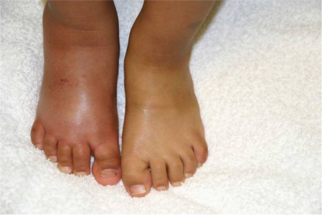
Figure 50-1 Insect bite. A 2-year-old child with swelling and erythema of the right foot and ankle because of an insect bite. A pustule and vesicles are noted over the dorsum of the foot. (Courtesy of Julie A. Boom, MD.)
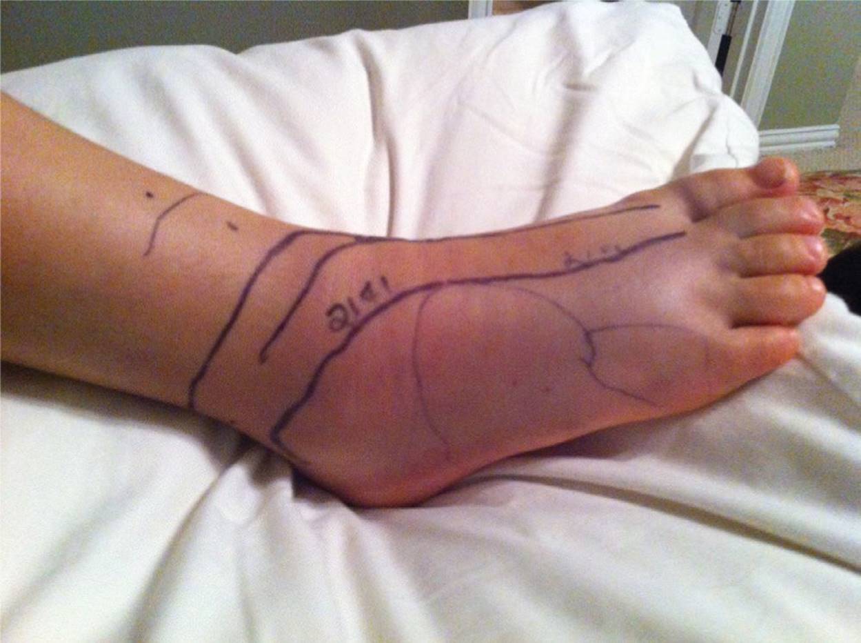
Figure 50-2 Snake bite with envenomation. A 9-year-old with swelling, erythema, and ecchymosis of the right foot and ankle 2 days after a Southern Copperhead bite. The fang marks are visible on the lateral aspect of the foot, and the area of swelling and erythema has been marked with a pen. (Courtesy of Lisa E. De Ybarrondo, MD.)

Figure 50-3 Pitting edema of the foot. An edematous foot with evidence of pitting following firm pressure. (Used with permission from Bickley LS, Szilagyi P, eds. Bates Guide to Physical Examination and History Taking. 8th ed. Philadelphia, PA: Lippincott Williams & Wilkins; 2003:455.)

Figure 50-4 Lower extremity and foot edema. Clinical picture of lower extremities showing marked edema. (Used with permission from Gold DH, Weingeist TA. Color Atlas of the Eye in Systemic Disease. Baltimore, MD: Lippincott Williams & Wilkins; 2001:643.)

Figure 50-5 Congenital lymphedema. Bilateral foot swelling noted by the mother of a newborn, 4 days following hospital discharge. (Courtesy of Jan Edwin Drutz, MD.)
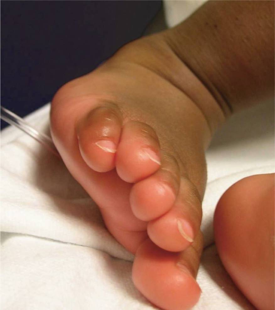
Figure 50-6 Foot edema and erythema in a child with Kawasaki disease. (Courtesy of Esther K. Chung, MD, MPH.)
DIFFERENTIAL DIAGNOSIS

Other Diagnoses to Consider
• Juvenile idiopathic arthritis
• Deep venous thrombosis
• Glomerulonephritis, renal failure
• Burns
• Myxedema
• Kawasaki disease
• Primary lymphedema (Turner syndrome, Noonan syndrome, or Milroy disease)
• Secondary lymphedema (filariasis, lymphatic obstruction)
• Köhler disease (osteonecrosis of the navicular bone)
• Acute hemorrhagic edema of infancy
• Malnutrition (kwashiorkor, scurvy, beriberi)
• Hypoalbuminemia
When to Consider Further Evaluation or Treatment
• It is important to reexamine an ankle sprain 3 to 5 days following the injury to assess the extent of ligamentous injury (partial tear vs rupture). Consider referral to orthopedic surgery for a severe sprain that includes a ligament rupture.
• Compartment syndrome presents with tense swelling of the foot and pain out of proportion to the degree of injury. Palor, paresthesias, and pulselessness are late signs. If there is a suspicion of compartment syndrome, then a prompt surgical evaluation is required with intracompartmental pressure monitoring.
• Indications for referral to an orthopedic surgeon include fracture, dislocation, subluxation, tendon rupture, wound penetrating into the joint, soft-tissue tumors, or bone tumors (benign or malignant). Foot fractures involving the talar neck, tarsometatarsal or intra articular calcaneus warrant urgent orthopedic consultation.
• If cellulitis has not visibly improved within 72 hours and/or symptomatic improvement has not occurred within 24 to 48 hours after starting oral antibiotics, consider resistant pathogens or alternative diagnoses such as abscess, osteomyelitis, or other.
• Evaluation for a deeper soft-tissue infection should be considered in patients with underlying conditions such as diabetes or lymphedema, and in patients who are systemically ill.
• According to the Ottawa Ankle Rules, radiographs of the ankle should be obtained after an acute ankle injury in children (>5 years old) if there is any pain in the malleolar zone accompanied by any one of the following findings: bone tenderness along the distal 6 cm of the posterior edge of the tibia or tip of the medial malleolus, bone tenderness along the distal 6 cm of the posterior edge of the fibula or tip of the lateral malleolus, or an inability to bear weight both immediately and in the emergency department for four steps.
• According to the Ottawa Foot Rules, radiographs of the ankle should be obtained after an acute foot injury in children (>5 years old) if there is any pain in the midfoot zone and any one of the following findings: bone tenderness at the base of the fifth metatarsal, bone tenderness at the navicular bone, or inability to bear weight immediately and in the emergency department for four steps.
SUGGESTED READINGS
Barillas-Arias L, Adams A, Lehman A. Pediatric vasculitic syndrome: Henoch-Schönlein purpura. Consult. Pediatr. 2008;7(9):361–367.
Bibbo C, Lin SS, Cunningham FJ. Acute traumatic compartment syndrome of the foot in children. Pediatr Emerg Care. 2000;16(4):244–248.
Dowling S, Spooner CH, Liang Y, et al. Accuracy of Ottawa Ankle Rules to exclude fractures of the ankle and midfoot in children: a meta-analysis. Acad Emerg Med. 2009;16(4):277–287.
Fleisher GR, Ludwig S, eds. Textbook of Pediatric Emergency Medicine. 6th ed. Baltimore, MD: Williams & Wilkins; 2010:233–234.
Jarvis JG, Moroz PJ. Fractures and dislocations of the foot. In: Beaty JH, Kasser JR, eds. Fractures in Children. 6th ed. Philadelphia, PA: Lippincott Williams & Wilkins; 2006:1129.
Sardana N, Craig TJ. Recent advances in management and treatment of hereditary angioedema. Pediatrics. 2011;128(6):1173–1180.
Tiemstra JD. Update on acute ankle sprains. Am Fam Physicians. 2012;85(12):1170–1176.