
Approach to the Problem
Often resulting from stressors placed on them by normal weight-bearing activities and the restrictive setting of everyday footwear, the feet are prone to dermatologic conditions. Developing within the enclosed environment of a warm and moist shoe, a rash can easily spread or worsen. For example, allergic dermatitis on an excoriated toe can evolve readily into an extensive foot cellulitis. A foot lump, whether related to infection or an underlying bony abnormality, may continue to grow, potentially compromise normal foot dynamics, and readily recur if not properly diagnosed and treated. Therefore, a good understanding of common pediatric foot ailments is important.
Key Points in the History
• Origin (between toes or overlying pressure points), natural course (timing or spread from a specific focus), and symptomatology (itching, burning, pain, redness, or swelling) may aid in differentiation of a foot rash or lump as reactive or infectious (dyshidrotic eczema vs tinea pedis; callus vs plantar wart).
• Seasonality may be an important clue, with tinea pedis and dyshidrotic eczema more common in the warm, summer months, and juvenile plantar dermatosis more common during colder months.
• A correlation with new or overly restrictive footwear may support the diagnosis of a corn or callus.
• Communal bathing has been associated with infections, including tinea pedis and plantar warts.
• Epidemiology may also provide clues: infantile acropustulosis occurs between birth and 2 years, juvenile plantar dermatosis occurs more commonly among prepubertal children, and tinea pedis and plantar warts are present in older children and adolescents.
Key Points in the Physical Examination
• Delineation of dermatitis from an infectious rash may be based on appearance and distribution (warmth, tenderness, vesiculation, or crusting in a localized interdigital or global distribution).
• Juvenile plantar dermatosis tends to affect the balls of the feet bilaterally with a shiny, smooth appearance (“glazed doughnut”). The involvement of the interdigital spaces is more often seen in tinea pedis.
• Allergic contact dermatitis usually affects the dorsum of the feet, sparing the toe webs and soles.
• Dyshidrotic eczema tends to favor the lateral aspects of digits, palms, and soles.
• Plantar corns are more sensitive to direct pressure, whereas plantar warts are more sensitive to lateral compression or pinching.
• Plantar warts usually disrupt skin lines, with lines usually maintained in calluses.
• After paring, plantar warts will often show pinpoint black dots, which are thrombosed capillaries. Calluses will have a smooth, glassy, and homogenous surface.
• Infantile acropustulosis is characterized by pruritic, recurrent vesiculopustular lesions that are concentrated on the palms and soles, extending to the dorsum of the hands, feet, and ankles.
|
PHOTOGRAPHS OF SELECTED DIAGNOSES |

Figure 51-1 Callus. The skin over the head of the fifth metatarsal is thickened and slightly yellow, with skin lines maintained. (Courtesy of Julie A. Boom, MD.)

Figure 51-2 Plantar wart. Plantar wart on the medial surface of the heel. (Courtesy of Denise W. Metry, MD.)

Figure 51-3 Tinea pedis. The interdigital pattern of tinea pedis is common. Note the spread onto the dorsum of the foot. (Courtesy of Denise W. Metry, MD.)
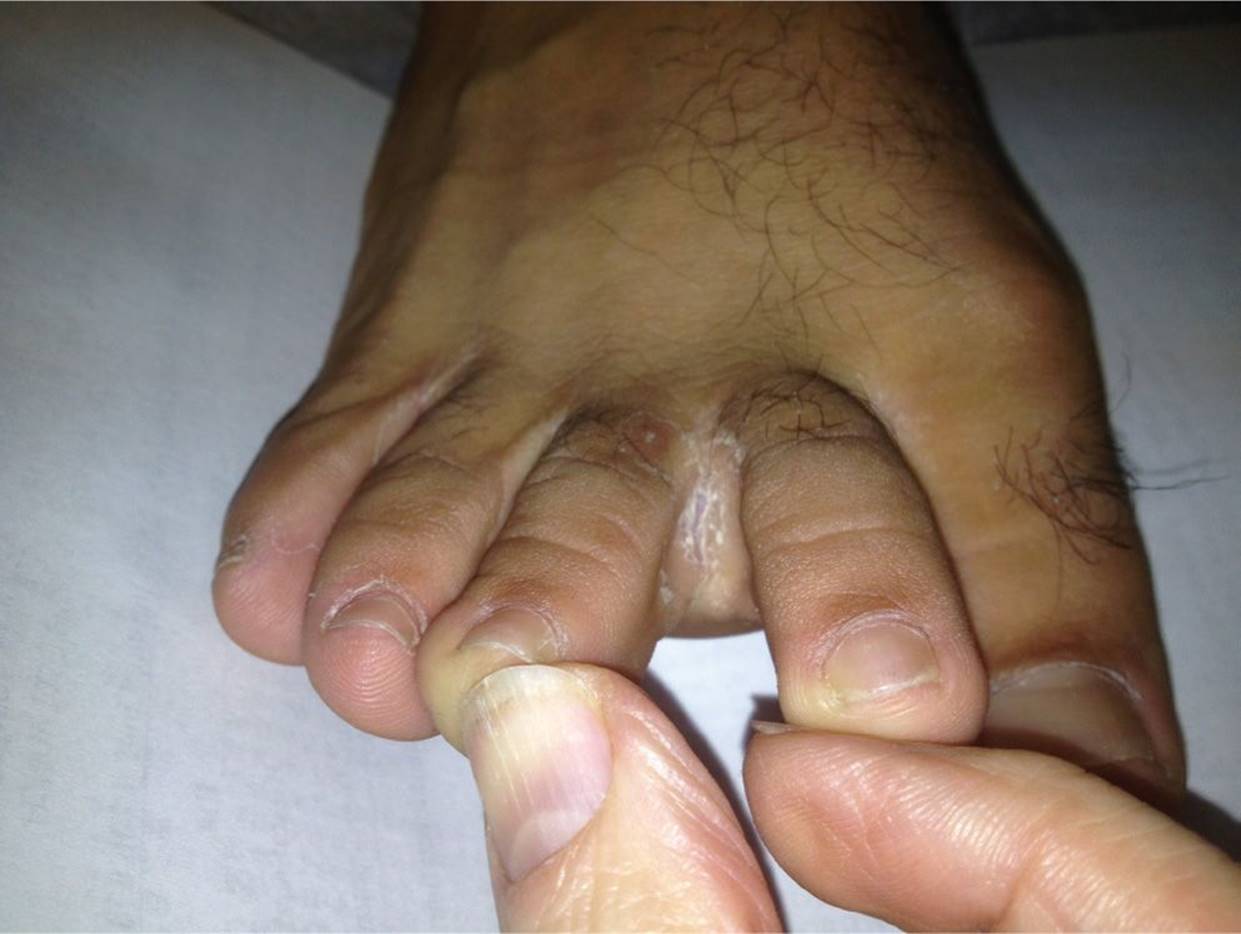
Figure 51-4 Tinea pedis. The interdigital pattern of tinea pedis is common. (Courtesy of Lisa E. De Ybarrondo, MD.)
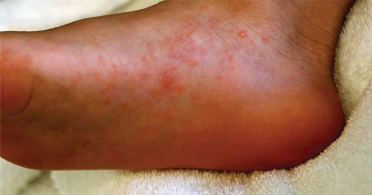
Figure 51-5 Dyshidrotic eczema. Multiple clear vesicles on the medial surface of the foot. (Courtesy of Julie A. Boom, MD.)
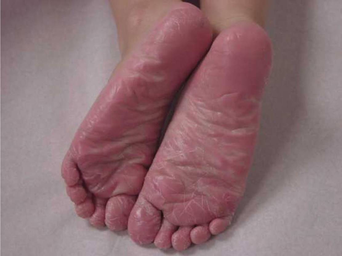
Figure 51-6 Juvenile plantar dermatosis. The skin on the soles has a smooth, shiny appearance with multiple fissures and cracks. (Courtesy of Denise W. Metry, MD.)

Figure 51-7 Impetigo. Classic crusted lesion on the great toe of a toddler with a history of eczema and similar lesions on her face and back. (Courtesy of Lisa E. De Ybarrondo, MD.)
DIFFERENTIAL DIAGNOSIS

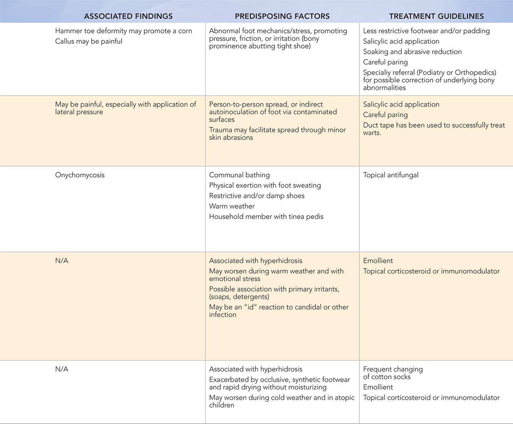
Other Diagnoses to Consider
• Allergic contact dermatitis
• Infantile acropustulosis
• Pustular psoriasis
• Cellulitis
• Pitted keratolysis
When to Consider Further Evaluation or Treatment
• Specialty referral for alternative medical or surgical approaches should be considered for painful or progressive warts, and corns or calluses that do not respond to traditional therapies such as topical salicylate.
• Severe tinea pedis may require treatment with a systemic antifungal agent if refractory to a topical approach or associated with onychomycosis.
• Severe dyshidrotic eczema or juvenile plantar dermatosis rarely may require systemic immunosuppressive therapy.
• Cellulitis due to a deep penetrating injury of the foot may require orthopedic evaluation for wound exploration if bone or joint infection or a retained foreign body is suspected.
SUGGESTED READINGS
Buescher ES. Infections associated with pediatric sport participation. Pediatr Clin North Am. 2002;49(4):743–751.
Freeman DB. Corns and calluses resulting from mechanical hyperkeratosis. Am Fam Physician. 2002;65(11):2277–2280.
Guenst BJ. Common pediatric foot dermatoses. J Pediatr Health Care. 1999;13(2):68–71.
Morelli JG. Principles of therapy. In: Kliegman RM, Stanton BF, Schor NF, St. Geme JW, Behrman RE, eds. Nelson Textbook of Pediatrics. 19th ed. Philadelphia, PA: Elsevier Saunders; 2011:2215–2218.
Omura EF, Rye B. Dermatologic disorders of the foot. Clin Sports Med. 1994;13(4):825–841.