
Approach to the Problem
The child who presents with a nonblanching rash requires careful evaluation. Purpuric lesions, including petechiae and ecchymoses, usually result from vascular injury or disorders of hemostasis. The underlying etiology may be trauma, a simple viral infection, or a more serious condition such as leukemia or a bleeding disorder. When a nonblanching rash is seen in association with fever, serious bacterial infection, including meningococcemia, must be considered.
Key Points in the History
• A history of fever makes an infectious etiology more likely.
• Acute presentation of a nonblanching rash is more concerning than a rash that has been present for more than a couple of weeks.
• The location and pattern of spread may give a clue to the diagnosis: Rocky Mountain spotted fever (RMSF) tends to begin peripherally; Henoch–Schönlein purpura tends to primarily involve the lower extremities and buttocks.
• The presence of photophobia, headache, or both, in association with a nonblanching rash, raises the suspicion for meningococcal or other bacterial meningitis.
• A history of trauma may be the cause of the nonblanching lesions: localized bruising may follow blunt trauma, and petechiae may be seen in areas of friction or scratching.
• Significant ecchymotic lesions in the absence of a history of trauma should raise suspicion for child physical abuse or a bleeding disorder.
• Forceful coughing or vomiting may cause petechiae, particularly on the face and upper chest.
• Accompanying fatigue may be caused by anemia because of bone marrow suppression or infiltration as seen with leukemia.
• A history of tick bites or travel or activities associated with tick exposure should raise suspicion for RMSF or ehrlichiosis.
• Mongolian spots are present at birth and, though they may fade, they generally do not undergo color changes over time. In contrast, ecchymoses change color over time and eventually resolve.
• A history of easy bruising or excessive bleeding in the patient, or a family history of a bleeding disorder, should raise suspicion for hemophilia or von Willebrand disease.
• Familiarity with home remedies found in certain Asian cultures, such as coining and cupping, is essential.
Key Points in the Physical Examination
• Petechiae are nonblanching macules up to 2 mm in diameter caused by the extravasation of blood from capillaries. Mucosal bleeding sometimes is referred to as “wet purpura.”
• Forceful coughing or vomiting may cause petechiae on the face and chest, above the nipple line.
• Purpura, seen with inflammatory injury to the smaller blood vessels, are elevated, firm, hemorrhagic plaques located predominantly on dependent surfaces.
• Ecchymoses are larger areas of bleeding into the skin. There is a characteristic change in color as they age, changing from red to purple to green to yellow-brown as the heme is degraded.
• Deep bleeding and hemarthroses are seen with clotting factor deficiencies, whereas petechiae are more commonly seen with thrombocytopenia.
• Ecchymoses that are not explained easily by accidental trauma should raise the suspicion of child physical abuse. Ecchymoses, uncommonly caused by infection, usually are indicative of trauma—accidental and nonaccidental—or a bleeding disorder. Bruising in normally active children is predominantly found on the pretibial surfaces.
• Cupping and coining are practices used by some Asian cultures to treat acute illnesses. Each has a characteristic appearance, and petechiae and ecchymoses may be seen in both.
|
PHOTOGRAPHS OF SELECTED DIAGNOSES |

Figure 69-1 Mongolian spots. Blue nevi in the typical sacral area. (Courtesy of Sidney Sussman, MD.)

Figure 69-2 Mongolian spot on the hand. (Courtesy of Esther K. Chung, MD, MPH.)
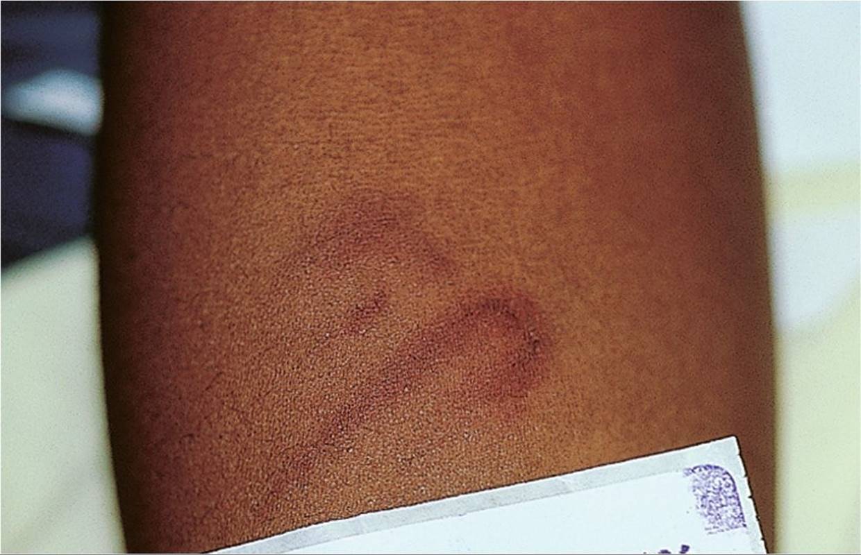
Figure 69-3 Child physical abuse. Curvilinear bruising from a looped cord. (Used with permission from Fleisher GR, Ludwig S, Baskin MN. Atlas of Pediatric Emergency Medicine. Philadelphia, PA: Lippincott Williams & Wilkins; 2004:425.)

Figure 69-4 Ecchymoses in a patient with hemophilia. (Courtesy of Sidney Sussman, MD.)

Figure 69-5 Henoch–Schönlein purpura. Note the palpable purpura on the posterior aspects of this child’s leg. (Courtesy of Steven Manders, MD.)

Figure 69-6 Rocky Mountain spotted fever. Note the multiple petechial lesions on the forearm. (Courtesy of Steven Manders, MD.)
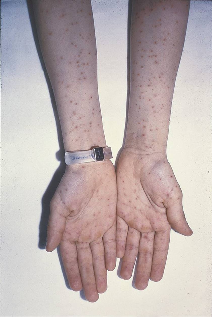
Figure 69-7 Rocky Mountain spotted fever. (Courtesy of Sidney Sussman, MD.)

Figure 69-8 Petechiae and ecchymoses in a patient with idiopathic thrombocytopenic purpura. (Courtesy of Sidney Sussman, MD.)

Figure 69-9 Purpura fulminans in a patient with meningococcemia. (Courtesy of Steven Manders, MD.)

Figure 69-10 Purpura fulminans. Purpura on the foot of the same patient in Figure 69.9. (Courtesy of Steven Manders, MD.)

Figure 69-11 Coining. Note the linear petechiae and ecchymoses over the back that are characteristic for this healing practice used by some Asian cultures. (Courtesy of Philip Siu, MD.)
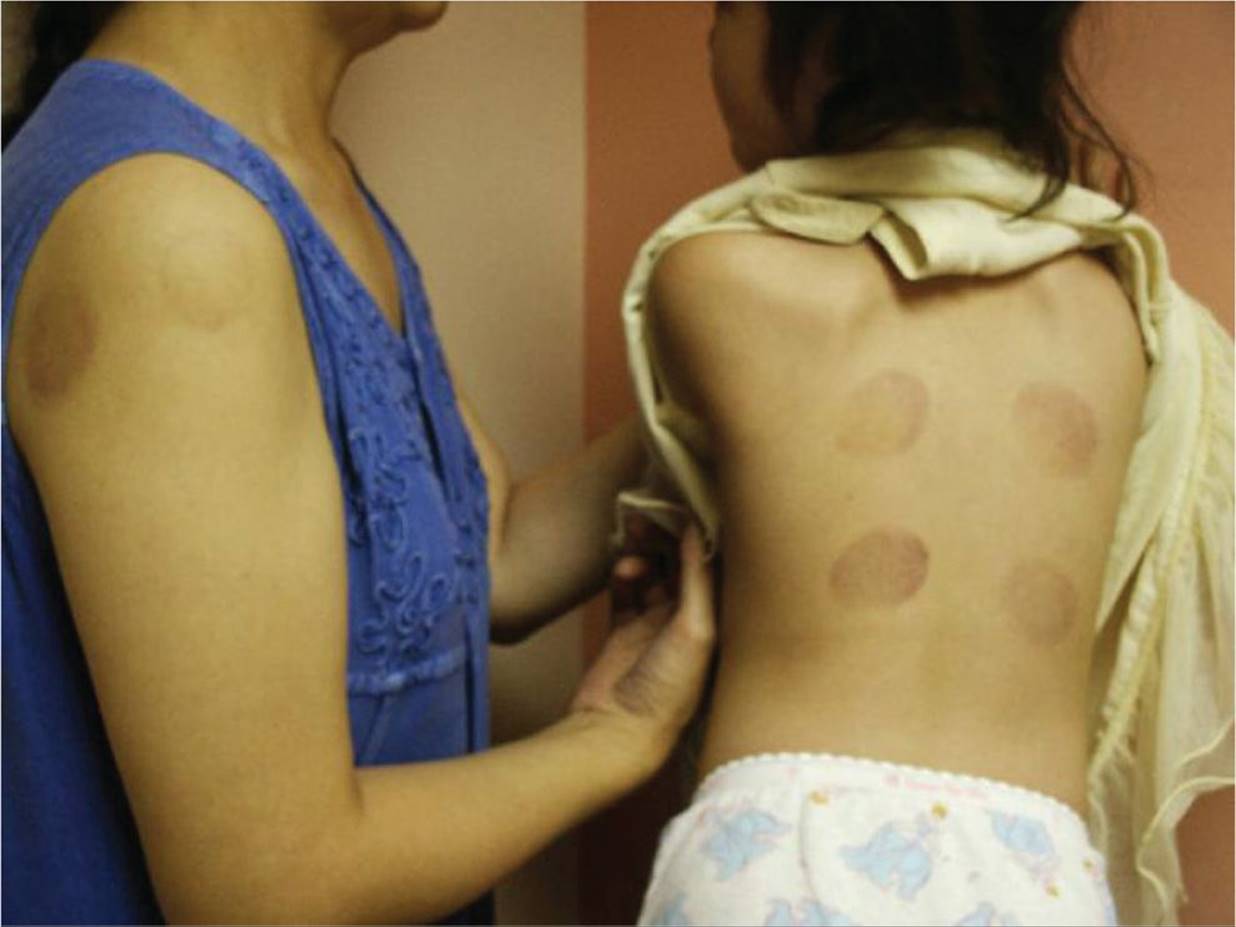
Figure 69-12 Cupping. Note the circular bruises on the mother’s arm and the child’s back that are the result of cupping, a healing practice used by some Asian cultures. (Courtesy of Philip Siu, MD.)
DIFFERENTIAL DIAGNOSIS


Other Diagnoses to Consider
• Leukemia
• Aplastic anemia
• Hemolytic uremic syndrome
• Systemic lupus erythematosus
• Liver disease
• Coagulation disorders
• Drug-induced thrombocytopenia
• Wiskott–Aldrich syndrome
When to Consider Further Evaluation or Treatment
• Patients with generalized petechiae or petechiae that are not easily explained by trauma should have a complete blood count. If the diagnosis of thrombocytopenia is established, further evaluation for a specific etiology should occur.
• An ill-appearing child with petechiae or purpura requires urgent evaluation and may need empiric treatment for an infectious etiology, such as bacterial sepsis or RMSF.
• Patients with ecchymoses suspicious for nonaccidental trauma should be evaluated further with coagulation studies and radiographic studies. Referral to child protective services should also be made.
• Patients with significant thrombocytopenia or involvement of other cell lines (anemia and/or white cell abnormalities) should be evaluated by a hematologist.
SUGGESTED READINGS
D’Orazio JA, Neely J, Farhoudi N. ITP in children: pathophysiology and current treatment approaches. J Pediatr Hematol Oncol. 2013;35(1):1–13.
Fleisher GR, Ludwig S, Baskin MN. Atlas of Pediatric Emergency Medicine. Philadelphia, PA: Lippincott Williams & Wilkins; 2004:425.
Leung AKC, Chan KW. Evaluating the child with purpura. Am Fam Physician. 2001;64(3):419–428.
Mudd SS, Findlay JS. The cutaneous manifestations and common mimickers of physical child abuse. J Pediatr Health Care. 2004;18(3):123–129.
Reyes MA, Eichenfield LF. Purpura. In: Long SS. ed. Principles and Practice of Pediatric Infectious Diseases. 4th ed. New York, NY: Elsevier; 2012:441–444.
Singh-Behl D, LaRosa SP, Tomecki KJ. Tick-borne infections. Dermatol Clin. 2003;21(2):237–244.
Weiss PF. Pediatric vasculitis. Pediatr Clin North Am. 2012;59(2):407–423.