
Igor Mitrovic, MD
This chapter reviews the normal structure and function of the vascular component of the cardiovascular system and then considers the pathophysiology of three common conditions frequently seen by practicing physicians: atherosclerosis, hypertension, and shock.
NORMAL VASCULAR STRUCTURE & FUNCTION
ANATOMY & HISTOLOGY
The blood vessels are a closed system of conduits that carry blood from the heart to the tissues and back to the heart. All of the blood flows through the lungs, but the systemic circulation is made up of many different circuits in parallel (Figure 11-1). This permits wide variation in regional systemic blood flow without changing the total systemic flow.

FIGURE 11-1 Diagram of the circulation in the adult. (Redrawn, with permission, from Barrett KE et al, eds. Ganong’s Review of Medical Physiology, 24th ed. McGraw-Hill, 2012).
The characteristics of the various types of blood vessels in humans are summarized in Figure 11-2. Note that as the diameter of the vessels decreases, their number in the body increases so that the total cross-sectional area increases.
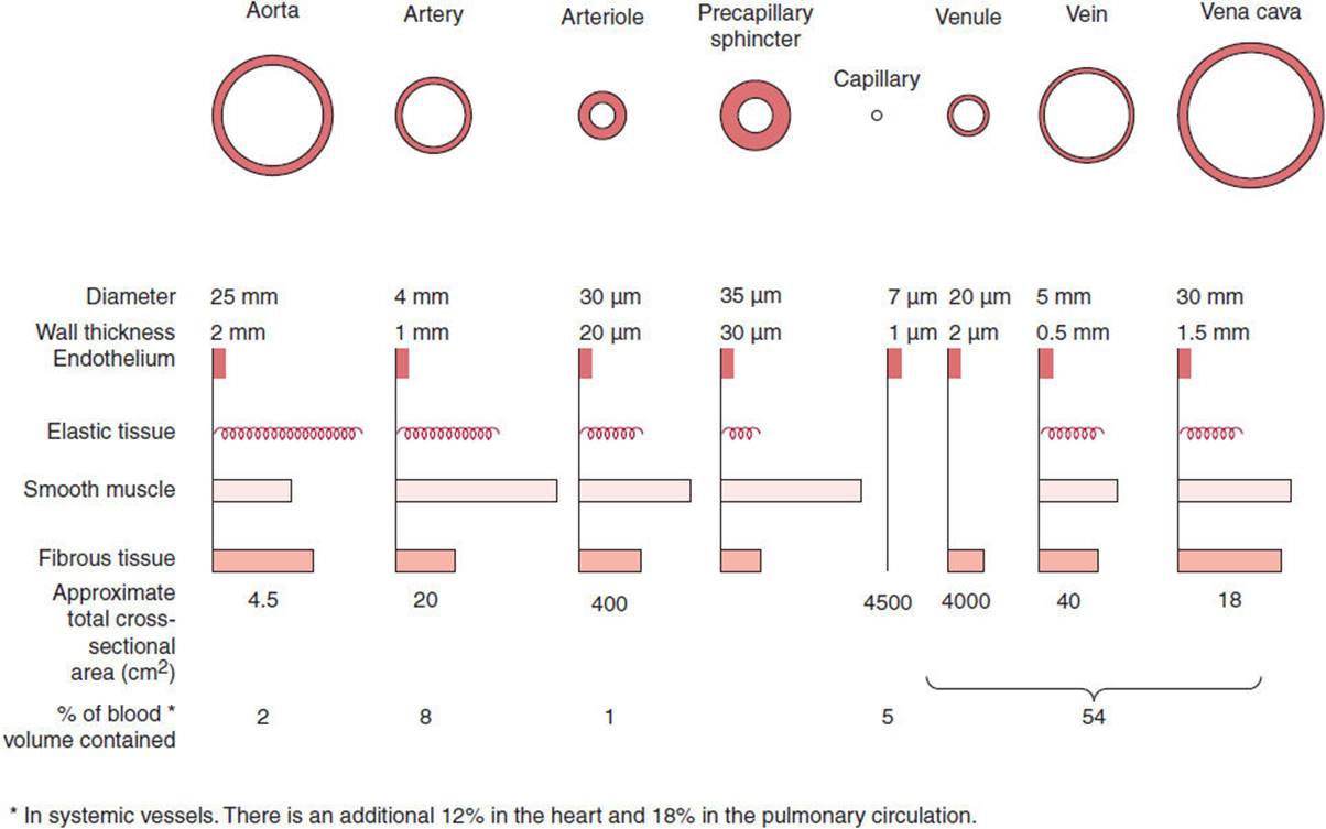
FIGURE 11-2 Characteristics of systemic blood vessels. Cross sections of the vessels are not drawn to scale because of the huge range in size from aorta and vena cava to capillaries. (Redrawn from Burton AC. Relation of structure to function of the tissues of the wall of blood vessels. Physiol Rev. 1954;34:619.)
All the blood vessels are lined by a single layer of endothelial cells. Collectively, the endothelial cells constitute a remarkable organ that secretes substances that affect the diameter of the vessels and provide for their growth, their repair when injured, and the formation of new vessels that carry blood to growing tissues.
Arterial Vessels
The aorta, the large arteries, and the arterioles are made up of an outer layer of connective tissue, the adventitia; a middle layer of smooth muscle, the media; and an inner layer, the intima, containing the layer of endothelial cells and some subendothelial connective tissue. The walls of the aorta and the large arteries contain abundant elastic tissue, much of it concentrated in the internal elastic lamina, a prominent band between the intima and the media, and another band, the external elastic lamina, between the media and the adventitia (Figure 11-3). The vessels are stretched by the force of cardiac ejection during systole, and the elastic tissue permits them to recoil during diastole. This maintains diastolic pressure and aids the forward motion of the blood. The walls of the arterioles contain less elastic tissue than the arteries but proportionately more smooth muscle (Figure 11-2). The muscle is extensively innervated by noradrenergic nerve fibers, which are constrictor in function. In some instances, there is a cholinergic innervation, which is vasodilator in function. The arteries and the arterioles offer considerable resistance to the flow of blood and are known as the resistance vessels.
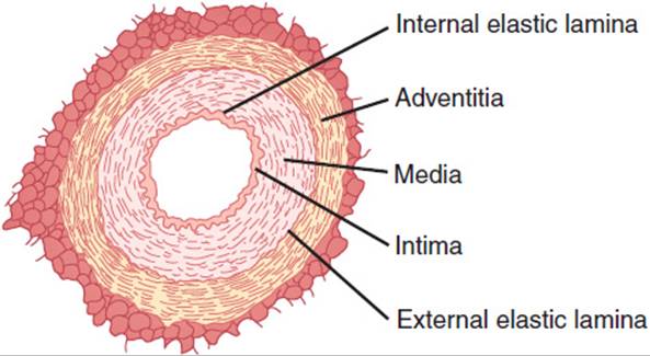
FIGURE 11-3 Cross section of a small artery. (Redrawn, with permission, from Ganong WF. Review of Medical Physiology, 22nd ed. McGraw-Hill, 2005.)
Capillaries
The terminal portions of the arterioles, sometimes called metarterioles, drain into the capillaries. On the upstream side, the openings of the capillaries are surrounded by smooth muscle precapillary sphincters. There is debate about whether the metarterioles and sphincters are innervated. The capillaries themselves are made up of a single layer of endothelial cells. Outside these cells there are occasional pericytes, fibrous cells whose function is unknown (Figure 11-4). The capillaries anastomose extensively, and although each capillary is only 5–9 μm in diameter, there are so many of them that the total cross-sectional area of all the capillaries is about 4500 cm2.
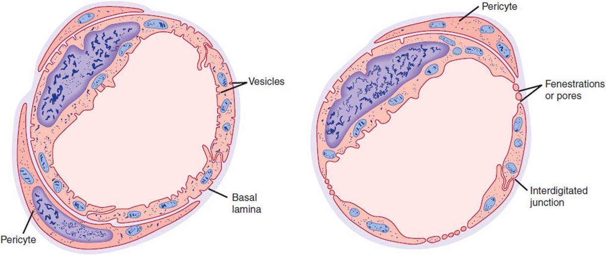
FIGURE 11-4 Cross sections of capillaries. Left: Continuous type of capillary found in skeletal muscle. Right: Fenestrated type of capillary. (Redrawn, with permission, from Orbison JL et al, eds. The Peripheral Blood Vessels.Williams & Wilkins, 1962.)
Some substances cross capillary walls by vesicular transport, a process that involves endocytosis of plasma, movement of the vesicles formed in this way across the endothelial cell cytoplasm, and exocytosis on the tissue side. However, relatively little material is moved in this fashion, and most fluid and solute exchange occurs at the junctions between endothelial cells. In the liver, there are large gaps between endothelial cells (Chapter 14). In endocrine tissues, the small intestine, and the kidneys, tissues in which there is bulk flow of material across capillary walls, the cytoplasm of the endothelial cells is attenuated to form gaps called fenestrations. These gaps appear to be closed by a discontinuous membrane, which permits the passage of substances up to approximately 600 nm in diameter. In skeletal muscle, cardiac muscle, and many other tissues, there are no fenestrations, but the junctions between endothelial cells permit the passage of substances up to 10 nm in diameter. Finally, in brain capillaries, there are tight junctions between the endothelial cells. These tight junctions permit very little passive transport and are a key component of the blood-brain barrier. Water and CO2 enter the brain with ease, but movement of most other substances in and out of brain tissue is mainly via transport proteins in the endothelial cells.
Venules & Veins
The venules are very similar to capillaries; they are about 20 μm in diameter, and their approximate total cross-sectional area is 4000 cm2. They drain into veins that have modest amounts of smooth muscle and elastic tissue in their relatively thin walls and average 5 mm in diameter. The veins drain into the superior and inferior vena cavae, which in turn drain into the right atrium of the heart. The walls of the veins, unlike those of the arteries and arterioles, are easily distended and can expand to hold more blood without much increase in intravascular pressure. Therefore, they are known as capacitance vessels. They are innervated, and their smooth muscle can contract in response to noradrenergic stimulation, pushing blood into the heart and the arterial side of the circulation. The intima of the limb veins is folded at intervals to form the venous valves that prevent retrograde flow.
Lymphatics
The smallest lymphatic vessels are made up of endothelial tubes. Fluid appears to enter them through loose junctions between the endothelial cells. They drain into larger endothelial tubes that have valves and contractile walls containing smooth muscle, so that the fluid they contain moves centrally. The central lymphatics drain into the right and left subclavian veins. Thus, the lymphatic system drains excess fluid in the tissues back into the vascular system.
CHECKPOINT
1. How does the composition of the wall of an arteriole differ from that of an artery?
2. What are the modes of transport across the capillary wall? In what organ is transport greatest?
3. Why are veins termed capacitance vessels?
PHYSIOLOGY
Biophysical Considerations
In any system made up of a pump and a closed system of pipes such as the heart and the blood vessels, the flow of fluid between the two ends of the system depends on the pressure difference generated by the pump and the resistance to flow in the pipes:

In the cardiovascular system, this translates into:
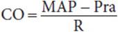
where CO is cardiac output, MAP is mean arterial pressure, and Pra is the pressure in the right atrium. Since Pra is normally close to 0 mm Hg, this expression has the following corollary:

Thus, mean arterial pressure increases when there is an increase in cardiac output or when the diameter of the blood vessels (principally the arterioles) is decreased.
Flow in blood vessels is laminar (ie, an infinitely thin layer of blood next to the vessel wall does not move, the next layer moves slowly, and the next layer moves more rapidly, with the fastest flow in the center). Usually the flow is smooth, and no sound is generated. However, if flow is accelerated, it becomes turbulent when a critical velocity is reached. Constriction of a blood vessel or a heart valve causes faster flow in the constricted region because the kinetic energy of flow is increased and the potential energy is decreased (the Bernoulli principle). Therefore, critical velocity is more often reached. The turbulence causes noise. The examining physician hears this noise through the stethoscope as a bruit or murmur. The two terms are often used interchangeably, although the term “murmur” is more commonly applied to noise heard over the heart and the term “bruit” to noise heard over blood vessels. The sounds of Korotkoff heard over an artery below a blood pressure cuff (discussed later) are an example.
The main factors that determine flow in a blood vessel are the pressure difference between its two ends, the radius of the vessel, and the viscosity of the blood. The relation can be expressed mathematically by the Poiseuille-Hagen formula:

where
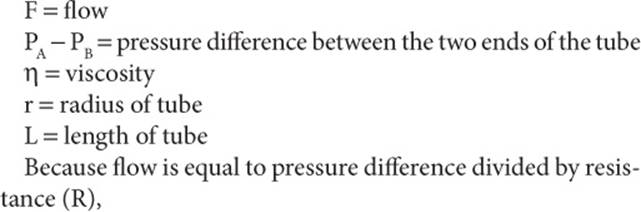

Note that flow varies directly and pressure inversely with the fourth power of the radius of the vessel. This is why small changes in the diameter of the arterioles, the principal resistance vessels, cause large changes in pressure. For example, when the radius of a vessel is doubled, resistance is decreased to 6% of its previous value. Conversely, a small decrease in arterial diameter produces a relatively marked increase in blood pressure. Viscosity also has an effect, but, except at very high or very low values, the effect is small. Viscosity is high in polycythemia and low in anemia.
The relation between distending pressure and wall tension is shown in Figure 11-5. This relation is called the law of Laplace. It states that the wall tension (T) in a hollow viscus is equal to the product of the transmural pressure (P) and the radius (r) divided by the thickness of the wall (W):

FIGURE 11-5 Law of Laplace. In a hollow object (eg, viscus, blood vessel), the distending pressure (P) equals the wall tension (T). (Redrawn, with permission, from Barrett KE et al, eds. Ganong’s Review of Medical Physiology,24th ed. McGraw-Hill, 2012.)

In thin-walled structures, wall thickness is negligible, but in structures such as arteries it becomes a significant factor. The transmural pressure is the pressure inside the viscus minus the pressure outside the viscus, but in the body the latter is negligible. Therefore, in a distensible hollow viscus, transmural pressure at equilibrium is equal to wall tension divided by the two principal radii of curvature of the object (r1 and r2):
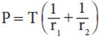
The operation of this law in the lungs is discussed in Chapter 9. In a cylinder such as a blood vessel, one radius is infinite, so

Thus, the smaller the radius of a vessel, the lower the wall tension that is necessary to balance the distending pressure. For example, the wall tension in the aorta is about 170,000 dynes/cm, whereas in capillaries it is about 16 dynes/cm. This is why the thin-walled, delicate capillaries do not collapse. The law of Laplace also applies to the heart. When the heart is dilated, it must develop more wall tension to function. Consequently, its work is increased.
With these principles and Figure 11-2 in mind, plus the fact that the major sites of vascular resistance are the arterioles, it is possible to understand the pressures in the various parts of the vascular system (Figure 11-6) and the velocity of flow in them. Systolic and diastolic pressures in the aorta and large arteries are stable, and there is a large pulse pressure. Normal pressure is about 120/80 mm Hg in healthy young adults. In the arterioles there is a sharp drop, so that pressure at the entrances to the capillaries is about 37 mm Hg and pulse pressure has disappeared. At the ends of the capillaries, it is about 17 mm Hg and falls steadily in the venous system to about 5 mm Hg at the entrance of the vena cavae into the right atrium. Velocity falls in the arterioles, is low in the capillaries because of the large total cross-sectional area, and increases again in the large veins.

FIGURE 11-6 Diagram of the changes in pressure and velocity as blood flows through the systemic circulation. (TA, total cross-sectional area of the vessels, which increases from 4.5 cm2 in the aorta to 4500 cm2 in the capillaries [Figure 11-2]; RR, relative resistance, which is highest in the arterioles.) (Redrawn, with permission, from Barrett KE et al, eds. Ganong’s Review of Medical Physiology, 24th ed. McGraw-Hill, 2012.)
The pressures mentioned previously are, of course, those recorded with patients in the supine position.
Because of the weight of the blood, there is a pressure increase in the standing position in both arteries and veins of 0.77 mm Hg for each centimeter below the heart it is measured and a corresponding decrease of 0.77 mm Hg for each centimeter above the heart. Thus, when the mean arterial pressure at the level of the heart is 100 mm Hg, the mean arterial pressure in a large artery in the foot of a standing averaged-sized adult is about 180 mm Hg; and in the head, it is about 62 mm Hg.
Measurement of Arterial Pressure
Arterial pressure can be measured directly by inserting a needle into an artery. Alternatively, it can be measured by the auscultatory method. The familiar inflatable cuff attached to a manometer is placed around the upper arm at the level of the heart and a stethoscope is placed over the brachial artery below the cuff. The cuff is inflated to well above the suspected systolic pressure and then deflated slowly. At the systolic pressure, a faint tapping sound is heard as blood first begins to pass beyond the cuff. With further lowering of the pressure, the sound becomes louder and then dull and muffled before finally disappearing. These are the sounds of Korotkoff, which are produced by turbulent flow in the brachial artery. The change from staccato to muffled sound occurs when blood first passes under the cuff continuously, even though the artery is still partially constricted. Continuous flow has a different auditory quality than interrupted flow. Finally, at the diastolic pressure, the sound disappears. Although diastolic pressure measured directly with a catheter in the brachial artery correlates best with disappearance of sound in normal adults, in children and after exercise it correlates better with the change to a muffled sound.
Normal Arterial Pressure
Normal blood pressure in the brachial artery at heart level in healthy young adults is about 120/80 mm Hg. It is affected by many factors, including emotion and anxiety, and in some individuals blood pressure is higher when taken by a physician in the clinic than it is during normal activities at home (“white-coat hypertension”). Systolic and diastolic pressures normally fall by as much as 20 mm Hg during sleep. Therefore, normal subjects are called “dippers.” In individuals with hypertension, the fall during sleep is reduced or absent (ie, hypertensives are “nondippers”).
There is general agreement that blood pressure rises with advancing age, but there has been uncertainty about the magnitude of this rise because hypertension is a common disease whose incidence increases with advancing age. However, individuals who have systolic blood pressures <120 mm Hg at age 50–60 years and never develop clinical hypertension still have systolic pressures that rise throughout life (Figure 11-7). This rise may be the closest approximation to the rise in normal individuals. Individuals with mild hypertension that is untreated show a significantly more rapid rise in systolic pressure. In both groups, diastolic pressure also rises but then starts to fall in middle age as the stiffness of arteries increases. Consequently, pulse pressure rises with advancing age.
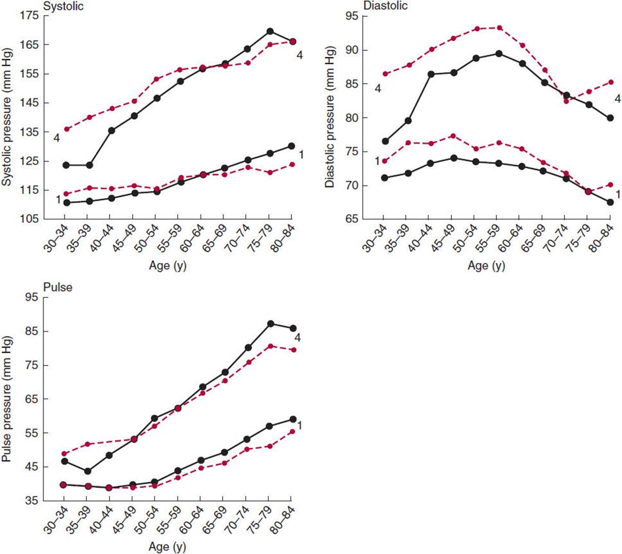
FIGURE 11-7 Effects of age and sex on systolic, diastolic, and pulse pressure in humans. Data are from a large group of individuals who were studied every 2 years throughout their adult lives. Group 1: individuals who had systolic blood pressures <120 mm Hg at age 50–60. Group 4: Individuals who had systolic blood pressure ≥160 mm Hg at age 50–60 and had not received treatment for hypertension (ie, individuals with mild, untreated hypertension). Values for females are shown in the solid black lines and those for males are shown in the dashed red lines. (Redrawn, with permission, from Franklin SS et al. Hemodynamic patterns of age-related changes in blood pressure: the Framingham Heart Study. Circulation. 1997;96:308.)
It is interesting that systolic and diastolic blood pressures are lower in young women than in young men until the age of 55–65 years, after which they become comparable. Because there is a positive correlation between blood pressure and the incidence of heart attacks and strokes (discussed later), the lower blood pressure before menopause in women may be one reason why, on average, women live longer than men.
Capillary Circulation
In the capillaries, the velocity of blood flow is decreased because, although single-vessel diameter is small, there is a large total cross-sectional area. It is in the capillary bed that nutrients leave and wastes enter the circulation. The forces producing movement of solute and solvent across capillary walls are called Starling forces after the physiologist who first described them and analyzed their function. They are the hydrostatic pressure difference across the capillary wall (capillary pressure minus tissue pressure) and the osmotic pressure gradient across the capillary wall (capillary oncotic pressure minus tissue oncotic pressure). The hydrostatic pressure gradient is outward because tissue pressure is low, and the oncotic pressure gradient is inward because large molecules in the blood do not cross the capillary wall. Obviously, most of the net movement of substances out of a typical capillary occurs at its arteriolar end, where the net pressure gradient is outward primarily because hydrostatic pressure in the capillary (about 37 mm Hg, see Figure 11-8) is greater than the oncotic pressure. As the capillary resistance and the filtration progressively cause a decrease in the hydrostatic pressure along the length of the vessel, the inwardly directed oncotic pressure gradient becomes greater than the hydrostatic pressure gradient so that, at the venular end, fluid is reabsorbed. Thus, net flow is out of the capillary at the arteriolar end and into the capillary at the venular end. Any excess solute and solvent in the tissues is picked up by the lymph vessels and moved to the venous circulation by the main lymphatic ducts. Flow in the small lymphatics is passive, but in the larger lymphatic ducts there are valves and the walls contract.
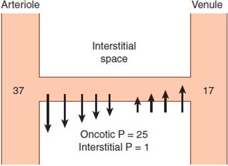
FIGURE 11-8 Schematic representation of pressure (P) gradients across the wall of a muscle capillary. The numbers at the arteriolar and venular ends of the capillary are the hydrostatic pressures in millimeters of mercury at these locations. The arrows indicate the approximate magnitude and direction of fluid movement. In this example, the pressure differential at the arteriolar end of the capillary is 11 mm Hg ([37 − 1] − 25) outward; at the opposite end, it is 9 mm Hg (25 − [17 − 1]) inward. (Redrawn, with permission, from Barrett KE et al, eds. Ganong’s Review of Medical Physiology, 24th ed. McGraw-Hill, 2012.)
REGULATION OF THE CARDIOVASCULAR SYSTEM
Given the vital nature of the cardiovascular system in maintaining blood flow to vital organs and adjusting flow so that it is increased in active tissues and decreased in inactive tissues, it is not surprising that multiple cardiovascular regulatory mechanisms have evolved. Cardiovascular adjustments are effected by altering the output of the pump (the heart), changing the diameter of the resistance vessels (chiefly the arterioles), and altering the amount of blood pooled in the capacitance vessels (the veins).
Regulation of cardiac output is discussed in Chapter 10. The caliber of the arterioles is regulated by vasodilator metabolites produced in metabolically active tissues, by the process of autoregulation, by a variety of vasoregulatory substances produced by endothelial cells, by circulating vasoactive hormones, and by a system of vasomotor nerves to the blood vessels and the heart. Discharge in the vasomotor nerves is regulated in feedback fashion by carotid sinus and aortic arch baroreceptors that monitor pressure in the arteries (high-pressure baroreceptor system) and baroreceptors in the cardiac atria and great veins (low-pressure baroreceptor system).
Vasodilator Metabolites
Various metabolic changes occurring in active tissues produce substances that dilate vessels supplying the tissues. This helps ensure the increased blood flow necessary to support the increased tissue activity. One important vasodilator is CO2. Another is K+, and adenosine dilates blood vessels in some tissues. In addition, the rise in temperature and the fall in pH that occur in some metabolically active tissues have a vasodilator effect.
Autoregulation
Many tissues have the ability to maintain a relatively constant blood flow during changes in perfusion pressure; this process is called autoregulation. The physiologic basis of autoregulation is unsettled. One factor is the myogenic response to stretch of the smooth muscle in arterioles; as pressure inside a vessel rises, its smooth muscle is stretched, and its response is to contract. Smooth muscle contracts in the absence of extrinsic innervation. Another factor may be accumulation of vasodilator metabolites; when flow to a tissue is reduced, the metabolites are not washed away, and they accumulate even in the absence of increased activity.
Substances Secreted by the Endothelium
The blood vessels are lined by a continuous layer of endothelial cells, and these cells play a vital role in the regulation of vascular function. They respond to flow changes (shear stress), stretch, a variety of circulating substances, and inflammatory mediators. In response to these stimuli, they secrete growth regulators and vasoactive substances. The growth factors regulate vascular development and are important in a number of diseases. The vasoactive substances produced by the endothelium generally act in a paracrine fashion to regulate local vascular tone. They include prostaglandins such as prostacyclin and also thromboxanes, nitric oxide, and endothelins.
A. Prostaglandins & Thromboxanes
Prostacyclin is produced by endothelial cells and thromboxane A2 by platelets from their common precursor, arachidonic acid. Thromboxane A2 causes platelet aggregation and vasoconstriction, whereas prostacyclin promotes vasodilation. The balance between the two is one of the mechanisms favoring local vasoconstriction and clot formation at sites of vascular injury while keeping the clot from extending, thereby maintaining normal flow in neighboring uninjured areas. The balance between platelet thromboxane A2 and endothelial prostacyclin can be shifted by administration of low doses of aspirin. Thromboxane A2 and prostacyclin are both produced from arachidonic acid by the cyclooxygenase pathway. Aspirin produces irreversible inhibition of cyclooxygenase. However, endothelial cells make more cyclooxygenase within a few hours, whereas circulating platelets do not, and new platelet cyclooxygenase appears only as new platelets enter the circulation over a period of days. Therefore, chronic administration of small doses of aspirin reduces intravascular clotting for prolonged periods and is of value in preventing myocardial infarctions, unstable angina, transient ischemic attacks, and stroke.
B. Nitric Oxide
The production of a potent vasodilator by endothelial cells was first suspected when it was noted that removal of the endothelium from rings of arterial tissue converted the normal dilator response to acetylcholine into a constrictor response. The responsible agent was first called endothelium-derived relaxing factor, but it is now known to be nitric oxide (NO). NO is produced from arginine (Figure 11-9) in a reaction catalyzed by nitric oxide synthase(NOS). Three forms of NOS have been cloned: NOS1, found in the nervous system; NOS2, found in macrophages and related immune cells; and NOS3, found in endothelial cells. NOS1 and NOS3 are activated by agents that increase intracellular Ca2+, including the vasodilators acetylcholine and bradykinin, whereas NOS2 is activated by cytokines. The NO that is formed in endothelial cells diffuses to adjacent vascular smooth muscle cells, where it activates soluble guanylyl cyclase, producing cyclic guanosine monophosphate (cGMP; Figure 11-9). The cGMP mediates relaxation of vascular smooth muscle.
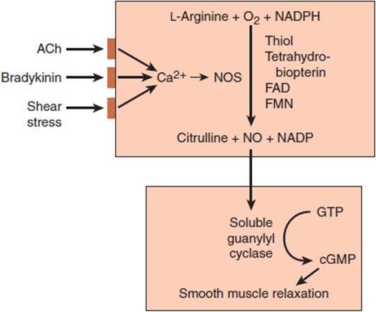
FIGURE 11-9 Synthesis of nitric oxide (NO) from arginine in endothelial cells and its action via stimulation of soluble guanylyl cyclase and generation of cyclic guanosine monophosphate (cGMP) to produce relaxation in vascular smooth muscle cells. The endothelial form of nitric oxide synthase (NOS) is activated by increased intracellular Ca2+, and an increase in Ca2+ is produced by acetylcholine (ACh), bradykinin, or shear stress acting on the cell membrane. Thiol, tetrahydrobiopterin, flavin adenine dinucleotide (FAD), and flavin mononucleotide (FMN) are requisite cofactors. GTP, guanosine triphosphate. (Redrawn, with permission, from Barrett KE et al, eds. Ganong’s Review of Medical Physiology, 24th ed. McGraw-Hill, 2012.)
The vasodilators that act by way of NO in vivo include not only acetylcholine and bradykinin but vasoactive intestinal polypeptide (VIP), substance P, and some other polypeptides. In addition, various substances that produce vasoconstriction in vivo would have a much greater constrictor effect if they did not simultaneously release NO. Consequently, NO is a major local regulator of blood flow. Its widespread role in regulation of the vascular system is indicated by the fact that infusion of amino acid analogs of arginine that inhibit NOS cause blood pressure to rise. Thus, it appears that NOS is acting in a chronic fashion to keep the vascular system dilated.
NO is responsible in large part for reactive hyperemia, the vasodilation and increased blood flow that occur in tissues and organs after a transient obstruction of their blood supply is removed. It can be seen in the forearm after occlusion of the blood supply above the elbow, and it can be quantitated by measuring the increase in forearm volume by plethysmography. NO-dependent vasodilation can also be measured clinically by determining the dilator response to graded doses of acetylcholine injected intra-arterially.
Recent advances in the field of NO research have led to identification of asymmetric-dimethylarginine (ADMA), an endogenous inhibitor of NOS enzymes. Data are emerging linking ADMA to endothelial dysfunction, cardiovascular mortality and chronic kidney disease.
NO is present in many tissues in addition to the vascular system. Its function in some of these tissues is discussed in other chapters of this book.
C. Endothelins
Endothelial cells also produce endothelin-1 (ET-1), the most potent vasoconstrictor agent yet discovered. Three closely related endothelins have been identified in mammals: ET-1, endothelin-2 (ET-2), and endothelin-3 (ET-3). All are polypeptides related to the sarafotoxins, polypeptides found in snake venoms. They contain 21 amino acid residues and two disulfide bonds (Figure 11-10). All are cleaved from larger prohormones (big endothelins) by endothelin-converting enzymes. The most widely expressed endothelin, ET-1, is found in vascular endothelial cells, vascular smooth muscle cells, macrophages, fibroblasts, myocardiocytes, brain neurons, pancreatic and intestinal epithelial cells, among others. Alternatively, ET-2 expression is restricted to intestinal epithelial cells and ovarian cells, whereas ET-3 expression is observed only in vascular endothelial cells and intestinal epithelial cells.
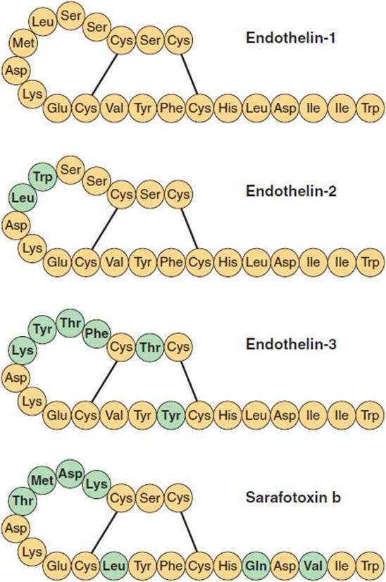
FIGURE 11-10 Structure of human endothelins and one of the snake venom sarafotoxins. The amino acid residues that differ from endothelin-1 are indicated in blue. (Redrawn, with permission, from Ganong WF. Review of Medical Physiology, 22nd ed. McGraw-Hill, 2005.)
Over the past several years, our understanding of endothelin physiology and pathophysiology (particularly related to ET-1) has increased tremendously. Two G protein–coupled receptors—A and B—that mediate endothelin effects have been identified. Endothelin A receptor has the greatest affinity for ET-1, whereas endothelin B receptor has the same affinity for all three polypeptide isoforms. Interestingly, vascular smooth muscle cells express both endothelin receptors, and their activation leads to vasoconstriction. Endothelial cells, however, express only endothelin B receptor, which stimulates endothelial NOS, leading to NO-dependent smooth muscle relaxation. Recent animal data suggest that the activation of the endothelin B receptor in collecting ducts leads to a similar NO-dependent increase in sodium excretion. Moreover, there are indications that ET-1 may contribute to the extracellular matrix remodeling in vascular, cardiac, and kidney disease.
Circulating Hormones That Affect Vascular Smooth Muscle
Hormones in the circulation that have general effects on the vascular system include vasoconstrictors and vasodilators. The principal vasoconstrictors are norepinephrine and epinephrine (see Chapter 12), vasopressin (Chapter 19), and angiotensin II (Chapter 21). The principal vasodilators are vasoactive intestinal peptide (VIP; see Chapter 13), kinins, and natriuretic peptides.
A. Kinins
The kinins are two related vasodilator polypeptides called bradykinin and lysyl-bradykinin (Figure 11-11). The decapeptide lysyl-bradykinin can be converted to the nonapeptide bradykinin by aminopeptidase. Both are metabolized to inactive fragments by the carboxypeptidase kininase I or the dipeptidylcarboxypeptidase kininase II. Kininase II and angiotensin-converting enzyme are the same enzyme, so inhibition of angiotensin-converting enzyme for the treatment of hypertension or heart failure increases plasma and tissue kinins.
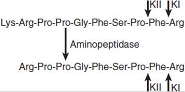
FIGURE 11-11 Kinins. Lysyl-bradykinin (top) can be converted to bradykinin (bottom) by aminopeptidase. The peptides are inactivated by kininase I (KI) or kininase II (KII) at the sites indicated by the short arrows. (Redrawn, with permission, from Barrett KE et al, eds. Ganong’s Review of Medical Physiology, 24th ed. McGraw-Hill, 2012.)
Kinins are formed from two kininogens: high-molecular-weight (HMW) kininogen and low-molecular-weight (LMW) kininogen. These kinin precursor proteins are products of a single gene produced by alternative splicing. The proteases responsible for cleavage of kininogens are kallikreins, a family of enzymes encoded in humans by three genes situated on chromosome 19.
Lysyl-bradykinin and bradykinin are primarily tissue hormones produced, for example, by the kidneys and actively secreting glands, but small amounts are also found in the circulating blood. They act on two receptors, B1 and B2, both coupled to G proteins. Kinins increase blood flow to actively secreting glands by producing vasodilation, and when injected systemically they are relatively potent vasodilators.
B. Natriuretic Hormones
Atrial natriuretic peptide (ANP) is a polypeptide containing 28 amino acid residues that is secreted from the atria when atrial myocytes are stretched. Brain natriuretic peptide (BNP) was originally isolated from the brains of experimental animals, but in humans it is secreted by the ventricular myocytes and is commonly known as β-type natriuretic peptide. CNP, a third type of natriuretic peptide, is also found in humans. These peptides cause natriuresis, probably by increasing the glomerular filtration rate, which in turn causes excretion of salt and water, reducing blood volume and relieving the stretch on the atrial myocytes. They antagonize the pressor effects of angiotensin II and other pressor hormones. They act by increasing intracellular cGMP. All three have vasodilatory activity, but CNP differs in apparently having a greater effect on veins than arterioles. Their physiologic function is still unsettled. However, their circulating levels are increased in heart failure, and measurement of circulating β-type natriuretic peptide is seeing increased use in the differential diagnosis and evaluation of heart failure. All three of these natriuretic peptides are found in various tissues other than the heart.
An additional natriuretic hormone that acts by inhibiting Na+-K+ adenosine triphosphatase (ATPase) is present in the circulation, but it raises rather than lowers blood pressure (see sections below concerning hypertension and salt sensitivity). There is substantial evidence that this hormone is actually ouabain and that it is secreted by the adrenal glands in response to increased dietary sodium intake.
Neural Control Via the Sympathetic Vasomotor System
Factors affecting the caliber of the arterioles in the body and hence peripheral resistance and tissue blood flow are summarized in Table 11-1. This list includes the factors discussed previously plus a few additional polypeptides that have minor or special effects. It also includes the control of blood pressure by noradrenergic and in some instances cholinergic sympathetic vasomotor nerves to the arterioles. In addition to the extensive nerve supply to these resistance vessels, there is a moderate innervation of the capacitance vessels.
TABLE 11-1 Summary of factors affecting the caliber of the arterioles.

Discharge of the noradrenergic vasomotor nerves causes constriction of the arterioles innervated by the nerves, and if the discharge is general rather than local, there is an increase in blood pressure. In addition, discharge of sympathetic noradrenergic nerves innervating the heart increases blood pressure by increasing the force and rate of cardiac contraction (inotropic and chronotropic effects), increasing stroke volume and cardiac output. Noradrenergic stimulation also inhibits the effect of vagal stimulation, which normally slows the heart and decreases cardiac output.
The main control of vasomotor discharge is feedback regulation via the baroreceptors in the high-pressure and low-pressure portions of the circulatory system (Figure 11-12). The baroreceptors are stretch-sensitive nerve endings located in the carotid sinuses and aortic arch on the arterial side and in the walls of the great veins and the cardiac atria on the venous side. The nerve fibers relay impulses in cranial nerves IX and X to the medulla oblongata, where the fibers end in the nucleus tractus solitarius (Figure 11-13). From the nucleus, second-order neurons pass to the caudal portion of the ventrolateral medulla and environs. From there, third-order inhibitory neurons pass to the rostral ventrolateral medulla, the location of the cell bodies of the neurons that control blood pressure. The axons of these neurons descend into the spinal cord and innervate the cell bodies of the blood pressure-regulating preganglionic sympathetic neurons in the intermediolateral gray column of the spinal cord. The axons of the preganglionic neurons leave the spinal cord and synapse on the postganglionic neurons in the ganglionic chain and collateral ganglia as well as on the catecholamine-secreting cells in the adrenal medulla. The axons of the postganglionic noradrenergic neurons innervate the blood vessels and the heart. These pathways and the probable synaptic mediator at each synapse in the chain are shown in Figure 11-13. Note in particular that increased activity in the baroreceptor afferents produced by increases in blood pressure inhibits sympathetic vasomotor outflow, whereas decreased baroreceptor afferent discharge stimulates sympathetic vasomotor outflow. This is brought about by the inhibitory γ-aminobutyric acid–secreting neuron link between the caudal portion of the ventrolateral medulla and the rostral ventrolateral medulla. In addition, increased baroreceptor discharge stimulates afferents from the nucleus tractus solitarius to the dorsal motor nucleus of the vagus and the nucleus ambiguus. This increases vagal discharge to the heart, slowing the cardiac rate and decreasing cardiac output.

FIGURE 11-12 Feedback regulation of systemic blood pressure by baroreceptors. (Redrawn, with permission, from Barrett KE et al, eds. Ganong’s Review of Medical Physiology, 24th ed. McGraw-Hill, 2012.)
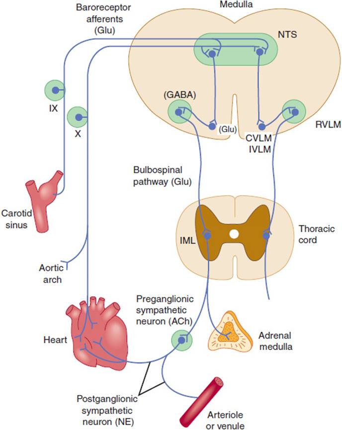
FIGURE 11-13 Basic pathways involved in the medullary control of blood pressure. The vagal efferent pathways to the heart are not shown. The probable neurotransmitters in the pathways are indicated in parentheses. (ACh, acetylcholine; GABA, γ-aminobutyric acid; Glu, glutamate; NE, norepinephrine; CVLM, IVLM, and RVLM, caudal, intermediate, and rostral ventrolateral medulla, respectively; IML, intermediolateral gray column; IX, glossopharyngeal nerve; NTS, nucleus tractus solitarius; X, vagus nerve.) (Redrawn from Reis DJ et al. Role of adrenaline neurons of the ventrolateral medulla [the C group] in the tonic and phasic control of arterial pressure. Clin Exp Hypertens [A]. 1994;6:221.)
There are ancillary reciprocal circuits between the nucleus tractus solitarius and more dorsal portions of the brainstem and the hypothalamus that smooth and adjust the response of the baroreceptor pathway, but the primary neural regulation of blood pressure is mediated by the baroreceptor pathway in the medulla oblongata.
In addition to direct effects on vasomotor discharge, the baroreceptor pathway causes changes in endocrine function that augment the homeostatic value of baroreceptor responses. Adrenal medullary secretion is increased by discharge of the sympathetic nervous system, although the contributions of circulating catecholamines to the increase in blood pressure are relatively small. Increased sympathetic discharge also increases renin secretion from the kidneys, and the resultant increase in circulating angiotensin II not only acts directly on vascular smooth muscle to cause constriction but also increases aldosterone secretion, which in turn increases Na+ retention, expanding intravascular volume. Associated with increased vasomotor discharge, there is also an increase in antidiuretic hormone (ADH, also referred to as vasopressin) secretion from the posterior pituitary. ADH expands total body water by increasing free water retention in the kidney (acting through the V2 vasopressin receptor). Although the primary role of ADH is to facilitate the lowering of osmolality, ADH also facilitates the expansion of intravascular volume. Although the volume expansion resulting from ADH is relatively small, ADH release increases with the severity of the effective circulating volume loss. Moreover, activation of the lower affinity V1 vasopressin receptor on vascular smooth muscle results in a marked increase in vascular tone.
Baroreceptor function can be tested in experimental animals and judiciously in humans by infusing the pressor drug phenylephrine at different doses and at each dose measuring the slowing of the heart rate by determining the interval between the R waves (RR interval) of the ECG. An example of results of this type of testing is shown in Figure 11-14.

FIGURE 11-14 Baroreflex-mediated lowering of the heart rate during infusion of phenylephrine in a human subject. Note that the values for the RR interval of the ECG, which are plotted on the vertical axis, are inversely proportionate to the heart rate. (Redrawn, with permission, from Kotrly K et al. Effects of fentanyl-diazepam-nitrous oxide anaesthesia on arterial baroreflex control of heart rate in man. Br J Anaesth. 1986;58:406.)
CHECKPOINT
4. Why do small changes in the diameter of the arterioles have relatively large effects on blood pressure?
5. Why does the velocity of blood flow decrease greatly in the capillaries and then increase in the veins?
6. What categories of factors are involved in regulating the diameter of arterioles?
7. By what mechanism does NO, produced by endothelial cells, act as a vasodilator?
8. What are the principal hormonal vasoconstrictors and vasodilators?
9. What is the role of baroreceptors in the feedback regulation of the high- and low-pressure portions of the circulatory system?
PATHOPHYSIOLOGY OF SELECTED VASCULAR DISORDERS
ATHEROSCLEROSIS
Prevalence & Significance
A condition that afflicts the large and medium-sized arteries of almost every human, at least in societies in which cholesterol-rich foodstuffs are abundant and cheap, is atherosclerosis. This condition begins in childhood and, in the absence of accelerating factors, develops slowly until it is widespread in old age. However, it is accelerated by a wide variety of genetic and environmental factors (see later discussion). It is characterized by localized fibrous thickenings of the arterial wall associated with lipid-infiltrated plaques that may eventually calcify. Old plaques are also prone to ulceration and rupture, triggering the formation of thrombi that obstruct flow. Therefore, atherosclerosis leads to vascular insufficiency in the limbs, abnormalities of the renal circulation, and dilations (aneurysms) and even rupture of the aorta and other large arteries. It also leads to common severe and life-threatening diseases of the heart and brain because of formation of intravascular clots at the site of the plaques.
In the United States and most other developed countries, it has been calculated that atherosclerosis is the underlying cause of about 50% of all deaths. Almost all patients with myocardial infarction—and most of those with stroke resulting from cerebral thrombosis—have atherosclerosis. The incidence of ischemic heart disease and strokes has been declining in the United States since 1963, but atherosclerosis is still very common. Thus, atherosclerosis underlies and is fundamentally responsible for a large portion of the clinical problems seen by physicians caring for adult patients.
Pathogenesis
The initial event in atherosclerosis is infiltration of low-density lipoproteins (LDLs) into the subendothelial region. The endothelium is subject to shear stress, the tendency to be pulled along or deformed by flowing blood. This is most marked at points where the arteries branch, and this is where the lipids accumulate to the greatest degree.
The LDLs are oxidized or altered in other ways. Thus, altered LDLs activate various components of innate immune system including macrophages, natural antibodies, and innate effector proteins such as C-reactive protein and complement. Altered LDLs are recognized by a family of scavenger receptors expressed on macrophages that cooperate with toll-like receptors to stimulate inflammation and drive atherogenesis. The scavenger receptors mediate uptake of the oxidized LDL into macrophages and the formation of foam cells (Figure 11-15). The foam cells form fatty streaks. The streaks appear in the aorta in the first decade of life, in the coronary arteries in the second decade, and in the cerebral arteries in the third and fourth decades.
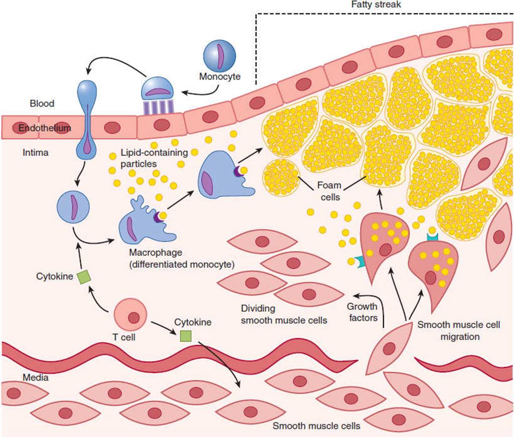
FIGURE 11-15 Formation of a fatty streak in an artery. After vascular injury, monocytes bind to the endothelium, then cross it to the subendothelial space, and become activated tissue microphages. The macrophages take up oxidized low-density lipoproteins (LDL), becoming foam cells. T cells release cytokines, which also activate macrophages. In addition, the cytokines cause smooth muscle cells to proliferate. Under the influence of growth factors, the smooth muscle cells then move to the subendothelial space where they produce collagen and take up LDL, adding to the population of foam cells. (Redrawn, with permission, from Hajjar DP et al. Atherosclerosis. Am Scientist. 1995;83:460.)
Oxidized LDLs have a number of deleterious effects, including stimulation of release of proinflammatory cytokines (such as macrophage migration inhibitory factor and type I interferon) and inhibition of NO production. Vascular smooth muscle cells in the vicinity of foam cells are stimulated and move from the media to the intima, where they proliferate, lay down collagen and other matrix molecules, and contribute to the bulk of the lesion. Smooth muscle cells also take up oxidized LDL and become foam cells. Lipids accumulate both intracellularly and extracellularly.
The intercellular “soup” in the plaques contains a variety of cell-damaging substances, including ozone. In addition, the “loading” of macrophages with cholesterol can be lipotoxic to the endoplasmic reticulum, resulting in macrophage apoptosis and plaque necrosis. Cholesterol crystals associated with necrotized macrophages further stimulate inflammation and lead to the recruitment of neutrophils. As the atherosclerotic lesions age, T cells of the immune system and monocytes are attracted to them, creating a vicious cycle of necrosis and inflammation.
As plaques mature, a fibrous cap forms over them. The plaques with defective or broken caps are most prone to rupture. The lesions alone may distort vessels to the point that they are occluded, but it is usually rupture or ulceration of plaques that triggers thrombosis, blocking blood flow.
Atherosclerotic lesions have been shown to have many of the characteristics of a low-grade infection. A number of investigators have searched for bacteria in plaques, and in a significant number Chlamydophila pneumoniae—an organism usually associated with respiratory infection—has been found. However, other organisms have also been found, and it is too early to say whether the chlamydiae are causative agents or merely coincidental tenants in the lesions.
A characteristic of atherosclerosis that is currently receiving considerable attention is its association with deficient release of NO and defective vasodilation. As noted, oxidized LDLs inhibit NO production. If acetylcholine is infused via catheter into normal coronary arteries, the vessels dilate; however, if it is infused when atherosclerosis is present, the vessels constrict. This indicates that endothelial secretion of NO is defective.
Interestingly, recent experimental evidence indicates that the activation of the vasculature endothelial receptor for endothelin B both stimulates eNOS and exerts antiproliferative effects on vascular smooth muscle cells. It has been speculated that the disrupted signaling via this receptor can be an additional contributing factor in pathophysiology of atherosclerosis.
Relation to Dietary Cholesterol & Other Lipids
Transforming a monocyte into a lipid-ingesting macrophage involves the appearance on its surface of a unique type of oxidized LDL receptor, the scavenger receptor, and monocytes are stimulated to produce these receptors by the action of macrophage colony-stimulating factor secreted by endothelial cells and vascular smooth muscle cells. When oxidized LDL-receptor complexes are formed, they are internalized and the receptors recycle to the membrane while the lipid is stored.
Obviously, accumulation of lipid in foam cells is a key event in the progression of atherosclerotic lesions, and it is well established that lowering plasma cholesterol slows the progress of atherosclerosis. The main pathways for the metabolism of ingested lipids are summarized in Figure 11-16. Because lipids are relatively insoluble, they are transported as special lipoprotein particles that increase their solubility. Dietary cholesterol and triglycerides are packaged in the protein-coated chylomicrons in intestinal epithelial cells. Under the influence of lipoprotein lipase, these particles release triglycerides to fat depots and muscles, and the resulting chylomicron remnants are taken up by the liver. The liver also synthesizes cholesterol and packages it with specific proteins to form very-low-density lipoproteins (VLDLs). These lipoprotein particles enter the circulation and under the influence of lipoprotein lipase donate triglycerides to tissues. In this way, they become cholesterol-rich intermediate-density lipoproteins (IDLs) and low-density lipoproteins (LDLs). The LDL supply cholesterol to the tissues. They provide all cells with the cholesterol for production of cell membranes and other uses. They also provide most of the cholesterol that is the precursor for all steroid hormones. As noted, oxidized LDLs are taken up by macrophages and smooth muscle cells in atherosclerotic lesions. On the other hand, high-density lipoproteins (HDLs) take cholesterol from peripheral cells and transport it to the liver where it is metabolized, keeping plasma and tissue cholesterol low. For this reason, it is referred to as “good cholesterol” as opposed to LDL cholesterol, which is “bad cholesterol.” Efforts are being made to increase HDL by pharmaceutical means in the treatment of atherosclerosis.

FIGURE 11-16 Simplified diagram of lipoprotein systems for transporting lipids in humans. In the exogenous system, chylomicrons rich in triglycerides of dietary origin are converted to chylomicron remnants rich in cholesteryl esters by the action of lipoprotein lipase. In the endogenous system, very low-density lipoproteins (VLDL) rich in triglycerides are secreted by the liver and converted to intermediate-density lipoproteins (IDL) and then to low-density lipoproteins (LDL) rich in cholesteryl esters. Some of the LDL enter the subendothelial space of arteries, are oxidized, and then taken up by macrophages, which become foam cells. LCAT, lecithin-cholesterol acyltransferase. The letters on the chylomicrons, chylomicron remnants, VLDL, IDL, and LDL identify the primary apoproteins found in them.
Clinical Manifestations
Because atherosclerosis is an abnormality of arterial blood vessels, it can affect almost any organ in the body. Calcified atherosclerotic plaques are occasionally detected on x-ray film, and angiographic visualization of deformed arterial walls is possible. In general, however, atherosclerosis is asymptomatic until one of its complications develops.
In coronary arteries, atherosclerotic narrowing that reduces the lumen of a coronary artery more than 75% causes angina pectoris, the chest pain that results when pain-producing substances accumulate in the myocardium. Typically, the pain comes on during exertion and disappears with rest, as the substances are washed out by the blood. When atherosclerotic lesions cause clotting and occlusion of a coronary artery, the myocardium supplied by the artery dies (myocardial infarction). Myocardial infarction is also discussed in Chapter 10.
In the cerebral circulation, arterial blockage at the site of atherosclerotic plaques causes thrombotic strokes. Strokes are discussed in Chapter 7. In the abdominal aorta, extensive atherosclerosis can lead to aneurysmal dilation and rupture of the vessel. In the renal vessels, localized constriction of one or both renal arteries causes renovascular hypertension (see later discussion). In the circulation to the legs, vascular insufficiency causes intermittent claudication (fatigue and usually pain on walking that is relieved by rest). If the circulation of a limb is severely compromised, the skin can ulcerate, producing lesions that are slow to heal. Frank gangrene of the extremities may also occur. Less frequently, clot formation and obstruction may occur in vessels supplying the intestines or other parts of the body.
Risk Factors
As noted, the progression of atherosclerosis is accelerated by a wide variety of genetic and environmental factors (risk factors). These are summarized in Table 11-2. Obviously, treating the accelerating conditions that are treatable and avoiding those that are avoidable should reduce the incidence of myocardial infarctions, strokes, and other complications of atherosclerosis.
TABLE 11-2 Conditions that accelerate the progression of atherosclerosis and the mechanisms responsible.
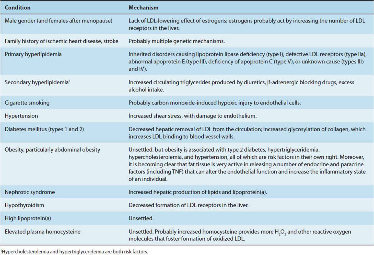
Estrogen increases cholesterol removal by the liver, and the progression of atherosclerosis is less rapid in premenopausal women than in men. In addition, epidemiologic evidence shows that estrogen replacement therapy protects the cardiovascular system in postmenopausal women. On the other hand, large doses of estrogens increase the incidence of blood clots, and even small doses produce a slight increase in clotting. In addition, in several studies, estrogen treatment of postmenopausal women failed to prevent second heart attacks. The reason for the discrepancies between the epidemiologic and experimental data is currently unsettled.
The effect of increased plasma levels of homocysteine and related molecules such as homocystine and homocysteine thiolactone, a condition sometimes called hyperhomocystinemia, deserves emphasis. These increases are associated with accelerated atherosclerosis, and the magnitude of the plasma elevation is positively correlated with the severity of the atherosclerosis. Markedly elevated levels resulting from documented mutations of relevant genes are rare, but mild elevations occur in 7% of the general population. The mechanism responsible for the accelerated vascular damage is unsettled, but homocysteine is a significant source of H2O2 and other reactive forms of oxygen, and this may accelerate the oxidation of LDL.
Homocysteine is an intermediate in the synthesis of methionine. It is metabolized by enzymes that are dependent on vitamin B6, vitamin B12, and folic acid. Supplementation of the diet with these vitamins reduces plasma homocysteine, usually to normal. Determining whether such supplements also reduce the incidence of the accelerated atherosclerosis will require prolonged, careful clinical trials, and the results of such studies to date are inconclusive.
Evidence is now overwhelming that lowering plasma cholesterol and triglyceride levels and increasing plasma HDL levels slows, and in some cases reverses, the atherosclerotic process. The desired decrease in lipids can sometimes be achieved with dietary restriction of cholesterol, saturated and trans fat alone, even though dietary restriction initiates a compensatory increase in cholesterol synthesis in the body. When dietary treatment is not adequate, reducing conversion of mevalonate to cholesterol with statins, drugs that inhibit hepatic 3-methylglutaryl coenzyme A (HMG-CoA) reductase, the enzyme which catalyzes this reaction, is beneficial. The currently available HMG-CoA reductase inhibitors include atorvastatin, lovastatin, pitavastatin, pravastatin, simvastatin, fluvastatin, and rosuvastatin.
In cases in which there is severe hypercholesterolemia because of congenitally defective LDL receptors, gene therapy may be an option. However, despite promising preliminary results, gene therapy in humans appears to be unachievable until better means for gene transfer are developed. Other approaches to slowing or preventing development of atherosclerosis by molecular biologic techniques are under development.
Antioxidant treatment with agents such as α-tocopherol, vitamin E, and β-carotene has been used to inhibit oxidation of LDL, and this reduces the incidence of atherosclerotic changes in experimental animals. However, the results of antioxidant treatment in humans have generally been disappointing or negative.
Men who smoke a pack of cigarettes a day have a 70% increase in death rate from ischemic heart disease compared with nonsmokers, and there is also an increase in women. Smoking cessation lessens the risk of death and of myocardial infarction. The deleterious effects of smoking include endothelial damage caused by carbon monoxide–induced hypoxia. Other factors may also be involved. Thus, stopping smoking is a major way to slow the progress of atherosclerosis.
Because of the increased shear stress imposed on the endothelium by an elevated blood pressure, hypertension is another important modifiable risk factor for atherosclerosis. Lowering blood pressure has its greatest effect in reducing the incidence of stroke, but there are beneficial effects on ischemic heart disease as well. With modern methods of treatment, blood pressure in hypertensives can generally be reduced to normal or near-normal values, and the decrease in strokes, myocardial infarctions, and renal failure produced by such treatment is clear testimony to the value of reducing or eliminating this risk factor.
In diabetics, there are microvascular complications and macrovascular complications (see Table 18–6). The latter are primarily related to atherosclerosis. There is a twofold increase in the incidence of myocardial infarction compared with nondiabetics; severe circulatory deficiency in the legs with gangrene is relatively common; there are more thrombotic strokes; and renal failure is a serious problem (see Chapter 18). It is interesting in this regard that rigorous control of blood pressure in diabetics has been shown to be more efficacious in reducing cardiovascular complications than rigorous control of blood glucose.
The nephrotic syndrome and hypothyroidism also accelerate the progression of atherosclerosis and are treatable conditions.
Although local inflammation clearly plays a direct role in the pathogenesis of atherosclerosis, the possibility that indirect mechanisms associated with autoimmune diseases, infections (including gum disease and gastric infections), or exposure to various pollutants contribute to (or even initiate) atherosclerosis remains controversial.
CHECKPOINT
10. What is the most common cause of death in the United States among individuals older than 45 years?
11. What is the hypothesized mechanism of atherosclerotic plaque formation?
12. What are some ways in which atherosclerotic plaques can cause cardiovascular disease?
13. Name five treatable risk factors that accelerate the progression of atherosclerosis.
HYPERTENSION
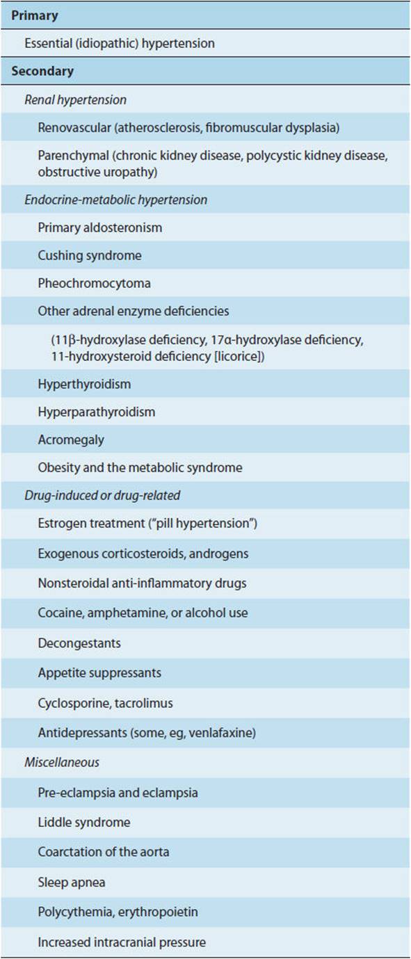
Hypertension is not a single disease but a syndrome with multiple causes. In most instances, the cause remains unknown, and the cases are lumped together under the term essential hypertension (Table 11-3). However, mechanisms are continuously being discovered that explain hypertension in new subsets of the formerly monolithic category of essential hypertension, and the percentage of cases in the essential category continues to decline. Essential hypertension is often called primary hypertension, and hypertension in which the cause is known is called secondary hypertension, although this separation seems somewhat artificial. This chapter discusses the pathogenesis of hypertension and its complications in general terms and then discusses the specific causes of the currently defined subgroups and the unique features, if any, that each adds to the general findings in patients with high blood pressure.
TABLE 11–3 Primary and secondary causes of hypertension.
Pathogenesis
Current guidelines of the Joint National Committee on Prevention, Detection, Evaluation, and Treatment of High Blood Pressure define normal blood pressure as systolic pressure of less than 120 mm Hg and diastolic pressure of less than 80 mm Hg. Hypertension is defined as an arterial pressure greater than 140/90 mm Hg in adults on at least three consecutive visits to the doctor’s office. People whose blood pressure is between normal and 140/90 mm Hg are considered to have pre-hypertension and people whose blood pressure falls in this category should appropriately modify their lifestyle to lower their blood pressure to below 120/80 mm Hg. As noted (Figure 11-7), systolic pressure normally rises throughout life, and diastolic pressure rises until age 50–60 years but then falls, so that pulse pressure continues to increase. In the past, emphasis has been on treating individuals with elevated diastolic pressure. However, it now appears that, particularly in elderly individuals, treating systolic hypertension is equally important or even more so in reducing the cardiovascular complications of hypertension. Moreover, some studies indicate that overly aggressive treatment (particularly of diastolic hypertension) may be associated with adverse cardiac events (primarily myocardial infarctions) in patients with coronary artery disease or chronic heart failure. The explanation may be that because the coronary arteries fill during diastole, in individuals with coronary artery disease or heart failure, adequate cardiac muscle perfusion is dependent on a somewhat higher diastolic blood pressure.
The most common cause of hypertension is increased peripheral vascular resistance. However, because blood pressure equals total peripheral resistance times cardiac output, prolonged increases in cardiac output can also cause hypertension. These are seen, for example, in hyperthyroidism and beriberi. In addition, increased blood volume causes hypertension, especially in individuals with mineralocorticoid excess or renal failure (see later discussion); and increased blood viscosity, if it is marked, can increase arterial pressure.
Clinical Presentation
Hypertension by itself does not cause symptoms. Headaches, fatigue, and dizziness are sometimes ascribed to hypertension, but nonspecific symptoms such as these are no more common in hypertensives than they are in normotensive controls. Instead, the condition is discovered during routine screening or when patients seek medical advice for its complications. These complications are serious and potentially fatal. They include myocardial infarction, heart failure, thrombotic and hemorrhagic strokes, hypertensive encephalopathy, and renal failure (Figure 11-17). This is why hypertension is called “the silent killer.”

FIGURE 11-17 Pathogenesis of the complications produced by arterial hypertension. LVH, left ventricular hypertrophy. (Redrawn, with permission, from Deshmukh R et al. Chapter 13 in Lilly LS et al, eds. Pathophysiology of Heart Disease: A Collaborative Project of Medical Students and Faculty, 3rd ed. Williams & Wilkins, 2003.)
Physical findings are also absent in early hypertension, and observable changes are generally found only in advanced severe cases. These may include hypertensive retinopathy (ie, narrowed arterioles seen on funduscopic examination) and, in more severe cases, retinal hemorrhages and exudates along with swelling of the optic nerve head (papilledema). Prolonged pumping against an elevated peripheral resistance causes left ventricular hypertrophy, which can be detected by echocardiography, and cardiac enlargement, which can be detected on physical examination. It is important to listen with the stethoscope over the kidneys because in renal hypertension (see later discussion) narrowing of the renal arteries may cause bruits. These bruits are usually continuous throughout the cardiac cycle. It has been recommended that the blood pressure response to rising from the sitting to the standing position be determined. A blood pressure rise on standing sometimes occurs in essential hypertension presumably because of a hyperactive sympathetic response to the erect posture. This rise is usually absent in other forms of hypertension. Most individuals with essential hypertension (60%) have normal plasma renin activity, and 10% have high plasma renin activity. However, 30% have low plasma renin activity. Renin secretion may be reduced by an expanded blood volume in some of these patients, but in others the cause is unsettled, and low-renin essential hypertension has not yet been separated from the rest of essential hypertension as a distinct entity.
In many patients with hypertension, the condition is benign and progresses slowly; in others, it progresses rapidly. Actuarial data indicate that on average untreated hypertension reduces life expectancy by 10–20 years. Atherosclerosis is accelerated, and this in turn leads to ischemic heart disease with angina pectoris and myocardial infarctions (Chapter 10), thrombotic strokes and cerebral hemorrhages (Chapter 7), and renal failure (Chapter 16). Another complication of severe hypertension is hypertensive encephalopathy, in which there is confusion, disordered consciousness, and seizures. This condition, which requires vigorous treatment, is probably due to arteriolar spasm and cerebral edema.
In all forms of hypertension regardless of cause, the condition can suddenly accelerate and enter the malignant phase. In malignant hypertension, there is widespread fibrinoid necrosis of the media with intimal fibrosis in arterioles, narrowing them and leading to progressive severe retinopathy, heart failure, and renal failure. If untreated, malignant hypertension is usually fatal in 1 year.
MANAGEMENT
A discussion of disease treatment is beyond the scope of this book. However, it should be noted that in all forms of hypertension modern treatment with β-adrenergic blocking drugs, inhibitors of the renin-angiotensin system, Ca2+channel inhibitors, and diuretics reduce blood pressure, usually to normal levels. In addition, these treatments delay or prevent complications and lengthen life expectancy. However, they are not curative and must be continued indefinitely. Thus, essential hypertension is like diabetes mellitus: It can be controlled but not cured. If a cause of hypertension can be identified, its treatment may result in a cure. Consequently, it is important to identify such cases.
Etiology
A. Coarctation of the Aorta
Congenital narrowing of the aorta usually occurs just distal to the origin of the left subclavian artery. Peripheral resistance is increased above the constriction. Therefore, blood pressure is elevated in the arms, head, and chest but lowered in the legs. However, because the constriction is proximal to the renal arteries, renin secretion is increased in most cases of coarctation as a result of the reduction in arterial pressure in the renal arteries. This tends to increase blood pressure throughout the body. Elimination of the constriction by resecting the narrowed segment of the aorta usually cures the condition.
B. Salt Sensitivity
Through selective inbreeding, Dahl was able to develop two strains of rats: salt-sensitive rats that showed an increase in blood pressure when fed a high-salt diet and salt-resistant rats that did not. The genetic mechanisms responsible for these strain differences are currently under investigation. There may be a similar division of humans into salt-sensitive and salt-resistant groups, although obviously the lines between the groups are less distinct. As shown in Table 11-4, about 30% of whites with normal renal function and normal blood pressure are salt sensitive compared with 55% of whites with essential hypertension. For unknown reasons, a larger percentage of black hypertensives are salt sensitive. These figures have obvious significance in terms of recommendations about salt intake in hypertension.
TABLE 11–4 Salt sensitivity in humans.

It should be emphasized that the figures just cited refer to individuals with normal renal function and normal (or reduced) secretion of mineralocorticoid hormones. When renal function is reduced, mineralocorticoid secretion is increased, or the effects of mineralocorticoids are enhanced, there is abnormal retention of salt and water, and hypertension is produced on this basis (see later discussion).
Although the genetic mechanisms responsible for the differences in salt sensitivity are still unknown, recent studies have shed new light on our understanding of salt-mediated hypertension. Salt appears to activate three pathways that may lead to vascular smooth muscle contraction: 1) salt stimulates a subset of G proteins (G12-13) that are responsible for the activation of myosin light chain kinase, which phosphorylates myosin to initiate contraction; 2) salt stimulates the Rho/Rho kinase pathway, which inhibits myosin light chain phosphatase to prevent smooth muscle relaxation; 3) short-term increases in dietary salt intake stimulate release of endogenous ouabain (whose effect on the vascular smooth muscle parallels the effects of the cardiac glycoside) to inhibit Na+-K+ ATPase with a consequent decrease in Na+-Ca+ exchanger activity, ultimately elevating intracellular calcium levels and increasing smooth muscle tone. Experimental evidence suggests that individual differences in these signaling pathways may indeed contribute to salt-related hypertension.
Finally, animal studies point at new possible mechanisms for salt-sensitive hypertension, including aldosterone-independent activation of mineralocorticoid receptors, as well as sympathetically mediated activation of sodium reabsorption in the distal renal tubule.
C. Renal Abnormalities
Goldblatt’s observation that renal artery constriction increased blood pressure in experimental animals was rapidly followed by demonstration of the same event in humans. However, disappointment followed when it was found that renal hypertension resulting from constriction of one or both renal arteries accounted for only a very small percentage of cases of clinical hypertension. The narrowing can be due to atherosclerosis, fibroelastic overgrowth of the wall of the renal artery, or external pressure on the vessel. The initial constriction decreases renal arteriolar pressure, and this leads to increased renin secretion. The renin-angiotensin system is discussed in Chapters 16 and 21. However, in many cases, some other mechanism takes over chronically to maintain the hypertension. The nature of this other mechanism is unknown.
Ureteral obstruction can cause hypertension in animals and probably in humans by increasing renal interstitial pressure and thus decreasing the pressure gradient across the renin-secreting juxtaglomerular cells.
Acute and chronic glomerulonephritis and other forms of diffuse kidney disease can cause hypertension when loss of the ability to excrete salt is severe enough that Na+ and water are retained and blood volume is expanded.
D. Hormonal Disorders (see Chapters 12 & 21)
A remarkable number of adrenal abnormalities cause hypertension. These include mainly conditions in which mineralocorticoids are secreted in excess, but excess secretion of cortisol also causes hypertension, as does excess secretion of catecholamines by tumors of the adrenal medulla. These disorders are covered in detail in Chapters 12 and 21.
One particular contributing factor to hypertension in women is estrogen. Secretion of angiotensinogen from the liver is under endocrine control and is uniquely stimulated by estrogens. Consequently, it is increased in women taking contraceptive pills containing large amounts of estrogens. When circulating angiotensinogen is increased, more angiotensin II is formed and blood pressure rises. The normal compensation for this response is decreased secretion of renin because angiotensin II feeds back directly on the juxtaglomerular cells to reduce renin secretion. However, in some women, the compensation is incomplete and the estrogens cause a significant increase in blood pressure. Some of the women with the condition have underlying essential hypertension, which is triggered by the estrogens, but in others the hypertension is cured by stopping estrogen treatment.
In view of the fact that Na+ retention resulting from mineralocorticoid excess causes hypertension, it may seem surprising that a natriuretic hormone is also a suspected cause of hypertension. ANP and other natriuretic peptides of cardiac origin cause sodium loss in the urine and generally lower blood pressure. However, there is, in addition, a digitalis-like natriuretic substance in the circulation. Its source seems to be the adrenals, although it has also been claimed that it is secreted by the hypothalamus. This substance, which may be naturally occurring ouabain, inhibits Na+-K+ ATPase. This results in loss of Na+ in the urine, but Ca2+ accumulates in cells because of the decrease in Na+ gradient across the cell membrane. The increase in intracellular Ca2+ causes vascular smooth muscle to contract. Consequently, blood pressure is increased. However, the physiologic and pathophysiologic significance of this natriuretic hormone remains unsettled, and hypersecretion of it cannot as yet be considered a proved cause of clinical hypertension.
E. Neurologic Disorders
The nervous system plays a key role in maintaining blood pressure in normal individuals (see prior discussion). Clonidine and other drugs lower blood pressure by acting on the brain to decrease sympathetic discharge, and several of the most effective treatments for chronic hypertension act peripherally to reduce the effect of vasomotor sympathetic discharge to the blood vessels and heart. These and other observations suggest that clinical hypertension could be caused by CNS abnormalities. Interruption of the afferent input from the baroreceptors to the CNS in experimental animals causes increased blood pressure. However, emphasis has been placed on the variability of the blood pressure in such animals rather than on any consistent elevation of mean arterial pressure. There is some evidence that chronic pressure on the rostral ventrolateral medulla (Figure 11-13) caused by minor anatomic abnormalities can cause hypertension in humans. However, this evidence is controversial, and as yet it cannot be said that this is an established cause of hypertension.
F. Nitric Oxide
An intriguing observation in experimental animals is that administration of drugs that inhibit the production of NO increase blood pressure. Furthermore, there is a sustained elevation in blood pressure in knockout mice in which the genetic expression of the endothelial form of NOS has been disrupted. These observations suggest that there is a chronic blood pressure-lowering effect of NO and raise the possibility that inhibition of the production or effects of NO could be a cause of hypertension in humans.
G. Facilitation of Na+-H+ Exchange
In approximately 50% of patients with essential hypertension, the function of a ubiquitous pH-regulating Na+-H+ exchanger in cell membranes is enhanced. Evidence indicates that this is associated with a polymorphism in the gene for one of the β subunits of a G protein that facilitates the function of the G protein. However, the overall significance of this abnormality remains to be determined.
H. Relation to Insulin Resistance
There is a higher incidence of insulin resistance, hyperinsulinemia, hyperlipidemia, and obesity in patients with essential hypertension and in their normotensive relatives than in the general population or in patients with hypertension from known causes. This combination of abnormalities is sometimes called the metabolic syndrome (previously syndrome X). There has been speculation that insulin resistance causes increased insulin secretion and that the resulting hyperinsulinemia stimulates the sympathetic nervous system, causing hypertension. However, correlation does not prove cause and effect, and patients with insulin-secreting pancreatic tumors (insulinomas) do not have an increased incidence of hypertension. Furthermore, in dogs and normal humans, prolonged infusions of insulin have a slight vasodilator rather than a vasoconstrictor effect, and in a careful study of obese patients with essential hypertension, prolonged infusion of insulin caused a small decrease rather than an increase in blood pressure. Thus, although the cause of the insulin resistance, hyperinsulinemia, obesity, and hyperlipidemia in hypertension remains unsettled, it seems unlikely that increased insulin resistance is a major cause of essential hypertension.
CHECKPOINT
14. Describe five physical findings in long-standing or severe hypertension.
15. Name 10 known causes of hypertension and a means by which each could be identified as the cause of hypertension in a patient.
16. What is the effect on blood pressure of disrupting the gene for the endothelial cell form of NOS in mice?
SHOCK
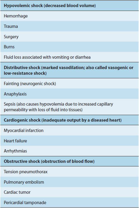
The term “shock” is used to denote various conditions, including the response to the passage of electric current through the body; the state that follows immediately after interruption of the spinal cord; and the stunned reaction to bad news. In the current context, it refers to an abnormality of the circulatory system in which there is inadequate tissue perfusion because of a relatively or absolutely inadequate cardiac output. The causes are divided into four groups: inadequate volume of blood to fill the vascular system (hypovolemic shock); increased size of the vascular system produced by vasodilation in the presence of a normal blood volume (distributive, vasogenic, or low-resistance shock); inadequate output of the heart as a result of myocardial abnormalities (cardiogenic shock); and inadequate cardiac output as a result of obstruction of blood flow in the lungs or heart (obstructive shock). Examples of the conditions or diseases that can cause each type are set forth in Table 11-5.
TABLE 11–5 Types of shock, with examples of conditions or diseases that can cause each type.
Hypovolemic Shock
Hypovolemic shock is characterized by hypotension; a rapid, thready pulse; cold, pale, clammy skin; intense thirst; rapid respiration; and restlessness or, alternatively, torpor. Urine volume is markedly decreased. However, none of these findings are invariably present. Hypovolemic shock is commonly subdivided into categories on the basis of cause. The use of terms such as hemorrhagic shock, traumatic shock, surgical shock, and burn shock is of some benefit because although there are similarities between these various forms of shock, there are important features that are unique to each.
In hypovolemic and other forms of shock, inadequate perfusion of the tissues leads to increased anaerobic glycolysis, with production of large amounts of lactic acid. In severe cases, the blood lactate level rises from a normal value of about 1 mmol/L to 9 mmol/L or more. The resulting lactic acidosis depresses the myocardium, decreases peripheral vascular responsiveness to catecholamines, and may be severe enough to cause coma.
Multiple compensatory reactions come into play to defend extracellular fluid volume (Table 11-6). The large number of reactions that have evolved indicates the importance of maintaining blood volume for survival.
TABLE 11-6 Compensatory reactions activated by hypovolemia.
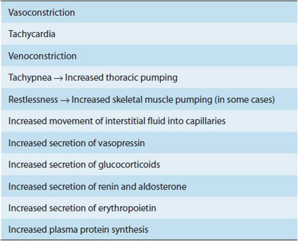
A decrease in pulse pressure or mean arterial pressure decreases the number of impulses ascending to the brain from the arterial baroreceptors, resulting in increased vasomotor discharge. The resulting vasoconstriction is generalized, sparing only the vessels of the brain and the heart. The coronary vessels are dilated because of the increased myocardial metabolism secondary to an increase in heart rate. Vasoconstriction in the skin accounts for the coolness and pallor, and vasoconstriction in the kidneys accounts for the shutdown in renal function.
The immediate cardiac response to hypovolemia is tachycardia. With more extensive loss of volume, tachycardia can be replaced by bradycardia, whereas with very severe hypovolemia, tachycardia reappears. Bradycardia may be due to unmasking of a vagally mediated depressor reflex, perhaps related to limiting blood loss.
Vasoconstriction in the kidney reduces glomerular filtration. This reduces water loss, but it reaches a point at which nitrogenous products of metabolism accumulate in the blood (prerenal azotemia). If hypotension is prolonged, there may be severe renal tubular damage, leading to acute kidney injury.
The fall in blood pressure and the decreased O2-carrying power of the blood caused by the loss of red cells results in stimulation of the carotid and aortic chemoreceptors. This not only stimulates respiration but increases vasoconstrictor discharge. In severe hypovolemia, the pressure is so low that there is no longer any discharge from the carotid and aortic baroreceptors. This occurs when the mean blood pressure is about 70 mm Hg. Under these circumstances, if the afferent discharge from the chemoreceptors via the carotid sinus and vagus nerves is stopped, there is a paradoxic further fall in blood pressure rather than a rise.
Hypovolemia causes a marked increase in the circulating levels of the pressor hormones angiotensin II, epinephrine, norepinephrine, and vasopressin. ACTH secretion is also increased, and angiotensin II and ACTH both cause an acute increase in aldosterone secretion. The resulting retention of Na+ and water helps reexpand blood volume.
Forms of Hypovolemic Shock
Hemorrhagic shock is probably the most carefully studied form of shock because it is easily produced in experimental animals. With moderate hemorrhage (5–15 mL/kg body weight), pulse pressure is reduced but mean arterial pressure may remain normal. With more severe hemorrhage, blood pressure always falls.
After hemorrhage, the plasma protein lost in shed blood is gradually replaced by hepatic synthesis, and the concentration of plasma proteins returns to normal in 3–4 days. The increase in circulating erythropoietin increases red blood cell formation, but it takes 4–8 weeks to restore red cell counts to normal.
Traumatic shock develops when there is severe damage to muscle and bone. This is the type of shock seen in battle casualties and automobile accident victims. Bleeding into the injured areas is the principal cause of such shock. The amount of blood that can be lost into a site of injury that appears relatively minor is remarkable; the thigh muscles can accommodate 1 L of extravasated blood, for example, with an increase in the diameter of the thigh of only 1 cm.
Breakdown of skeletal muscle is a serious additional problem when shock is accompanied by extensive crushing of muscle (crush syndrome). When pressure on tissues is relieved and they are once again perfused with blood, free radicals are generated, which cause further tissue destruction (reperfusion-induced injury). Increased Ca2+ in damaged cells can reach toxic levels. Large amounts of K+ enter the circulation. Myoglobin and other products from reperfused tissue can accumulate in kidneys in which glomerular filtration is already reduced by hypotension, and the tubules can become clogged, causing anuria.
Surgical shock is due to combinations, in various proportions, of external hemorrhage, bleeding into injured tissues, and dehydration.
In burn shock, there is loss of plasma from burn surfaces and the hematocrit rises rather than falls, producing severe hemoconcentration. There are, in addition, complex metabolic changes. For these reasons, plus the problems of easy infection of burned areas and kidney damage, the mortality rate when third-degree burns cover more than 75% of the body is close to 100%.
Distributive Shock
In distributive shock, most of the symptoms and signs described previously are present. However, vasodilation causes the skin to be warm rather than cold and clammy. Anaphylactic shock is a good example of distributive shock. In this condition, an accelerated allergic reaction causes release of large amounts of histamine, producing marked vasodilation. Blood pressure falls because the size of the vascular system exceeds the amount of blood in it even though blood volume is normal.
A second type of distributive shock is neurogenic shock, in which a sudden loss of sympathetic autonomic activity (as seen in head and spinal cord injuries) results in vasodilation and pooling of blood in the veins. The resulting decrease in venous return reduces cardiac output and frequently produces fainting, or syncope, a sudden transient loss of consciousness. More benign and much more common form is postural syncope, which occurs on rising from a sitting or lying position. This is common in patients taking drugs that block sympathetic discharge or its effects on the blood vessels. Falling to the horizontal position restores blood flow to the brain, and consciousness is regained. Pressure on the carotid sinus produced, for example, by a tight collar can cause sufficient bradycardia and hypotension to cause fainting (carotid sinus syncope). Fainting caused by a variety of activities has been given appropriate names such as micturition syncope, cough syncope, deglutition syncope, and effort syncope.
Syncope resulting from neurogenic shock is usually benign. However, it must be distinguished from syncope resulting from other causes and, therefore, merits investigation.
Another form of distributive shock is septic shock. This condition is discussed in detail in Chapter 4. It is now the most common cause of death in ICUs in the United States. It is a complex condition that includes elements of hypovolemic shock resulting from loss of plasma into the tissues (“third spacing”) and cardiogenic shock resulting from toxins that depress the myocardium. It is associated with excess production of NO, and therapy with drugs that scavenge NO may be beneficial.
Streptococcal toxic shock syndrome is a particularly severe form of septic shock in which group A streptococci infect deep tissues; the M protein on the surface of those bacteria has an antiphagocytic effect. It also is released into the circulation, where it aggregates with fibrinogen.
Cardiogenic Shock
About 25% of syncopal episodes are of cardiac origin and are due either to transient obstruction of blood flow through the heart or to sudden decreases in cardiac output caused by various cardiac arrhythmias. In addition, fainting is the presenting symptom in 7% of patients with myocardial infarctions.
Cardiogenic shock results whenever the pumping function of the heart is impaired to the point that blood flow to tissues is no longer adequate to meet resting metabolic demands; most commonly, it is due to extensive infarction of the left ventricle. The incidence of shock in patients with myocardial infarction is about 10%, and the mortality rate is 60–90%.
However, cardiogenic shock can also be caused by other diseases (heart failure, arrhythmias) that severely compromise normal ventricular function. The symptoms are those of hypovolemic shock plus congestion of the lungs and viscera resulting from failure of the heart to put out all the venous blood returned to it. Consequently, the condition is sometimes called “congested shock.”
Obstructive Shock
The picture of congested shock is also seen in obstructive shock. Causes include massive pulmonary emboli, tension pneumothorax with kinking of the great veins, and bleeding into the pericardium with external pressure on the heart (cardiac tamponade). In the latter two conditions, prompt surgery is required to prevent death. Pulsus paradoxus occurs in cardiac tamponade. Normally, blood pressure falls about 5 mm Hg during inspiration. In pulsus paradoxus, this response is exaggerated, and blood pressure falls 10 mm Hg or more as a result of increased pressure of the fluid in the pericardial sac on the external surface of the heart. However, pulsus paradoxus also occurs with labored respiration in severe asthma, emphysema, and upper airway obstruction.
Refractory Shock
Some patients with hypovolemia or septic shock die soon after the onset of the condition, and others recover as compensatory mechanisms gradually restore the circulation to normal. In an intermediate group of patients, shock persists for hours and gradually progresses. It eventually reaches a state in which there is no longer any response to vasopressor drugs and in which, even if the blood volume is returned to normal, cardiac output remains depressed. This condition is known as refractory shock. It used to be called irreversible shock, and patients still die despite vigorous treatment. However, more and more patients are saved as understanding of the pathophysiologic mechanisms increases and treatment is improved. Therefore, “refractory shock” seems to be a more appropriate term.
Various factors appear to make shock refractory. Precapillary sphincters are constricted for several hours but then relax while postcapillary venules remain constricted. Therefore, blood flows into the capillaries and remains there. Various positive feedback mechanisms contribute to the refractory state. For example, cerebral ischemia depresses vasomotor and cardiac discharge, causing blood pressure to fall and making the shock worse. This, in turn, causes a further reduction in cerebral blood flow. In addition, myocardial blood flow is reduced in severe shock. Myocardial failure makes the pumping action of the heart less effective and consequently makes the shock worse and further lowers myocardial blood flow.
A complication of shock that has a very high mortality rate is pulmonary damage with production of acute respiratory distress syndrome. The cause appears to be capillary endothelial cell damage and damage to alveolar epithelial cells with the release of cytokines (see Chapter 9).
CHECKPOINT
17. What are the four major pathophysiologic forms of shock?
18. Name three pathophysiologic consequences of lactic acidosis in shock.
19. Describe five specific forms of hypovolemic shock.
20. Name three specific forms of distributive shock and distinguish them from hypovolemic shock.
21. Name three factors that tend to make shock refractory.
CASE STUDIES
Yeong Kwok, MD
(See Chapter 25, p. 721 for Answers)
CASE 58
A 65-year-old woman presents to the clinic to establish care. Her past medical history is notable for type 2 diabetes and hypertension. She has a 45-pack-year smoking history. A few weeks ago, she was shoveling her driveway when she had to stop due to tightness in her chest. She does not get any regular exercise because her calves become very painful after walking one block.
Questions
A. What is the likely diagnosis?
B. What is the pathogenesis of this condition?
C. What are this patient’s rick factors, and how do they contribute to the development of atherosclerosis?
CASE 59
A 56-year-old black man presents to the clinic for a routine physical examination. He has not seen a physician for 10 years. On arrival, he is noted to have a blood pressure of 160/90 mm Hg.
Questions
A. Does this man have hypertension? Why or why not?
B. What physical findings might be present if he has had long-standing hypertension?
C. What are some of the important complications of hypertension?
D. What are some causes of hypertension?
CASE 60
A young woman is brought to the emergency department by ambulance after a severe motor vehicle accident. She is unconscious. Her blood pressure is 64/40 mm Hg; heart rate is 150 bpm. She is intubated and is being hand-ventilated. There is no evidence of head trauma. The pupils are 2 mm and reactive. She withdraws to pain. Cardiac examination reveals no murmurs, gallops, or rubs. The lungs are clear to auscultation. The abdomen is tense, with decreased bowel sounds. The extremities are cool and clammy, with thready pulses. Despite aggressive blood and fluid resuscitation, the patient dies.
Questions
A. What are the four major pathophysiologic causes of shock? Which was likely in this patient?
B. What pathogenetic mechanism accounts for this patient’s unresponsiveness? For the cool, pale extremities?
C. What forms of hypovolemic shock may have been present in this patient? Why?
REFERENCES
General
Baylis C. Nitric oxide synthase derangements and hypertension in kidney disease. Curr Opin Nephrol Hypertens. 2012 Jan;21(1):1–6. [PMID: 22048724]
Meyers KE et al. Endothelin antagonists in hypertension and kidney disease. Pediatr Nephrol. 2013 May;28(5):711–20. [PMID: 23070275]
Atherosclerosis
Bandeali S et al. High-density lipoprotein and atherosclerosis: the role of antioxidant activity. Curr Atheroscler Rep. 2012 Apr;14(2):101–7. [PMID: 22441969]
Ntaios G et al. Adipokines as mediators of endothelial function and atherosclerosis. Atherosclerosis. 2013 Apr;227(2):216–21. [PMID: 23332774]
Ohkita M et al. Pathophysiological roles of endothelin receptors in cardiovascular diseases. J Pharmacol Sci. 2012 Aug 18;119(4):302–13. [PMID: 22863667]
Rosenfeld ME. Inflammation and atherosclerosis: direct versus indirect mechanisms. Curr Opin Pharmacol. 2013 Apr;13(2):154-60. [PMID: 23357128]
Tousoulis D et al. Pathophysiology of atherosclerosis: the role of inflammation. Curr Pharm Des. 2011 Dec;17(37):4089–110. [PMID: 22204371]
Tsoumani ME et al. Platelet-mediated inflammation in cardiovascular disease. Potential role of platelet-endothelium interactions. Curr Vasc Pharmacol. 2012 Sep;10(5):539–49. [PMID: 22338568]
Wang JC et al. Aging and atherosclerosis: mechanisms, functional consequences, and potential therapeutics for cellular senescence. Circ Res. 2012 Jul 6;111(2):245–59. [PMID: 22773427]
Zernecke A et al. Improving the treatment of atherosclerosis by linking anti-inflammatory and lipid modulating strategies. Heart. 2012 Nov;98(21):1600–6. [PMID: 23086996]
Hypertension
Chrysant SG et al. Effectiveness of lowering blood pressure to prevent stroke versus to prevent coronary events. Am J Cardiol. 2010 Sep 15;106(6):825–9. [PMID: 20816123]
Cohen DL et al. Hypertension and kidney disease: what do the data really show? Curr Hypertens Rep. 2012 Oct;14(5):462–7. [PMID: 22814743]
De Leo M et al. Subclinical Cushing’s syndrome. Best Pract Res Clin Endocrinol Metab. 2012 Aug;26(4):497–505. [PMID: 22863391]
Hamlyn JM et al. Endogenous ouabain: a link between sodium intake and hypertension. Curr Hypertens Rep. 2011 Feb;13(1):14–20. [PMID: 20972650]
Ito Y et al. Subclinical primary aldosteronism. Best Pract Res Clin Endocrinol Metab. 2012 Aug;26(4):485–95. [PMID: 22863390]
Mannelli M et al. Subclinical phaeochromocytoma. Best Pract Res Clin Endocrinol Metab. 2012 Aug;26(4):507–15. [PMID: 22863392]
Santos PC et al. Renin-angiotensin system, hypertension, and chronic kidney disease: pharmacogenetic implications. J Pharmacol Sci. 2012;120(2):77–88. [PMID: 23079502]
Shimosawa T et al. The kidney and hypertension: pathogenesis of salt-sensitive hypertension. Curr Hypertens Rep. 2012 Oct;14(5):468–72. [PMID: 22752520]
Van Buren PN et al. The pathogenesis and management of hypertension in diabetic kidney disease. Med Clin North Am. 2013 Jan;97(1):31–51. [PMID: 23290728]
Shock
Buerke M et al. Pathophysiology, diagnosis, and treatment of infarction-related cardiogenic shock. Herz. 2011 Mar;36(2):73–83. [PMID: 21424345]
Cole JN et al. Molecular insight into invasive group A streptococcal disease. Nat Rev Microbiol. 2011 Sep 16;9(10):724–36. [PMID: 21921933]
De Bisschop MB et al. Anaphylaxis. Curr Opin Crit Care. 2012 Aug;18(4):308–17. [PMID: 22732436]
De Kock I et al. Sepsis and septic shock: pathophysiological and cardiovascular background as basis for therapy. Acta Clin Belg. 2010 Sep–Oct;65(5):323–9. [PMID: 21128559]
Huet O et al. Oxidative stress and endothelial dysfunction during sepsis. Front Biosci. 2011 Jan 1;16:1986–95. [PMID: 21196278]
Jozwiak M et al. Management of myocardial dysfunction in severe sepsis. Semin Respir Crit Care Med. 2011 Apr;32(2):206–14. [PMID: 21506057]
Khan BQ et al. Pathophysiology of anaphylaxis. Curr Opin Allergy Clin Immunol. 2011 Aug;11(4):319–25. [PMID: 21659865]
Lappin E et al. Gram-positive toxic shock syndromes. Lancet Infect Dis. 2009 May;9(5):281–90. [PMID: 19393958]
Levinson AT et al. Reducing mortality in severe sepsis and septic shock. Semin Respir Crit Care Med. 2011 Apr;32(2):195–205. [PMID: 21506056]
Levy MM et al. The Surviving Sepsis Campaign: results of an international guideline-based performance improvement program targeting severe sepsis. Intensive Care Med. 2010 Feb;36(2):222–31. [PMID: 20069275]
Martin GS. Sepsis, severe sepsis and septic shock: changes in incidence, pathogens and outcomes. Expert Rev Anti Infect Ther. 2012 Jun;10(6):701–6. [PMID: 22734959]
Moranville MP et al. Evaluation and management of shock states: hypovolemic, distributive, and cardiogenic shock. J Pharm Pract. 2011 Feb;24(1):44–60. [PMID: 21507874]
Nduka OO et al. The pathophysiology of septic shock. Crit Care Clin. 2009 Oct;25(4):677–702. [PMID: 19892247]