
Goran Augustin1, 2
(1)
Department of Surgery Division of Gastrointestinal Surgery, University Hospital Center Zagreb, Zagreb, Croatia
(2)
School of Medicine University of Zagreb, Zagreb, Croatia
Abstract
Small bowel perforation is extremely rare during pregnancy. There are several causes, the most common being perforation due to intestinal endometriosis which is the disease found in the reproductive age group.
11.1 Small Bowel Perforation
Small bowel perforation is extremely rare during pregnancy. There are several causes, the most common being perforation due to intestinal endometriosis which is the disease found in the reproductive age group.
11.1.1 Intestinal Perforation Due to Intestinal Endometriosis
11.1.1.1 Introduction
Endometriosis is defined by the presence of endometrium outside the uterus and usually affects pelvic structures including the bowel. Intestinal involvement occurs in 3–37 % of patients with endometriosis [1] and may affect the ileum, appendix, sigmoid colon, and rectum [2]. The most common nongenital manifestation is in the rectosigmoid [3, 4]. The peritoneal implantation of endometrium by retrograde menstruation or the possible metaplasia of peritoneal cells is still the most accepted etiological theory of endometriosis [1]. Intestinal endometriosis may be found in every layer of the bowel wall, but it is most commonly found within the subserosa as superficial serosal implants [5, 6]. Under cyclical hormonal influences, these implants may proliferate and infiltrate the intestinal wall and cause a fibrotic reaction with formation of strictures and adhesions, which may lead to bowel obstruction and recurrent abdominal pain [2, 7]. On the other hand, transmural bowel wall involvement is not so common and the intestinal mucosa usually remains intact, and perforation of the affected intestinal tract is a very rare complication [3, 6].
11.1.1.2 Incidence
Up till now and including the present report, only 22 cases of intestinal perforation from endometriosis have been reported, and among these, 45.5 % (10/22) occurred in pregnancy (Table 11.1); of these ten patients, 20 % (2/10) presented postpartum. One of these postpartum presenting patients with cecal perforation had a past medical history of terminal ileal and cecal Crohn’s disease diagnosed 9 years previously. The histology confirmed that cecal perforation was due to decasualized endometriosis. There was no evidence of Crohn’s disease [12]. Sigmoid colon is the most common site of perforation from endometriosis in general population and accounted for 55 % (12/22) published cases [12, 16].
Table 11.1
Summary of cases of bowel perforation secondary to endometriosis related to pregnancy
|
Author |
Year |
Site of perforation |
Duration of pregnancy |
|
Haufler [8] |
1931 |
Jejunal |
6 months |
|
Gini et al. [9] |
1981 |
Appendix |
35 weeks |
|
Nakatani et al. [10] |
1987 |
Appendix |
26 weeks |
|
Faucheron et al. [11] |
2008 |
Appendix |
|
|
Beamish et al. [12] |
2010 |
Cecum |
Postpartum |
|
Clement [13] |
1977 |
Sigmoid |
37 weeks |
|
Rud [14] |
1979 |
Sigmoid |
|
|
Floberg et al. [15] |
1984 |
Sigmoid |
Postpartum (41 weeks) |
|
Schweitzer et al. [2] |
2006 |
Sigmoid |
Term pregnancy |
|
Pisanu et al. [16] |
2010 |
Rectum |
33 weeks |
Intestinal endometriosis typically takes the form of asymptomatic serosal implants that occasionally result in intestinal obstruction with recurrent abdominal pain [5]. Transmural involvement is not as common, and spontaneous perforation of intestinal endometriosis is a rare complication that occurred in pregnancy in almost 50 % of published cases (Fig. 11.1) [2, 3, 5].
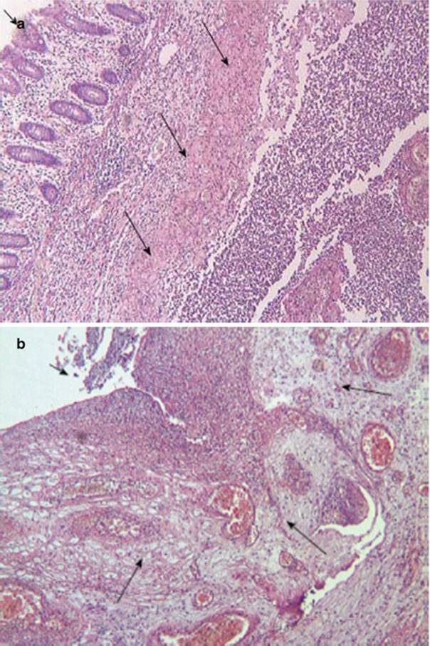
Fig. 11.1
The rectal wall. (a) Decidualization of the rectal wall (long arrows); mucosa side of the rectal wall (short arrow) (HE, ×40); (b) Decidualized endometriosis around the rectal perforation (long arrows); rectal perforation with necrosis at the peritoneal side of the rectal wall (short arrow) (HE, ×100) [16]
11.1.1.3 Pathophysiology
In the third trimester and postpartum, endometrial lesions tend to contract as observed by McArthur and Ulfelder, the mechanism of which remains obscure. This contraction in an area already weakened by endometrial stromal infiltration can cause perforation as suggested by both McArthur and Ulfelder in 1965 [17] and Garg et al. in 2009 [5]. In those patients with perforation, the entire intestinal wall is replaced by endometriotic tissue. In pregnancy, under the effect of progesterone, the area of ectopic endometrium becomes decidualized with a progressive reduction in size [11, 17]. Actually, the reduction in size of a transmural endometriotic nodule may lead to perforation, by weakening of the intestinal wall [5], particularly in the third trimester, which is the time of perforation in most reported cases [5, 16, 17]. Moreover, decidualization causes a severe inflammatory response with an increased number of natural killer cells and decidual changes, which are responsible for a higher risk of perforation [11, 18]. Additionally perforation was also facilitated by the progressive traction of the enlarged uterus on the strictly adherent sigmoid colon and, consequently, on the decidualized and weakened area of the anterior rectal wall [16]. Moreover, the absence of endometriotic foci in the cul-de-sac at final laparoscopy made rectal perforation unpredictable. Although endometriosis improves during pregnancy, the current report shows the potential occurrence of serious and unexpected complications of the disease.
11.1.1.4 Clinical Presentation
If the perforation is present, then symptoms and signs of acute abdomen are present. Before the occurrence of perforation, abdominal pain could be cyclical as is present in endometriosis. Enlargement of endometriotic nodules could aggravate constipation or initiate abdominal distension due to impaired bowel peristalsis.
11.1.1.5 Differential Diagnosis
Both the rareness of the perforation and the symptoms that are suggestive of pyelonephritis or diverticulitis may be misleading and could delay the diagnosis especially before the perforation ensue.
11.1.1.6 Diagnosis
If acute abdomen is found, clinically standard laboratory findings and plain abdominal X-rays are made. Pneumoperitoneum is found when the bowel perforation is present.
11.1.1.7 Therapy
The appropriate management of these patients may be challenging, and a good outcome is absolutely dependent on a multidisciplinary approach. The best approach is to resect, en bloc, endometriosis and perforated bowel. If the perforation is on the small bowel, anastomosis could be made (or ileostomy if prolonged peritonitis is present), and if sigmoid colon perforates, then the Hartmann’s operation is the procedure of choice.
11.1.2 Spontaneous Small Bowel Perforation
Morton and Hubbard in 1959 reported seven cases of nontraumatic rupture of the small bowel during pregnancy with 85 % mortality [19].
11.2 Perforation of the Colon and Rectum
11.2.1 Colorectal Carcinoma in Pregnancy
Due to the extreme rarity of perforated colorectal carcinomas in pregnancy, an overview of colorectal carcinoma in general during pregnancy is presented. Few cases of perforated colorectal carcinoma are additionally discussed.
11.2.1.1 History
Many reports of rectal and colonic carcinoma occurring in pregnancy appear in the literature, the first being in 1835, and Cruveilhier (Fig. 11.2) reported the first case of rectal carcinoma in pregnancy in 1842.

Fig. 11.2
Jean Cruveilhier 1837
These early reports were concerned mainly with the obstetric problems of delivering the fetus in the presence of a pelvic tumor. Robert Greenhalgh (St. Bartholomew’s Hospital; consulting physician to the Samaritan Hospital for Women and City of London Lying In Hospital) in 1866 described a Cesarean section done under an ether spray on a woman with obstructed labor from a rectal carcinoma [20]. Interest in this subject lies in two effects, that of the pregnancy on the carcinoma and that of the complications of the carcinoma on the management of the pregnancy. Warren in 1957 came to the conclusion that pregnancy does not adversely affect the course of carcinoma of the rectum [21].
11.2.1.2 Incidence
Incidence of colorectal cancer in pregnancy in 1981 was 0.002 % due to the fact that incidence rises with age [22]. Up to 1949, there were 75 cases published [23, 24]. Approximately 275 cases of colon cancer associated with pregnancy have been reported in the literature up to 1998 [25]. A review of 205 of these cases performed by Bernstein et al. demonstrated that 85 % of these cancers were located below the peritoneal reflection and mean age of 31 years (range 16–48) in a review of 42 pregnant patients with colon cancer [26].
First two published cases of perforated low rectal and sigmoid cancer were by Nash in 1967 [27]. The first patient was aged 38, in her fifth pregnancy (38 weeks). Artificial rupture of the membranes was carried out, and an hour later an 8 lb live boy was born. The second patient was aged 28; in her second pregnancy, in 32nd week, she had exteriorization of the sigmoid tumor.
Most cases of CRC are discovered in late pregnancy, and 60–80 % of the patients have rectal carcinoma [28].
11.2.1.3 Pathogenesis
The main pathogenesis of CRC in pregnancy is still associated with lots of unanswered questions. Some factors including pregnancy hormones, the enzyme cyclocoxygenase-2 (Cox-2), and tumor suppressor protein p53 were mentioned to be associated with CRC. A majority of CRC cases have been found to be positive for estrogen (20–54 %) [29] and progesterone receptors (10–100 %) [30]. Maybe the increased levels of estrogen and progesterone during pregnancy stimulate the growth of tumor cells with such receptors; however, all reports did not support this hypothesis. Slattery et al. in a study found only one case with positive progesterone receptor among 156 pregnant cases with CRC [29]. The elevated amounts of Cox-2 in CRC patients have raised the hypothesis of its association with colorectal cancer; however, there are little evidences to elucidate its carcinogenic role.
11.2.1.4 Clinical Presentation
Common presenting signs and symptoms of CRC include abdominal pain, anemia, nausea, vomiting, and rectal bleeding. Because these signs and symptoms are also frequently found in pregnancy, physicians and patients often attribute them to the usual complications of pregnancy [28]. The diagnostic challenge for clinicians is distinguishing pregnancy symptoms from the warning signs of colorectal cancer (Table 11.2) [25]. Clinicians must be aware of these potential warning signs and symptoms in order to make the diagnosis at an early stage of the disease. Rectal bleeding is particularly an ominous sign and should never be attributed solely to pregnancy without a proper evaluation.
Table 11.2
Commonly confused signs, symptoms, and laboratory results between pregnancy and colorectal cancer [25]
|
Signs, symptoms, lab |
Normal pregnancy |
Pregnancy with colorectal cancer |
|
Weight loss |
In general weight gain, but women can experience weight loss in the first trimester |
Pregnancy can obscure weight loss secondary to CRC, primarily in second and third trimesters |
|
Rectal bleeding |
Common in pregnancy secondary to high incidence of hemorrhoids |
Often attributed to hemorrhoids without pursuing appropriate workup |
|
Nausea and vomiting |
Common in pregnancy, particularly during the first trimester |
Often attributed to pregnancy, delaying workup |
|
Constipation |
Common in pregnancy |
Often attributed to pregnancy, delaying workup |
|
Abdominal mass |
Natural process in pregnancy |
Potential palpable masses secondary to CRC often missed secondary to changes of pregnancy |
|
Anemia |
Physiological finding in pregnancy |
Anemia of pregnancy masks blood loss from CRC |
Presentation of perforated CRC is sudden and severe pain, with signs and symptoms of acute abdomen.
11.2.1.5 Diagnosis
Elective Presentation
Serum CEA is an important laboratory test used in the evaluation of pregnant and nonpregnant patients. CEA levels during pregnancy are usually normal but may be slightly elevated [31]. CEA levels obtained prior to surgery provide a baseline to monitor the response to treatment. CEA levels also have prognostic value since increased levels prior to surgery are associated with disseminated disease and increased recurrence rates [32]. However, CEA is not useful as a tool for screening due to the low sensitivity and specificity [32].
Acute Abdomen (Perforation)
Laboratory findings show elevated levels of WBC and CRP, possible microcytic anemia, and other parameters according to the duration of the perforation. The patient can present with prerenal insufficiency due to the shift of fluid in the third space. Plain abdominal X-ray shows pneumoperitoneum if the perforation is above the peritoneal fold.
11.2.1.6 Therapy
Gestational Age: Elective Presentation
Gestational age and CRC stage are important to select treatment modality. If tumor is resectable, surgical excision is recommended especially in those diagnosed in early pregnancy (before 20 weeks of gestation). In cases of more advanced pregnancy, surgery can be postponed at the earlier possible date at which fetus can be viable (around 32 weeks). In advanced stages, when adjuvant therapy is needed, elective abortion would help to save mother’s life, while in greater gestational ages, it is possible to pursue adjuvant therapy after early delivery. It is important that the mother be fully informed of possible risks of each choice prior to her decision. In religious countries like Iran, there is another extra challenge for parents and clinician, since due to religious beliefs, the legal abortion is only permitted up to the week 16, and after this time there would be problems to perform the abortion legally.
One-Stage or Two-Stage Procedure: Elective Presentation
If operation is indicated in early pregnancy, then tumor resection and termination of pregnancy should be performed. It can be done in a single act or as a two-stage procedure [33]. In the first stage, termination of pregnancy is performed. After 2–3 weeks, when the uterus returns to a normal size and the pelvic venous congestion related to pregnancy decreases, oncologic resection of the colorectal cancer is performed.
Ovarian Transposition: Elective Presentation
The primary benefit of ovarian transposition is prevention or delay of premature menopause, not preservation of fertility. In fact, with “curative” doses in the range of 8,500 cGy with external beam plus intracavitary brachytherapy, the resultant endometrial damage essentially precludes successful pregnancy, either spontaneously or with in vitro technique.
Doses of radiation delivered to the ovary following successful laparoscopic oophoropexy have been determined and reported by several authors. Covens et al. [34] studied three patients in whom ovarian transposition was performed and determined the radiation dose expected to be received by the ovaries after delivery by either an external beam radiation dose of 4,500 cGy or via brachytherapy. The mean dose of radiation received by the transposed ovary was 175 cGy (range, 40–370 cGy) after a mean follow-up of 2 years. The iatrogenic menopause rate was 10 % overall, but iatrogenic menopause did not occur in any of the patients younger than 40 years of age.
Numerous studies have attempted to specify radiation therapy tolerance doses for the various tissues and structures of the body. Common dose definitions that are used to describe various tissue tolerances are the minimal tolerance dose, TD 5/5, and the maximal tolerance dose, TD 50/5, which refer to a severe complication rate of 5 and 50 %, respectively, with 5 years of radiation completion. For the ovary, these values are approximately 300 cGy and 1,200 cGy, with sterility being the endpoint of severe complication. Tolerance doses for other tissues are significantly higher. In clinical practice, however, the tolerance doses for the ovary and other tissues may actually be lower, given the now common treatment regimens that typically include chemotherapy or altered radiation fractionation schemes. In general, if the ovaries can be appropriately shielded from the direct radiation beam via an oophoropexy maneuver, a dose below the TD 50/5 (300 cGy) can be achieved with a reasonable expectation of ovarian function preservation posttreatment. Simply the rule of thumb for radiation and ovarian function could be applied, meaning 10 cm distance to radiation field = 10 % dose of radiation [35].
The rule of thumb for radiation and ovarian function means 10 cm distance to radiation field = 10 % dose of radiation.
The typical outcome measure in ovarian transposition following radiation therapy is ovarian function. This is often measured by quantitative analysis of ovary-stimulating or ovary-producing hormones as well as fertility outcomes. Spontaneous pregnancies are possible if tubal function is preserved as part of the oophoropexy. Morice et al. [36] reported on 37 consecutive cases of ovarian transposition. In these cases, 43 % (16/37) of pregnancies occurred spontaneously; of these, 75 % (12/16) did not have the ovaries repositioned from the adnexa. Quantitative analysis of ovarian function has been reported by Covens et al. [34] in three patients undergoing ovarian transposition. Serum FSH was found to be normal in 67 % (2/3) of patients menstruating regularly 24–32 months after radiation. Treissman et al. [37] reported on a patient in whom laparoscopic ovarian transposition was performed before definitive treatment for an anal carcinoma. Tulandi and Al-Took [38] reported that normal menstruation returned after irradiation in a 34-year-old woman who underwent laparoscopic ovarian transposition before radiation for treatment of rectal carcinoma. Although postmenopausal symptoms and elevation of serum gonadotropins initially indicated ovarian failure, normal menstruation resumed and correlated with normal FSH levels 8 months after treatment.
The timing depends on the treatment algorithm of rectal cancer. If the operation is indicated, it can be performed during that operation for rectal cancer. If neoadjuvant chemoradiotherapy is indicated, then laparoscopic transposition can be performed prior to neoadjuvant chemoradiotherapy.
Preservation of ovarian function by laparoscopic transposition of the ovaries before pelvic irradiation has been demonstrated to be a safe and effective procedure for patients with Hodgkin’s disease as well as in the treatment of a variety of gynecologic malignancies. Historically, surgical exploration of the abdomen or pelvis as part of staging or resection procedures has allowed for access to the ovaries for direct-open transposition of the ovaries for planned subsequent radiation therapy.
Techniques for ovarian transposition using a laparoscopic approach vary according to the radiation field shape, size, and location.
Medial Ovarian Transposition
Tinga et al. [39] have described transposition of the ovaries in a fixed position behind the uterus (to lie beneath an external midline block) as well as a superior transposition to the level of the iliac crest. They contend that the disadvantage of the midline oophoropexy is a higher level of internal radiation scatter, as the area is generally surrounded by in-field radiation.
Lateral Ovarian Transposition
Morice et al. [40] reported a series of 24 patients who underwent ovarian transposition to the paracolic gutters, before radiation for gynecologic malignancies. The authors concluded that this procedure was a safe and effective method of preserving ovarian function. The peritoneum of both pelvic sidewalls is incised, and the retroperitoneal spaces developed. The common, external, and internal iliac vessels are identified. The ovarian vessels and ureters are traced on both sides. Under the direct vision of the ureters, the utero-ovarian ligaments are separated with an endoscopic linear cutting stapler (Endo-GIA). The peritoneum under and lateral to the ovarian vessels are incised to an area outside of the true pelvis, under direct vision of the ureters. At this point, the ovarian vessels could be turned laterally with a sufficient angle to maintain appropriate blood supply. The ovaries and tubes are fixed high in the paracolic gutters, below the spleen and the liver, with 2-0 silk sutures, in three points, to prevent torsion. The upper and lower poles of the ovaries are marked with hemoclips. At the end of the procedure, good blood supply to both ovaries is confirmed by a small incision of the Fallopian tubes, which are cauterized. The metal clips around the ovaries help to verify that they would be out of the radiation portals on radiation verification films [35].
Perforation
The first two published cases of perforated low rectal and sigmoid cancer were by Nash in 1967 [27]. After describing the first two patients, Nash defined two possible plans of action: if the pregnancy is sufficiently advanced, labor can be induced, the uterus emptied, and the colonic condition treated in the much easier operating conditions thus afforded; if, however, the patient’s condition is too poor or if she is not yet at full term, operation must be done in the presence of the gravid uterus. In these circumstances resection is very difficult, and I think exteriorization is the only possible treatment. Cesarean section via an infected peritoneal cavity seems very unwise and would appear to be indicated only if a perforated rectal carcinoma obstructs vaginal delivery.
11.2.1.7 Prognosis
Maternal Outcome
The delay leads to late diagnosis of the disease and subsequently poor prognosis. A majority of CRC cases in pregnancy present with Duck class C (44 %) in which adjutant therapies are needed to improve the surgical outcome [26]. The median survival in a review of 42 pregnant patients with CRC was less than 5 months and 56 % died by the time of the report [41].
Fetal Outcome
CRC in pregnancy represents a serious threat to both the mother and the fetus. Woods et al. reported that 78 % (25/32) of pregnancies in women with colonic tumors above the rectum resulted in healthy, live-born infants. Prematurity, intrauterine death, stillbirth, and termination were all contributors to the death of these infants [42].
Genetic Counseling
CRC occurs rarely in young patients, and as a result, this patient population is more likely to have strong predisposing factors compared to the general population of patients with CRC [43]. Such predisposing factors for CRC include hereditary nonpolyposis colorectal cancer (Lynch syndrome), familial adenomatous polyposis, Gardner’s syndrome, Peutz-Jeghers syndrome, and long-standing inflammatory bowel disease. However, these increased risk groups represent only a small portion of CRC diagnosed in pregnancy [25]. A review of 19 pregnant patients by Girard et al. from 1981 demonstrated that 21 % (4/19) of patients had one of these strong predisposing factors for CRC [22]. Despite the negative result for possible hereditary nonpolyposis colon cancer (Amsterdam II criteria), Bethesda criteria should be checked for microsatellite instability. If the microsatellite instability testing is positive, the genetic testing of the three genes associated with HNPCC should proceed. Once a mutation was identified in the family, other family members could consider predictive testing.
11.2.2 Spontaneous Colorectal Perforation
There are two causes of spontaneous perforation of the colon: stercoral and idiopathic.
11.2.2.1 Stercoral (Stercoraceous) Perforation
Incidence and Diagnostic Criteria
Stercoral perforation is rare, even in nonpregnant patients. The first case of stercoral perforation was described in 1894 [44]. A review by Maurer et al. identified only 88 cases from 1894 to 2000 [45]. These authors proposed diagnostic criteria:
· Round or ovoid perforation, ≥1 cm in diameter, antimesenteric location
· Fecaloma (hard, laminated, inspissated fecal mass) within the colon, protruding through the perforation site, or lying within the peritoneal cavity
· Typical pathohistological features (pressure necrosis or ulcer with chronic inflammatory reaction around the perforation site)
· Absence of other active colonic pathology, such as diverticulitis, carcinoma, and Hirschsprung’s disease
Stercoral perforation during pregnancy is extremely rare, and there are three cases published. Women presented at 22, 36, and 41 weeks of pregnancy [46–48].
Risk Factors
Chronic constipation is the main risk factor and is present in 81 % of patients. Use of antacids, codeine-containing narcotics, nonsteroidal anti-inflammatory agents, major tranquilizers, and tricyclic antidepressants has been linked to stercoral perforation [49, 50]. These medications are all known to cause constipation.
Treatment
Treatment is surgical, and resection of the diseased segment with proximal colostomy and closure of the rectal stump or mucous fistula are the procedures with the lowest mortality rate in general population [49]. This is the recommended procedure in pregnant population as was performed in two cases [47, 48].
Prognosis
The reported mortality rate among all cases in general population of stercoral perforation is 47 %, with a 35 % mortality rate among surgically treated cases [49]. Of three pregnant patients [46–48], one died (maternal mortality of 33 %). Perinatal mortality is 66 % probably due to prolonged peritonitis. The first patient presented at 41 weeks of gestation delivered a stillborn infant and died after delivery. Stercoral perforation was diagnosed on autopsy [46]. The second patient in 36 weeks of pregnancy was explored and Cesarean section performed, with live baby, before the Hartmann procedure [47]. The third had sigmoid perforation in 22 weeks of pregnancy. One day after the Hartmann procedure, the patient delivered vaginally a dead female infant [48].
11.2.2.2 Idiopathic Perforation
The features of idiopathic perforation of the colon are as follows [51]:
· Linear perforation
· No feculent ulcer at microscopic examination
· Clear mucosal edge not extending to the serosa
· Regular broken ends of the muscular layer
There is no published case of idiopathic colorectal perforation.
11.2.2.3 Spontaneous Intestinal Perforation
Incidence
This is an extremely rare entity, and the incidence is unknown.
Risk Factors
All risk factors for “spontaneous” ischemic events in general population are also risk factors in pregnancy (Table 11.3). The difference is in incidence of different risk factors due to specific age of presentation. Also pregnancy per se is a risk factor for thromboembolic events. Combination of different risk factors could be present and should be kept in mind which aggravates the possibility of intestinal ischemia and subsequent perforation. Most spontaneous intestinal perforations are due to previously unknown but present risk factors. There are cases when women during early pregnancy (by mistake because unaware of pregnancy) [55] or early postpartum period by patient itself [56] take oral contraceptives with development of mesenteric vein thrombosis. These risk factors are the same and could include the perforation both on small and large bowel.
Table 11.3
Possible risk and combinations of factors for spontaneous intestinal perforation
|
Antiphospholipid syndrome (macroangiopathy) |
|
HELLP syndrome (microangiopathy) |
|
Syphilis |
|
Vasculitis |
|
Low-flow states |
|
Arrhythmia |
|
Sepsis |
|
Shock |
|
Disseminated intravascular coagulation |
|
Recent surgery/C-section |
|
Thromboembolism per se |
|
Ischemia caused by previous surgical manipulation [52] |
|
Cocaine abuse [53, 54] |
|
Valvular heart disease |
|
Infective endocarditis |
|
Diabetes mellitus |
|
End-stage renal disease |
|
Idiopathic hypertension |
|
Oral contraceptives |
Prevention
It is recommended that all women with antiphospholipid syndrome should maintain antithrombotic treatment throughout their entire pregnancy and during the postpartum period. The prevention of choice is combined low-dose aspirin and full antithrombotic doses of low-molecular-weight heparin. In high-risk groups, such as history of previous thrombotic events, warfarin may be used during pregnancy, but only after organogenesis (6th–12th week) because of high risks of fetal malformations. Despite the significant risks associated with pregnancy in patients with antiphospholipid syndrome, with the correct management the likelihood of a live birth is around 75–80 % [26].
Treatment
The type of the operation depends on the extent of peritonitis and underlying ischemia. If the perforation is present without visible ischemia, resection with anastomosis is the preferred method. If there are signs of ischemia, ischemic segment is resected and anastomosis with or without stoma is performed. In long-standing generalized peritonitis, stoma is the preferred option.
11.2.3 Bowel Perforation Secondary to (Illegally) Induced Abortion
11.2.3.1 Incidence
Bowel perforation secondary to illegally induced abortion though rare and uncommon in developed world is a significant and major cause of maternal morbidity and mortality in developing or especially undeveloped countries where abortion laws are still restrictive and most abortions are performed clandestinely and illegally by unqualified personnel [57, 58]. The incidence of abortion-related complications such as bowel injuries has been reported in most developing countries to be increasing at an alarming rate [59]. Ignorance and inability to take quick decision regarding termination of an unwanted pregnancy compel a large number of women to seek illegally induced abortion in the second trimester from unauthorized person in unrecognized places. The rate of bowel perforation as a complication of induced abortion has been reported in literature to range from 2 to 18 % of all abortion-related complications [60–64]. However, as in other iatrogenic surgical problems, many cases may have been unreported because of its medicolegal implications [65, 66].
11.2.4 Treatment
Surgical intervention is considered to be the gold standard treatment for patients with bowel perforation following induced abortion [65]. All patients underwent surgical treatment [27, 33, 34, 60, 61, 65, 67–70]. One of the many factors affecting the surgical outcome in patients with bowel perforation is time interval from perforation to laparotomy [10, 65]. Early surgery can minimize the complications, while delayed surgery leads to severe peritonitis and septic shock. The majority of patients in developing countries were operated more than 24 h after the onset of illness [4, 31, 61, 65]. Delayed definitive surgery may be attributed to late presentation due to lack of accessibility to health-care facilities and lack of awareness of the disease; as a result some patients with bowel perforation following induced abortion may decide to take medications in the prehospital period with the hope that the symptoms will abate. It is also possible that some clinicians managing the patients initially may not have considered perforation as a possible diagnosis leading to delayed referral to tertiary care hospital. The ileum and the sigmoid colon are the most common parts of the bowel affected [60, 61, 65, 67–70]. The relative fixity of these portions of the bowel has been suggested as a possible reason for this. Early surgical interference is the optimal treatment option for perforation. However, the type of surgery to be applied is controversial [65]. The surgical management of small intestinal injuries is fairly straightforward with minimal sequel. The practice in managing these patients is a simple closure in solitary perforations and segmental intestinal resection and primary anastomosis in multiple perforations or gangrenous bowel. The management of large bowel injury is more controversial [15, 65]. This is more so when the left colon is involved. A simple colostomy has been reported to be the safest approach in the management of these injuries. Other options include primary repair, resection and primary anastomosis, and repair with a proximal protective colostomy or ileostomy. A simple colostomy is easier and faster to accomplish in these poor surgical risk patients. However, the major drawback of colostomy is the need for a second operation to restore intestinal continuity, the specialized care before closure, and the attendant cost which reduces its popularity [36, 71]. The challenge is even more conspicuous in a developing country like Tanzania where resources for caring of patients with colostomy are limited. The management of stoma remains difficult in developing countries because of the shortage of suitable equipment in this respect, and peristomal ulceration remains a major problem [71]. Primary repair and resection and primary anastomosis are performed in case of viable bowel, whereas colostomy is reserved after resection of a gangrenous large bowel.
11.2.5 Prognosis
11.2.5.1 Complication Rate
The overall complications rate in the series by Mabula et al. [61] was 47.1 % which is higher compared to previous report by Thapa et al. [72]. High complication rate was also reported by Saleem and Fikree [73] in Pakistan. This difference in complication rates can be explained by differences in antibiotic coverage, meticulous preoperative care and proper resuscitation of the patients before operation, improved anesthesia, and somewhat better hospital environment. As reported by Rehman et al. [74], surgical site infection was the most common postoperative complication. High rate of surgical site infection may be attributed to contamination of the laparotomy wound during the surgical procedure. In the study by Mabula et al. [61], mortality rate was 10.3 % which is higher than that reported by Bhutta et al. [75]. High mortality rate is attributed to high gestational age at termination of pregnancy, late presentation, delayed surgical treatment, and postoperative complications. The overall median length of hospital stay was 18 days, which is lower than that reported by Rehman et al. [75].
Self-discharge against medical advice is a recognized problem and this is rampant, especially among patients with complications of illegally induced abortions [35]. Similarly, poor follow-up visits after discharge from hospitals remain a cause for concern. These issues are often the results of poverty, long distance from the hospitals, and ignorance.
11.3 Spontaneous Liver Rupture
Very exceptionally lesions of the liver are observed as the result of violent muscular exertion, as in the course of parturition or epileptic seizures. In such cases it is usually necessary to assume a diminished resistance of the organ as a necessary condition. Allesandri, 1927
11.3.1 Incidence
Spontaneous liver hemorrhage during pregnancy is uncommon and mostly associated with preeclampsia, eclampsia, or a HELLP syndrome (hemolysis, elevated liver enzymes, and low platelet count). The incidence is estimated at 1/45,000 [76], and the mortality of the mother and child is high (15 and 42 %, respectively) [77]. However, spontaneous rupture in uncomplicated pregnancy, without association with previous conditions, has also been reported but is extremely rare [78, 79]. Up to 1943, there were 29 cases (the case of Vesalius included) of true spontaneous rupture of the liver (only two in pregnancy, Abercrombie in 1844 and Readmaker in 1943) [80].
11.3.2 History
Spontaneous rupture of the liver resulting from insignificant or no trauma has been noted in the literature for more than a century. The first known case was reported by Vesalius at some undetermined date. Andral, in 1829, sketchily reported two cases complicating lesions which may have been gummata or carcinomas. In many of the earliest cases, there is some possibility that gastric hemorrhage, secondary to liver disease, may have been mistaken for actual rupture of the liver. Thus, a case quoted in Paris Medical, in 1847, by Fauconneau and Dufresne, as reported by Latour, was undoubtedly such an instance. Abercrombie, as communicated by James Copeland, reported a case of ruptured liver complicating pregnancy in 1844 [81]. In this delightfully naive and meticulously detailed report, Abercrombie described a 35-year-old woman who, to obtain relief from “gastrodynia,” took recourse to placing a silk handkerchief around her body and pulling it tight to give her relief. On this occasion a servant pulled the handkerchief so tightly that it made me fear some injury under existing circumstances. Labor followed and normal delivery occurred, followed by collapse some 50 min later. The crude stimulants of the time were administered, several famous physicians and surgeons were called in consultation, but the patient died 26 h after delivery. Autopsy revealed about two pounds of blood in the abdomen and two lacerated openings in the liver substance, about an inch apart. The liver itself presented a mottled appearance throughout and was unusually soft. Bleeding came from a torn branch of the portal vein. Devic and Beriel, in 1906, reviewed the literature of spontaneous rupture of the liver [82]. McEwan and McEvan and also Corriden have added more recent and excellent reviews [83, 84]. Spontaneous rupture of the liver is rare and exceptional. A number of causes for its occurrence have been described, and traumatic rupture is not rarity. Allessandri [85] stated: Very exceptionally lesions of the liver are observed as the result of violent muscular exertion, as in the course of parturition or epileptic seizures. In such cases it is usually necessary to assume a diminished resistance of the organ as a necessary condition [85]. Case by Rademaker, although complicating pregnancy, was not in labor [80]. Rademaker collected 28 cases in general population and found that causes other than violent muscular effort are multiple. Some associated disease or even minor trauma was present in all but three cases of 28 collected. It is no wonder that Sciacca came to the conclusion that when rupture of the liver occurs with minimal cause, the parenchyma is probably not normal [86].
As previously stated, the first known case is by Abercrombie in 1844 [81]. He described a woman aged 35 who suffered from epigastric pain, abdominal distension, and belching at the eighth month of pregnancy. Having had frequent attacks of gastrodynia on former occasions, she, of her own accord, had immediate recourse to pressure for relief; and placing a silk handkerchief round her body she desired one of her servants to pull it as tight as she possibly could. The wish was complied with, but to an extent that made the author fear of some injury under existing circumstances. Author therefore begged for its removal. Author prescribed for her a draft composed of calcined magnesia, Liquor Opii Sedativus, Spt. ether, sulfur, and Aq. cinnamomi, to be taken immediately, and ordered hot fomentations to the epigastrium. By these mean the pain gradually abated. A few hours later labor began and was soon followed by the successful breech delivery of the fetus. An hour after the expulsion of the placenta, the patient collapsed. Hemorrhage was suspected, but there was no evidence of its coming from the birth canal. Two days later the patient died, and a postmortem examination revealed a large unruptured subcapsular hematoma on the superior and anterior surfaces of the liver. Abercrombie was of the opinion that the damage to the liver might have been caused by the tight bandaging but that hemorrhage did not occur until the pressure of the gravid uterus on the upper abdomen had been removed. He also put forward an alternative suggestion that hemorrhage took place in a liver that was already so diseased that it was readily injured by the muscular compressions of labor.
Alessandri (quoted by Rademaker, 1943), writing on this subject, agrees that rupture of the liver may occur as the result of violent muscular activity during parturition or during epileptic seizures, but he gives no case report or reference to support his views.
Rademaker in 1943 describes the case of a woman aged 32 admitted to hospital in the eighth month of pregnancy with a blood pressure of 260/160 mmHg and albuminuria [80]. After a severe bout of vomiting she collapsed, and the diagnosis of a ruptured uterus was made. At operation a rupture of the liver (the right lobe of the liver contained a vertical tear 6 in. in length, with an area of degenerated tissue which appeared to be mush and about the size of a grapefruit), with blood in the peritoneal cavity, was found. The hemorrhage was controlled by a pack, and after a stormy convalescence the patient recovered. In this case hypertension was thought to be the cause. Rademaker submits the interesting suggestion that sudden death during eclampsia may sometimes be due to a ruptured liver. It is doubtful, however, whether this speculation can be sustained, for, although scattered areas of necrosis and small subcapsular hemorrhages are not infrequently found in eclampsia, severe hemorrhages of the liver are not a feature of this disease.
Another case was by Links in 1946 during the fourth month of pregnancy. The cause of the liver damage was obscure, but Links suggests that it may have been due to a transient hypertension. Burton-Brown and Shepherd described a case 17 h after parturition [87]. Injury was the result of trauma produced by violent contraction of the diaphragm and abdominal muscles during labor. But it is interesting to note that there was also an element of toxemia, evidenced by the raised blood pressure and albuminuria.
11.3.3 Etiopathogenesis
11.3.3.1 Historical Perspective
Pathology of rupture of the liver varies with the accompanying lesion of the liver. In the past, a gumma or carcinoma may easily erode into a blood vessel causing a discharge of blood into the liver parenchyma. Again an aneurysm or hemangioma may rupture with a similar result. A vessel may rupture by reason of arteriosclerosis, as was possibly the case in Bernard’s patient. A vessel may become occluded with resultant infarct, which could cause vessel rupture, as Mazel has suggested. Another mechanism, as in Rademaker’s case, is rapidly rising blood pressure, with toxemia of pregnancy causing rupture of a blood vessel [80]. Devic and Beriel [82] and, later, Mazel have formulated this theory of the pathology of spontaneous rupture of the liver: In traumatic rupture, the rupture is the cause of hemorrhage. In spontaneous rupture, the hemorrhage is the cause of rupture. The chain of events leading to the rupture is infarct, hypervascularization at the periphery, rupture of a vessel, intra-hepatic hemorrhage with resulting rupture of the tissue and production of a subcapsular hematoma which when rupturing the capsule permits escape of blood into the peritoneum [88].
11.3.3.2 Mechanism
In most cases, a liver rupture occurs in the third trimester of pregnancy or within the first 24 h postpartum. It occurs more often in the multiparous women above the age of 30 [89, 90]. In 75 % of the cases, it consists of a solitary injury of the right liver lobe, in 11 % of the left liver lobe, and a bilateral injury in 14 %, as shown by a study of Henny et al. [91]. The pathogenesis of a spontaneous liver rupture as a complication of hypertensive disorders of pregnancy such as preeclampsia, eclampsia, or HELLP syndrome remains unclear [92]. When hemorrhage follows delivery, one might suppose that the sudden decrease in intra-abdominal pressure or the stress of uterine contracture and the Valsalva maneuver, or both, may have encouraged the rupture. Clotting abnormalities and disseminated intravascular coagulation with focal areas of hepatic necrosis lead to spontaneous hepatic hemorrhage. Bleeding from the hepatic parenchyma results in a subcapsular hematoma that ruptures into the peritoneal cavity. Other uncommon underlying conditions associated with a liver hemorrhage are bleeding from a hemangioma, metastasis or hepatoma, trauma, infections (malaria, syphilis, rupture of amoebic abscesses), aneurysms and the use of cocaine during pregnancy.
Spontaneous hepatic hemorrhage of pregnancy is associated with the HELLP syndrome [93, 94]. In 1982, Weinstein introduced the acronym HELLP to describe a syndrome observed in severe preeclampsia consisting of hemolysis, elevated liver function tests, and low platelet counts [95]. Hemolysis is the result of shearing of erythrocytes by fibrin strands that are deposited in the microcirculation, producing schistocytes. The syndrome also has been called microangiopathic hemolytic anemia.
The histopathology of the liver in toxemia of pregnancy has been described consistently as showing fibrin plugs or strands in the sinusoids and hepatic arterioles with resultant areas of periportal necrosis [93, 96–99]. Vasospasm of the hepatic arterial circulation with resulting endothelial damage may lead to the platelet aggregation and fibrin deposition. Vascular disruption and occult parenchymal hemorrhage ensue. Coalescence of multiple focal areas of infarction and hemorrhage may progress to overt parenchymal hemorrhage and hematoma. A subcapsular hematoma, which can involve a large segment of the liver, ruptures with resultant intraperitoneal hemorrhage. In some cases, a hematoma may develop; however, the process may resolve, and the hepatic lesion may heal spontaneously without complete progression to the syndrome of spontaneous hepatic hematomas associated with pregnancy (SHHP). Several authors have reported successful nonoperative management of SHHP that have not ruptured [93, 100–103]. The spectrum of pathology is highly variable. Evidence suggests that if intraperitoneal rupture does not occur, the hepatic lesion may heal without sequel. At the minimum, these patients with hepatic hematomas that have not ruptured mandate close observation in the peripartum period for signs of hepatic rupture and the syndrome of SHHP.
11.3.4 Clinical Presentation
A study by Rinehart et al. showed that epigastric pain is present in 69.5 %, hypovolemic shock in 56 %, nausea and vomiting in 24.8 %, and shoulder pain in 20.5 % of patients with a spontaneous liver rupture [104]. On clinical examination, signs of peritonitis can be present and fetal heart sounds are usually bad or absent.
Hypovolemic shock within the first 24 h is mostly because of an excessive vaginal blood loss due to a failure of the uterus to contract after delivery of its contents. If the uterus is found to be contracted appropriately and no placental fragments are retained, a laceration of maternal soft tissues like a cervical or vaginal tear can be the cause of persistent vaginal blood loss. If all these gynecologic causes are excluded, intraperitoneal bleeding should be ruled out next.
11.3.5 Differential Diagnosis
Because a spontaneous hepatic rupture after a normal pregnancy is extremely rare, other more common causes for a postpartum acute abdomen and/or hypovolemic shock must be excluded. Cardiovascular instability without visible blood loss can also occur due to a traumatic laceration of the blood vessels resulting in a large vulvar, vaginal, or retroperitoneal hematoma. A large pulmonary embolism can also present with sudden cardiovascular instability without bleeding. It usually occurs after a deep venous thrombosis but can also occur primarily. The risk of embolism is tenfold higher after a Cesarean birth than after a vaginal delivery.
Another cause for a hypovolemic shock is a secondary postpartum hemorrhage. This is defined as vaginal blood loss occurring at least 24 h after the end of the third stage of labor and during the following 6 weeks. The spectrum of this condition can vary from inconvenient to fatal and occurs in almost 1 % of the patients who delivered vaginally. Almost 50 % of the patients have associated lower abdominal pain and uterine tenderness.
The underlying cause is also an inability of the uterus to contract due to retained products of pregnancy and/or an intrauterine infection [105, 106].
11.3.6 Diagnosis
Ultrasound is a simple and reliable method of confirming the diagnosis of spontaneous hepatic hemorrhage [93, 107]. The familiarity with and immediate availability of ultrasound to obstetricians make this the initial diagnostic procedure of choice in patients in whom the diagnosis is suspected either ante- or postpartum.
The hemodynamic status of the patient determines the investigations to be performed. In the hemodynamically stable patient, a CT scan with contrast is the most useful investigation. This allows the surgeon to quantify the liver injury, define the underlying hepatic disorders, and determine the treatment modalities (Fig. 11.3). If the patient is hemodynamically unstable, an urgent laparotomy must be performed. This can be preceded by an urgent ultrasound, if available.

Fig. 11.3
A 36-year-old woman with toxemia of pregnancy, right upper quadrant pain, and falling hematocrit (HELLP [hemolysis, elevated liver enzymes, low platelet count] syndrome). Axial contrast-enhanced CT section shows nonenhancing hepatic foci (white asterisk) due to infarction and hematoma, foci of active bleeding (white arrows), and subcapsular and perihepatic hemorrhage (black asterisks) [108]
11.3.7 Treatment
11.3.7.1 Historical Perspective
In 1943, Rademaker made a review of all published cases of spontaneous liver rupture in general and pregnant population. Here are significant facts that appeared to him [80]:
· Careful observation by the family physician; otherwise, the patient would have died within a few hours.
· Porro procedure was the quickest means of removal of the fetus and the uterus to prevent any further bleeding from that source.
· Death of fetus as result of hemorrhage of the mother.
· Pleural effusion from pressure of the pack on diaphragm.
· Prompt multiple blood and plasma transfusions to save these desperately ill patients.
11.3.7.2 Current Recommendations
Treatment of Underlying Pathology/Medical Treatment
There is not a specific treatment of the evolving hepatocellular pathology that occurs in the preeclampsia-eclampsia syndrome and that may lead to spontaneous hepatic hemorrhage. Systemic anticoagulation is contraindicated. The underlying preeclampsia-eclampsia syndrome should be treated by the usual methods, such as administration of magnesium sulfate and the use of antihypertensive agents. When the hepatic lesion is suspected, hypertension should be controlled aggressively to prevent further progression and hemorrhage. Intraperitoneal rupture of the subcapsular hematoma is accompanied by hemorrhage and hypovolemia. Blood volume replacement is requisite, with appropriate infusion of platelets and fresh frozen plasma. If the diagnosis is made antepartum, prompt termination of the pregnancy is mandatory, usually by Cesarean section [109].
In the postpartum period, the chosen therapy mainly depends on the hemodynamic status and the severity of the liver injury. If the patient is hemodynamically stable and there is evidence of a contained subcapsular hematoma, a conservative treatment can be started. Patient must be admitted to the intensive care unit for closely hemodynamic monitoring. Serial CT scans or ultrasounds must be performed in order to document expansion or rupture of the subcapsular hematoma. If the patient is hemodynamically unstable, two options are available: surgery or endovascular embolization of the hepatic arteries.
Interventional Techniques
Occlusion of the hepatic artery has been reported for treatment of hemorrhage due to a variety of conditions, including hepatic trauma, ruptured hepatoma, and spontaneous hepatic hemorrhage [110–116]. The hepatic artery can be occluded by surgical ligation or the interventional radiological percutaneous technique of angiographic embolization. The right and hepatic arteries, or both, can be occluded selectively by operative or radiological techniques.
Surgical Therapy
Attempts to control hemorrhage from the liver surgically, using local measures such as topical hemostatic agents and suture ligation of surface bleeders, are of limited value. Failure is predictable when dealing with hemorrhage from large areas of denuded and friable liver in patients with associated clotting deficiencies. The technique of angiographic embolization allows for the most precise localization of the site of hemorrhage and is highly successful in arresting hemorrhage (Fig. 11.4). The method applied depends mostly upon the severity of the shock and the availability of an experienced interventional radiologist. The major advantage of embolization is of course its less invasiveness.

Fig. 11.4
(a) Hepatic arteriogram of patient with bleeding subcapsular hematoma. Arrows identify multiple pseudoaneurysms with bleeding. (b) Arteriogram after embolization of right hepatic artery (arrow) [117]
Hepatic artery interruption has been well tolerated [112–116]. Transient elevations in the aspartate transferase and alanine transferase levels will result. In a liver with significant acute or chronic disease, the degree of hepatic dysfunction that follows hepatic artery occlusion may be accentuated. If the occlusion is proximal to the origin of the cystic artery, acute gangrenous cholecystitis may occur. Areas of focal hepatic necrosis can develop with or without secondary infection. Hypotension should be avoided after hepatic artery occlusion to maximize hepatic arterial flow. Supplemental oxygen may be administered on theoretical grounds. Hepatic perfusion through arterial collaterals may develop as little as 10 h after hepatic artery occlusion [114].
Stain et al. prefer hepatic artery occlusion as the primary therapy of SHHP rather than tamponade of the hepatic hemorrhage with abdominal gauze packing [117]. Smith et al. reviewed the available literature in the period 1976–1990 of abdominal packing for spontaneous hepatic rupture associated with pregnancy [76]. Including their four patients, they identified 27 cases, for an 82 % survival rate. Their recommendation was that abdominal packing should be the primary treatment for ruptured hepatic hematoma. The extensive experience by Feliciano et al. in treatment of traumatic hepatic hemorrhage by abdominal packing undoubtedly influenced their recommendation [118]. It should be noted that packing failed in both patients treated primarily by packing. Stain et al. could not compare their experience with Feliciano et al.’s series because their report does not provide details of the magnitude of the liver lesion or the morbidity associated with the two operations in each of their four patients. An analysis of the references cited by Smith et al. [76] in their collective review reveals that in at least six of the reports, significant numbers of the patients were not treated with perihepatic packing, but with local measures, including Surgicel (Johnson & Johnson Medical, Inc., Arlington, TX) or Gelfoam (Upjohn Company, Kalamazoo, MI) [94, 96, 119–122]. It is unclear if these patients exhibited the full spectrum of the syndrome, including rupture of the hepatic hematoma with life-threatening hemorrhage, or had contained nonbleeding hematomas. Packing should be reserved for patients in whom the diagnosis of SHHP is made at the time of Cesarean section and in whom a Pringle maneuver incompletely controls the hepatic hemorrhage. In this situation, hepatic packing may have a role as a temporizing measure en route to the angiography suite. It does require a second operative procedure for removal of the packs. However, if in the operating room the bleeding is controlled by clamping of the right, left, or common hepatic artery, hepatic arterial ligation is preferable as a definitive treatment. Figure 11.5 presents the algorithm for the treatment of spontaneous hepatic rupture in pregnancy proposed by Stein et al.
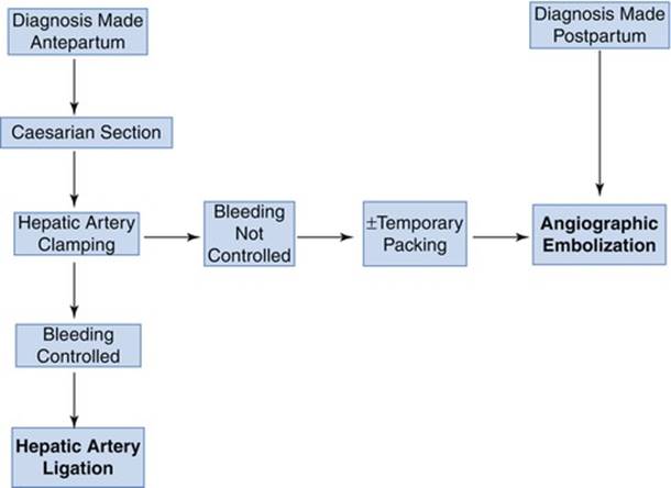
Fig. 11.5
Algorithm for the treatment of spontaneous hepatic rupture in pregnancy (see text for details) [117]
Obstetric Management
If a rupture occurs during pregnancy, delivery of the fetus is one of the first steps. A classical Pfannenstiel incision is, however, not suitable because the full abdomen cannot be visualized unless a second upper abdominal incision is made. A median laparotomy is therefore the recommended approach because it has the great advantage of visualizing the entire abdomen, speed of execution, and less blood loss [106].
11.3.8 Prognosis
Up to 1943, there were 29 cases (the case of Vesalius included) of true spontaneous rupture of the liver, and only two of these complicated pregnancy (Abercrombie 1844, the patient died, and Rademaker 1943, the patient recovered but the fetus died) [80]. In general, five of these came to operation, and four recovered making maternal mortality after surgical therapy 80 %. In that time if liver rupture was evident, the Porro procedure (excision of the Fallopian tubes, the ovaries, and the uterus at supravaginal level) was recommended after incision of the uterus and deliverance of the fetus.
In 1976, Bis and Waxman, in a collective review, reported 62 % fetal and 59 % maternal mortality rates of liver rupture during pregnancy [89]. In the collected reports of 15 patients treated with hepatic artery ligation/occlusion (radiologically or surgically), the mortality was 29–37 % [54, 110, 111, 117, 119, 123, 124].
11.4 Peptic Ulcer Bleeding
11.4.1 Introduction
Pregnancy seems to have a beneficial effect on peptic ulcer (see Chap. 4). Clark in 1953 investigated dyspeptic symptoms during 313 pregnancies in 118 women with the diagnosis of peptic ulcer before pregnancy [125]. He found that there was a remission of ulcer symptoms in 88 % (276/313) of the pregnancies. More than 50 % of these women claimed to have been completely symptom-free during the whole pregnancy; the remainder had minor symptoms which they regarded as unconnected with the ulcer. In the remaining 12 % (37/313), symptoms persisted which were indistinguishable from those of ulcer, and 14 women were admitted to hospital for treatment of the “indigestion.” In no case did hemorrhage or perforation occur during the pregnancy.
11.4.2 Incidence
Small hemorrhages from the upper part of the alimentary tract do not appear to be very rare in pregnancy, although Avery Jones in 1947, investigating for ulcer activity a series of 10,000 women attending an antenatal clinic, found only one case of mild hematemesis, and no ulcer could be demonstrated [126]. In a series of 587 cases of hematemesis and melena admitted to Dr Norris’s unit at Whittington Hospital during the 4-year period 1957–1960, there were four pregnant women (MacCaig JN, 1962, personal communication). These data show that only 0.7 % of bleeding peptic ulcers occur during pregnancy. It may be that the small number of proven cases is due to a reluctance to carry out radiological investigations during pregnancy before the era of endoscopy. If investigations were done after the birth of the child, the ulcer would probably have healed. Up to 1971, 31 perforations and 32 cases of hemorrhage from proved peptic ulcer during pregnancy have been reported [127]. The ratio of gastric and duodenal ulcers in pregnancy and also bleeding peptic ulcers during pregnancy is not known.
Peptic ulcer bleeding occurs in the late third trimester or during the first days postpartum [128].
There are only several case reports of operated bleeding peptic ulcers [129–132].
11.4.3 Risk Factors
In 1948, Bernstine and Friedman reported four cases of hemorrhage from peptic ulcer in pregnant women [133]. All of these women had been treated with progesterone before the onset of the gastrointestinal hemorrhage.
11.4.4 Diagnosis
Bleeding peptic ulcer is diagnosed by esophagogastroduodenoscopy (EGD). Before the era of endoscopy, peptic ulcers were diagnosed with radiological examination. Due to the ionizing radiation, it was contraindicated during pregnancy. Therefore, for many cases of peptic ulcer hemorrhage during pregnancy, the diagnosis was never confirmed [134].
11.4.5 Treatment
11.4.5.1 Conservative Treatment
Today, with potent acid suppression medications, most of peptic ulcer bleeding can be treated without operation. During long period of occurrence during pregnancy, most of these bleedings stopped with medical management [128]. If needed EGD intervention is performed. Endoscopic interventions are not contraindicated during pregnancy (see Chap. 2). Johnston, in a period between 1939 and 1953, found two with duodenal ulcer, one complicated by hemorrhage and one by perforation [135]. Bleeding ulcer responded to conservative management.
11.4.5.2 Surgical Treatment
In general population the type and location of bleeding peptic ulcer dictates the type of surgical procedure. Due to only several cases operated during pregnancy, these surgical principles should be applied in pregnant patients. Durst and Klieger in 1955 reported a case in which hemorrhage from a gastric ulcer occurred at term and continued after delivery; late consent was given for emergency surgery, and death occurred during the operation [129]. In 1957, Vasicka reported a case of massive gastrointestinal hemorrhage from peptic ulcer. Gastric resection with gastroduodenostomy was performed in the 20th week of gestation. This was the first reported case of surgical intervention for this complication during pregnancy. Hypertension, albuminuria, and vaginal hemorrhage developed in the 28th week, and a viable infant was delivered by Cesarean section during the 33rd week [132]. In the third case, a Spanish girl, already in labor, was bleeding from a gastric ulcer. Cesarean section was performed, but the fetus was dead. The operation was continued by performing partial gastrectomy, and the patient recovered [130]. Ulcer pain in aged 23 in 28 week pregnancy occurred only during her second and fourth pregnancies. It is also remarkable that the fetus survived such a massive hemorrhage in the mother and that the pregnancy continued to term, 8 weeks after emergency partial gastrectomy [131].
There are three known cases of surgical treatment for hemorrhage from duodenal ulcer occurring during pregnancy. In one case vagotomy and pyloroplasty were performed [136], and another two cases partial gastrectomy was made [127].
11.4.6 Prognosis
Maternal mortality from 1905 to 1955 was 83 % and from 1905 to 1962 lowered to 71 % with fetal mortality 60 and 67 %, respectively (one case did not report fetal outcome) [131]. Due to the extreme rarity of the disease, maternal mortality, in studies during the first half of the twentieth century, was also evaluated in cumulative pregnancy or puerperal deaths. MacNalty found in 770 puerperal deaths in 1937 only one death from hemorrhage due to gastric ulcer [137]. Hooker studied 350,000 pregnancies over a 3-year period (1930–1932) and found only one death from perforation of a gastric ulcer [138]. Sandweiss studied 70,310 pregnancies seen in the 10-year period (1928–1937) in various Detroit hospitals and found one death due to perforated duodenal ulcer [139]. In 1943, Sandweiss, in a review of the literature on deaths due to perforation and hemorrhage from gastroduodenal ulcer during pregnancy and the puerperium, found only 14 cases. In seven cases they were gastric ulcers, and in seven duodenal. All of the 14 patients died postpartum, and premature delivery occurred in four [140].
Carangelo reported four cases of massive gastrointestinal hemorrhage in pregnancy, with three deaths [134]. Three of the four patients had an eclamptic syndrome with hypertension, edema, convulsions, oliguria, and albuminuria, and all four had a septic course with fever. The onset of labor in three of the patients occurred between 24 and 48 h after the onset of hematemesis. There were three stillbirths and one premature but viable infant.
Reports of only seven cases of hemorrhage from proved ulcers could be found in the literature (Table 11.4). Two of the mothers survived, and not more than three of the pregnancies produced live births [131].
Table 11.4
Reported cases of hemorrhage from peptic ulcers during pregnancy 1905–1962 [131]
|
Authors |
Week of pregnancy |
Lesion |
Mother outcome |
Fetal outcome |
|
Le Play (1905) |
7th month |
Gastric ulcer |
Died |
Abortion |
|
Mulsow and Brown (1936) |
35th week |
Duodenal ulcer |
Died |
Lived (twins) |
|
Ministry of Health [137] |
Puerperium |
Gastric ulcer |
Died |
Not recorded |
|
Le Winn (1947) |
37th week |
Duodenal ulcer |
Lived |
Died |
|
Johnston [135] |
Labor |
Duodenal ulcer |
Died |
Died |
|
Durst and Klieger [129] |
Term |
Gastric ulcer |
Died |
Lived |
|
Stevenson [130] |
Term |
Gastric ulcer |
Lived |
Died |
11.5 Mesenteric Ischemia
11.5.1 Mesenteric Vein Thrombosis
… not everyone is aware that violent abdominal pain, ileus, and collapse may mean mesenteric vascular occlusion … Warren and Eberhard, 1935
11.5.1.1 History
Mesenteric vein thrombosis (MVT) was first summarized as a cause of intestinal infarction in 1895 by Elliot, who treated the infarcted bowel with resection, creating two stomas and reanastomosing those 2 weeks later. Elliot stated that the patient had thrombophlebitis. He collected 14 cases of MVT from the literature [141]. In 1898, Köster in his inaugural address at Gothenburg reviewed the literature and described three further cases of mesenteric venous occlusion [142]. Charles Hilton Fagge (Fig. 11.6), at Guy’s Hospital, London, published the first case in puerperium in 1876 [143]. A woman died a month after delivery in a few hours after the onset of severe abdominal symptoms. Autopsy showed thrombi in the superior mesenteric veins extending into the trunk of the portal vein nearly to the point where it breaks up into its branches. The thrombosis extended into the veins beyond the territory that was congested. There was no endocarditis or any evidence of peritonitis or any cause for internal strangulation found. The report by Warren and Eberhard [144] helped to establish MVT as a distinct clinical entity. It has been classified clinically as primary (idiopathic) or secondary [145].

Fig. 11.6
Charles Hilton Fagge (1838–1883) (Courtesy of the Gordon Museum, King’s College London)
11.5.1.2 Incidence
MVT is an extremely rare condition in general population and accounts for 0.002–0.06 % of all inpatient admissions [146], 0.01 % of all emergency surgical admissions, and <1/1,000 laparotomies for acute abdomen [147]. Abdu et al. found in their literature review of 372 cases of MVT in general population (1911–1984) that the condition was most common in the sixth and seventh decades of life [148]. In contrast, recent series in general population found the average age to be between 45 and 65 years [149–154]. In the study by Rhee and Gloviczki [155], MVT comprised only 6.2 % of all patients treated for mesenteric ischemia in general population. In autopsy studies, MVT is found in 0.2–2 % of the population in general [144, 146]. In a study by Ottinger and Austen, MVT was found in less than 1 % of patients with mesenteric ischemia [146].
In pregnancy and puerperium, up to 1963, there were 15 cases described [156–159]. Of these, 27 % (4/15) followed abortions, 13 % (2/15) occurred during pregnancy, and 53 % (8/15) during the puerperium. One case was not proved. After 1963, similar number of patients is described [55, 160–168].
In general population, Milch and Masotti in 1936 stated that in mesenteric arterial thrombosis, the superior mesenteric artery is involved 40 times more often than the inferior mesenteric artery [169], while Jackson et al. in 1904 stated that in cases with venous mesenteric occlusion alone, 99 % occur in the superior mesenteric vein [170]. The venous collateral circulation of the inferior mesenteric vein is more elaborate than that of the superior - therefore MVT of the inferior mesenteric vein, followed by infarction of the descending colon, it is exceedingly rare.
11.5.1.3 Risk Factors
Three factors, usually referred to as Virchow’s triad, are concerned in the formation of every thrombus. These are damage to vascular endothelium, changes in the velocity and character of the local bloodstream, and changes in the constituents of the blood. MVT is a very uncommon surgical pathology frequently associated with known coagulation defects [160, 162, 165, 166, 171–173]. The maternal risk of thromboembolic episodes is increased eight times in the presence of any one of the coagulopathies [174]. The incidence of the factor V Leiden mutation may be as high as 46 % in patients with a history of venous thromboembolism during pregnancy [175].
The presence of MVT is found also when there is no detectable coagulation error and is extremely rarely encountered in pregnancy [166, 176, 177]. This condition is attributed to physiological hypercoagulability which occurs during pregnancy due to multiple factors including rise in factors VII and VIII and fibrinogen and reduction in fibrinolytic activity [176]. But a rise in platelet count together with an increase in platelet stickiness maximal on the 12th day of puerperium has been demonstrated [178]. There are only four cases in the literature with no precipitating factor for the development of MVT and is called primary (idiopathic) MVT [166, 176, 179, 180].
Specific group of MVT has been reported in pregnancy in association with certain surgical and medical comorbidities [165] which include repeated abdominal surgery, Cesarean section, appendectomy for gangrenous appendicitis, elective laparoscopic cholecystectomy, mesenteric cyst excision, vesicoureteral reflux, and mistaken intake of oral contraceptives during pregnancy (Table 11.5) [168, 181]. MVT caused by postoperative stasis in general population was first described by Maylard in 1901 [186].
Table 11.5
Primary (idiopathic) and secondary MVT with known risk factors in pregnancy and puerperium
|
Hypercoagulopathies [160, 161] |
|
Oral pills during pregnancy or puerperium [55, 56, 181] |
|
Postoperative |
|
Single or repeated abdominal surgery [182, 183] |
|
Cesarean section [162] |
|
Cytomegalovirus infection (with complications) [163] |
|
Chronic idiopathic MVT [164] |
|
IVF pregnancy [184, 185] |
|
Hemoglobinopathy [165] |
|
Primary (idiopathic) [166, 176, 179, 180] |
|
Smoking |
|
Obesity |
|
β-thalassemia |
|
Antiphospholipid syndrome |
|
Abnormal fibrinogen |
|
Homocystinuria |
|
Paroxysmal nocturnal hemoglobinuria |
|
Thrombocythemia |
|
Abdominal trauma |
|
Abdominal sepsis |
|
Myeloproliferative disorders |
|
Cancer |
Reed and Coon reported the first case of MVT from oral contraception in 1963 [187]. Other cases have been infrequently described [181, 188–190]. Hoyle et al. discussed 21 cases, 9 had been taking 0.05 mg estrogen, and the rest had taken higher doses [188]. Twelve had been taking the agent for more than a year, and two had suffered MVT in the first cycle. The problem with association of risk factors and MVT is that patients sometimes have additional risk factors like heavy smoking [181]. Even in this case by Graubard and Friedman with two risk factors, BMI of the patient was not mentioned which is also a possible additional risk factor.
The anatomic location of venous thromboembolism associated with IVF cycles has been suggested to differ from those seen in the general population. In venous thrombosis associated with IVF, the veins of the upper extremities and neck are involved in 80 % of reported cases, whereas only 11 % of deep vein thrombosis diagnosed in the general population involves the upper extremities [191]. Further, 97 % of upper extremity deep vein thrombosis reported in the literature during pregnancy was associated with assisted reproductive technology [192].
11.5.1.4 Clinical Presentation
Clinical symptoms of acute MVT are variable, nonspecific, and difficult to differentiate from arterial occlusion. In most cases of acute MVT, the main symptom is abdominal pain lasting from several days to more than 3 months [157], first cramp-like before becoming continuous when peritonitis supervenes. They continued to have bowel movements initially, becoming constipated later. Patients had pain out of proportion to abdominal tenderness. This was noted by Berry and Bougas, who believed it as an important differential point in diagnosis [193]. Other symptoms are nausea/vomiting, fever, and abdominal distention. The tendency toward hemoconcentration was also seen in their patients and probably reflects slow progress of the disease, allowing sequestration of extracellular fluid in the gut. Donaldson and Stout state that occult blood can always be found in the stools [194]. On rectal examination, there may be blood on the examining finger. If hematemesis in acute form is found, then splenic vein thrombosis is present. On the other hand, splenic vein thrombosis is not necessarily followed by hematemesis [195].
Subacute or chronic MVT is usually asymptomatic due to the development of collateral vessels, but when it is combined with portal vein thrombosis, esophageal variceal bleeding with hematemesis may occur [150, 196].
Most patients in general population using oral contraception had at least 2 weeks of symptoms such as abdominal pain, discomfort, anorexia, vomiting, and change in bowel habits [189, 197, 198].
Sometimes the leading presentation can be spontaneous abortion or stillbirth [157].
11.5.1.5 Diagnosis
Clinical diagnosis of MVT is usually difficult, and it is frequently delayed due to lack of awareness among primary physicians and absence of active signs in early stages of the disease. Furthermore the early features of the disease get masked by the effects of pregnancy. Early diagnosis is possible only if high degree of suspicion is exercised in cases of severe abdominal pain with the absence of positive physical signs. In general population, in 1914, in only 4 % of patients the condition was diagnosed preoperatively [199].
Almost all patients present with leukocytosis with elevated hematocrit, but these are not helpful for diagnosis [200]. Grieshop et al. reported an 80 % incidence of elevated white cell count in cases of MVT in general population [201].
The diagnosis can be confirmed with color Doppler ultrasound [173] which is widely available, cheaper, feasible for emergency imaging, and applicable in pregnancy due to lack of need for X-rays or potentially harmful intravenous contrast, but this modality requires expert hands, and some studies have shown this modality to have only 70 % sensitivity [149].
Contrast-enhanced abdominal CT in general population currently holds a sensitivity of >90 % and is the most common diagnostic test of choice (Fig. 11.7). Magnetic resonance imaging offers no distinctive advantage over CT [150].
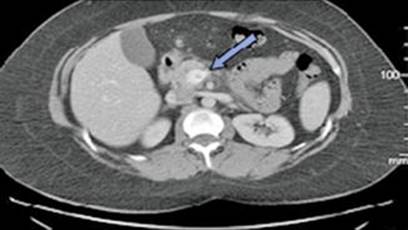
Fig. 11.7
CT scan with superior mesenteric vein thrombosis (blue arrow) in a patient on IVF program [185]
Some authors claim that angiography findings in general population of veno-occlusive disease are less sensitive. In recent studies, angiography showed only 55 % sensitivity and has not been suggested as a primary diagnostic modality [150, 202].
Often, definitive diagnosis is made during surgical exploration mostly due to acute abdomen. If there is no strangulation, volvulus, mesenteric thromboembolism, or cocaine abuse should be excluded [53].
11.5.1.6 Treatment
Management involves multiple disciplines including surgery, clinical hematology, obstetrics, and neonatology and depending on the presentation of a particular case. Most importantly, the treatment depends on the stage of the disease [150].
Surgery
Explorative laparotomy is indicated for patients with symptoms and signs of acute abdomen or suspected or confirmed intestinal infarction (Fig. 11.8) by diagnostic modalities. Pielliet is reported by Elliot as being the first to perform a successful resection for venous occlusion in general population and published his findings in 1895 [141]. During operation, fresh and organized thrombi are found extruding from the veins, while arteries are constricted but patent [182]. Consideration should be given to the use of intra-arterial papaverine to reverse (arterial) spasm [202] and thrombolytic agents via operatively placed catheters [203, 204] which, together with continued heparin, may prevent extension of infarction (see further paragraphs for pregnant population).
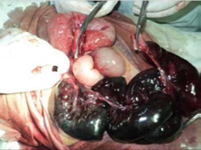
Fig. 11.8
Gangrenous ileal segment due to mesenteric vein thrombosis [177]
If there are segmental gangrenous segments, minimal resections should be performed to minimize the possibility of short bowel syndrome. Multiple resections with anastomoses with or without proximal stoma are recommended. Another option after multiple segmental resections is to close (potentially) viable bowel segments by stapling and abdomen left open with vacuum-assisted closure (VAC) with 125 mmHg continuous negative pressure (Fig. 11.9) in the aim of assessing the viability of remaining bowel after 24 and 48 h [168].

Fig. 11.9
Open abdomen with vacuum dressing [168]
At the second and third surgical looks, some intestinal segments could require subsequent additional resections. Eventually, after 48 h of open abdomen management, the intestinal continuity should be restored. Abdominal wall should be primarily closed without aponeurotic defect.
The rare cases of laparostomy use in pregnant women previously reported have not been successful in terms of fetus or mother issue [205]. There are two case reports of favorable outcome after laparostomy as a treatment of wound dehiscence in pregnant women [206] and second- and third-look operations due to mesenteric ischemia [168]. In both cases VAC was included as a part of treatment process. Among many techniques developed for open abdomen management, VAC allows currently the best results in terms of primary abdominal wall closure [207]. In some series of nonpregnant patients, using VAC protocols, complete fascial closure rate was achieved in 100 % [208]. In abdomen with constantly growing gravid uterus and low intra-abdominal pressure requirements, primary closure appears to be a particularly challenging task. It is nevertheless a key endpoint in a pregnant woman, in order to protect the fetus and to assure a vaginal delivery.
As the enlargement of the spleen in protein C deficiency and its gastric pressure could become prominent together with anemia and leukopenia in addition to mild-severe thrombocytopenia, splenectomy seems to be an appropriate choice together with the periodic use of protein C concentrate [209].
Thrombectomy
Other treatments such as peripheral or regional thrombolysis with or without surgical thrombectomy and a combination of surgical thrombectomy and regional heparinization have been reported in general population [210–213]. There are anecdotal reports of venous thrombectomy, but this has not shown improved outcome and is generally not recommended [214, 215]. In pregnancy, treatment with thrombolytics is not advocated [176].
Anticoagulation
In the patients without bowel infarction or peritonitis, anticoagulation with heparin followed by warfarin after delivery is the mainstream therapy [150]. Two of the first proponents of anticoagulation therapy in general population were Murray in 1940 [216] and Strohl and Lasner in 1948 [217]. Anticoagulation for MVT has been shown to be associated with improved outcome by Naitove and Weisman in 1965 when mortality was 50 % for those who did not receive anticoagulants compared with 0 % for those who received postoperative anticoagulation [218]. Heparin has been shown to prevent recurrence of thrombosis after intestinal resection (14 % vs. 26 % [148] and 0 % vs. 19 % [219]) and to be associated with lower mortality when recurrence does occur (22 % vs. 59 % [148]). In anticoagulated patients not undergoing surgery, most thrombosed veins will partially or completely recanalize over time. In one study, 80 % of anticoagulated patients with mesenteric and/or portal vein thrombosis showed vascular recanalization over a mean follow-up time of 5 months compared with <10 % of non-anticoagulated patients [220]. During pregnancy LMWH is recommended in order to avoid the possible side effects of warfarin on the fetus, especially its teratogenic and bleeding effects. There are no defined guidelines for the duration of anticoagulation after pregnancy, and decisions on a case-by-case basis need to be taken by expert clinical hematologists [176]. In patients with inherited hypercoagulable disorders (i.e., protein S, protein C, antithrombin III deficiencies, and factor V Leiden mutation), lifelong anticoagulation is warranted. For patients with reversible predisposing causes, at least 6 months of anticoagulation is recommended [221].
A review in Thrombosis Research published guidelines for anticoagulation management strategies for various clinical situations before and after controlled ovarian stimulation [222]. Patients with a history of prior MVT should be placed in the clinical classification of “previous episode(s) of venous thromboembolism receiving long-term anticoagulation.” It is recommended switching from oral anticoagulants to LMWH therapy (e.g., enoxaparin 1 mg/kg every 12 h) before controlled ovarian stimulation and continuing this regimen throughout pregnancy [222]. These authors recommend refraining from the administration of LMWH for 24 h before egg retrieval and restarting therapeutic anticoagulation 3 h after egg retrieval [222]. In the setting of IVF, associated MVT, and the absence of large studies, lifelong oral anticoagulation in patients at low risk for bleeding and full anticoagulation with LMWH per the above protocol during repeat IVF cycles is considered prudent.
However, controversy in management for the prevention of venous thrombosis still exists after delivery. Though the induction of oral anticoagulation in protein C deficiency has been widely used, it carries significant risks in a patient with esophageal varices and thrombocytopenia [209].
Obstetric Management
Continuation of Pregnancy
The decision on continuation or termination of pregnancy is taken as per the case and available facilities. While workers like Engelhardt et al. [166] and Fouad et al. [176] successfully continued the pregnancy, Lin et al. and Foo et al. terminated the pregnancy [179, 223]. In one recent case report with several operations due to gangrenous bowel and open abdomen, the pregnancy was uneventfully carried to full-term vaginal delivery [168].
Mode of Delivery
If the pregnant woman has proven PVT, every effort should be made to prevent further complications during and after delivery. The straining and bearing down that take place during vaginal delivery have also been reported to result in a marked but transient portal vein pressure in all pregnant women. Thus, the avoidance of vaginal delivery and the use of Cesarean section should minimize the portal vein pressure increase and reduce the possibility of variceal bleeding [209, 224].
11.5.1.7 Prognosis
The prognosis of MVT does not appear as ominous as that associated with arterial thrombosis in general population [150], but mortality in general population was 60 % in the beginning of the twentieth century, lowered to 34 % in 1935 [144]. Currently, the mortality in general population is in the range of 13–50 % [203, 225–227]. In general population, the subgroup with oral contraception as the cause had high rates of the mortality and morbidity in the review from 1977 – 50 % of the patients died and 50 % required at least two operations. Arterial thrombosis carried twice the mortality of venous thrombosis but was half as common [190].
In pregnancy, up to 1963, out of 15 described cases mortality was 87 % (13/15), and the two patients that survived were from the last year (1963) of the analyzed period [156, 157].
11.5.2 Portal Vein Thrombosis
11.5.2.1 Introduction
Portal vein thrombosis (PVT) shares the same or similar characteristics as MVT inclusion localization, risk factors, diagnosis, and therapy.
11.5.2.2 Incidence
PVT is even rarer in pregnancy than MVT with only few cases published [209, 228–230]. One case was in the first trimester [229], two in the second trimester [228, 230], and one in the third trimester [209].
11.5.2.3 Risk Factors
Risk factors are the same as for MVT (Table 11.5) which are the same as for MVT during pregnancy. Concurrence of several disorders seems to be relatively common in PVT in adults, and extensive investigation should be considered in these patients [231]. Probably the only risk factor more influential on the development of PVT than MVT is cirrhosis, but due to extremely low incidence of both conditions in this age group, the conclusions cannot be made.
11.5.2.4 Clinical Presentation
Clinical presentation is different from MVT. Mostly patients present with short history of acute right hypochondrial pain that lasts from hours to several days and fever of around 38 ºC [229]. The signs include tachycardia, fever, and tenderness in the upper right abdominal quadrant without peritoneal irritation.
11.5.2.5 Diagnosis
Abdominal Ultrasound/Doppler
Diagnosis is made by ultrasound or more specifically by Doppler. Unlike contrast CT and helical CT associated with CT angiography, color Doppler does not involve potentially harmful intravenous injection of contrast [232, 233]. Also, unlike MRI and 3D magnetic resonance portography with contrast, color Doppler is more widely available, cheaper, and feasible on emergency basis [234, 235].
Urgent abdominal ultrasound is sometimes performed to rule out acute cholecystitis but can demonstrate well-defined homogenous hyperechoic oval-shaped formation in the splenomesenteric confluence and/or branches of the portal vein. Color Doppler study confirms these findings by showing margination of the colored flow in the residual lumen. Splenic and mesenteric veins should be visualized because their thrombosis could lead to bowel ischemia. The hepatic artery shows increased compensatory flow. Collateral venous circulation should be verified or excluded because it helps establishing definitive diagnosis. The color Doppler sonographic features of acute thrombosis in portal vein or superior mesenteric vein are as follows:
1.
2.
When PVT is confirmed by Doppler, complete coagulation profile should be obtained.
11.5.2.6 Treatment
Anticoagulation
Prompt treatment of PVT is essential in order to prevent extension to superior mesenteric vein, splenic vein, and hepatic vein which could lead to the development of hepatic or mesenteric infarction [232, 237]. Prompt treatment could also prevent progression to portal hypertension with risk of variceal hemorrhage [209, 238, 239]. Furthermore, early treatment could result in complete lysis of the clot, and the resolution of the clot could be as early as 4 days, but could take up to 1 month.
Interventional Techniques
Some authors have reported the use of fibrinolytic infusion by the systemic route, transhepatically, or via the superior mesenteric artery with controversial results [240–242].
Percutaneous interventional techniques such as balloon dilatation and stenting have been effective in some cases with acute, recent PVT; however, these treatment modalities do not prevent rethrombosis [241]. Therefore, patients at risk should be kept on prophylactic anticoagulants for life in order to prevent rethrombosis.
Response to Therapy
Response to therapy in pregnant patients cannot be estimated due to extremely small number of patients, but color Doppler provided reliable means of monitoring response to therapy of the PVT [229].
11.6 Inferior Epigastric Artery Bleeding
11.6.1 Anatomy
The inferior epigastric artery (IEA) arises from the external iliac artery deep to the inguinal ligament. It both serves as a surgical landmark and constitutes a potential target for injury during inguinal hernia repair. The IEA divides deep to the rectus sheath into two branches: an ascending branch that anastomoses at the umbilicus, medial to the rectus sheath, with the abdominal branch (or superior epigastric artery) of the internal thoracic artery, and a descending branch that gives off obturator branches that course along the ischium and anastomose with the obturator artery and pubic branches, which in turn course along the pubic rami and reach the pubic symphysis [243].
11.6.2 Mechanisms of Injury
11.6.2.1 Direct Trauma
Direct injuries of the IEA include blunt trauma [244] or penetrating abdominal wall trauma. Direct blunt injuries as well as indirect injuries can cause damage to the artery probably by stretching of the artery over its elastic properties causing avulsion with subsequent bleeding [245]. Penetrating abdominal wall trauma during interventions including Cesarean section, paracentesis [246], and insertion of trocars during laparoscopy [247] causes direct injury to the IEA. Injuries to the ascending branch are well documented and usually occur after direct trauma to the abdominal wall, for instance, during laparoscopic surgery, subcutaneous injections, insertion of lumboperitoneal shunts, or ascites fluid aspiration [248–250]. In one of the Indian studies, 95.5 % of patients with injury of IEA had intrapartum Cesarean delivery, while 4.5 % had an elective operation. Therefore, emergency Cesarean section is a risk factor for IEA bleeding [251]. During Cesarean section, care during transverse cutting and suturing of lateral extension of rectus sheath is advised [252].
11.6.2.2 Spontaneous
Rarely, the ascending branch may rupture spontaneously, most notably in patients taking anticoagulant medications, after lifting heavy weights, coughing, or straining. Thus, tearing of the branches of the epigastric vessels is a well-known cause of rectus sheath hematoma (see Sect. 11.6.8) [253–255].
11.6.2.3 Blunt Pelvic Trauma/Fracture
In contrast, there have been only five reports in nonpregnant population of massive bleeding from the pubic branch of the IEA in patients with pubic rami fractures caused by blunt pelvic trauma [256–259].
11.6.3 Incidence
Early reoperation rate after Cesarean section in India is 0.45–0.6 % [251, 252]. In two Indian studies with early relaparotomy after Cesarean section, rectus sheath hematoma was found in 21–27 % of cases [251, 252]. In Western countries this is an extremely rare condition during pregnancy with only several case reports after Cesarean section [244, 260, 261]. Incidence of spontaneous IEA bleeding is lower due to several reasons:
· Pregnant women are more cautious and are not exposed to blunt or penetrating abdominal/pelvic trauma as general female population (more at home, less car driving).
· Avoidance of heavy lifting.
· Avoidance of closed spaces and people with infectious diseases mostly respiratory conditions.
· Lower incidence of surgical or other invasive interventions during surgery.
11.6.4 Clinical Presentation
Clinical presentation depends on the severity of the injury and the timing of the incident. If observed intraoperatively and IEA itself is injured/transected, then significant bleeding is present. If the peritoneum is intact, then large subperitoneal hematoma is found. If the peritoneum is damaged, significant bleeding into the abdominal cavity from the abdominal wall is found. If the bleeding is not observed intraoperatively, then the severity of the clinical presentation depends on the caliber of the artery that is injured. If IEA is damaged, then immediately after the operation there will be symptoms and signs of significant blood loss and hemorrhagic shock: tachycardia, lowered blood pressure, increased pulse rate, pallor, and cold sweat. Additionally the patient will have distended and painful abdomen. On inspection hematoma over the IEA is found.
11.6.5 Differential Diagnosis
Spontaneous rupture presents significant diagnostic problem because skin changes in smaller hematomas could be absent, and the patient presents with acute significant pain with peritoneal irritation which mimics intra-abdominal conditions such as appendicitis, diverticulitis, cholecystitis, tumors, and visceral injuries [255].
11.6.6 Diagnosis
The IEA represents a potentially overlooked source of pelvic arterial hemorrhage. The IEA should be considered as a possible source of arterial hemorrhage if arteriography of internal iliac artery branches does not yield a bleeding source [244].
11.6.7 Treatment
11.6.7.1 Transarterial Embolization
Almost all cases of bleeding IEA after Cesarean section are treated by transarterial embolization (Fig. 11.10) [244, 260, 262]. In only one case by Randall et al., the patient was treated conservatively (abdominal CT was used for the diagnosis) [261].
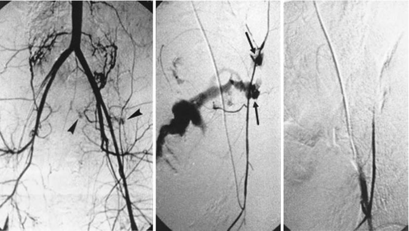
Fig. 11.10
Left, aortoiliac arteriogram showing suspicious abnormal stains (arrowheads) in the left side of the pelvis. Middle, selective left inferior epigastric arteriogram showing massive bleeding from two injured portions (arrows). Right, complete occlusion of bleeding points after embolization [260]
11.6.7.2 Analgesia
Most patients are treated with peroral NSAIDs and/or opioids for pain relief. There is one case of transversus abdominis plane block for severe pain eliminating large dosage of peroral pain killers [261].
11.6.8 Spontaneous Rectus Hematoma
11.6.8.1 History
Rectus sheath hematoma (RSH) is an ancient disorder first being accurately described by Hippocrates and mentioned by Galen. The first reported case in the United States was by Richardson in 1857 [263]. In 1882, Karl Maydl, of Vienna, wrote a most valuable series of articles on the subcutaneous tearing of muscles and tendons [264]. He devoted much attention to the tearing of the rectus abdominis muscle and tabulated his cases. In 1918, Emerson recorded a case of rupture of the deep epigastric artery due to muscle strain. Perman was the first to write a comprehensive article on hematoma in the sheath of the rectus abdominis muscle. In 1925, Carey Culbertson reported two cases of hematoma occurring spontaneously in the sheath of the rectus abdominis muscle, with consideration of its gynecological and obstetrical significance [265]. First Thomas Stephen Cullen (Fig. 11.11) in 1918 described bluish periumbilical discoloration due to ruptured ectopic pregnancy and then with Brödel in 1937, at Johns Hopkins Hospital, reported two cases of spontaneous hematoma of the rectus abdominis during pregnancy [266, 267]. Both were black patients. Resolution was uneventful in one with a 7-month pregnancy, but operative exploration was carried out on the other with success.

Fig. 11.11
Thomas Stephen Cullen (1868–1953); painting by Thomas C. Corner; oil on canvas, 48 by 40 in., 1907 (Courtesy of the Alan Mason Chesney Medical Archives of the Johns Hopkins Medical Institutions)
11.6.8.2 Incidence
Maxwell in 1929 found 11 cases [268]. Of these, 73 % (8/11) were during pregnancy and 27 % (3/11) during labor. Torpin in 1943 analyzed 27 reported cases in pregnancy [269]. In 1946, Teske reported a case and analyzed 100 cases from the literature in general population. He showed that 60 % are on the right side and more than 80 % in the lower quadrants [270]. Aird stated that only about 150 cases have been reported up to 1949 [271]. Riera et al. in 2009 claim that there are 52 cases of the condition during pregnancy described [272]. There are numerous case reports during the last several decades [273–283].
11.6.8.3 Mechanism
The condition occurs mainly in multipara and late in pregnancy. It has also been described in the puerperium and even within half an hour of delivery and expulsion of the placenta. In almost all cases in pregnancy, there was some evidence of trauma from labor, from coughing, or from a fall. Muscle degeneration from influenza or typhoid is a possible cause. The precipitating factor in most spontaneous cases seems to be inelasticity of the wall of an artery or vein which prevents the vessel from accommodating itself in a movement, a cough, or a sneeze or, in or after labor, to the remarkable variations in length which the rectus muscle undergoes between extreme contraction and extreme relaxation. According to Brodel, there is only one major vessel to take care of this long stretch of muscle, and in order to avoid damage to itself, it must keep away from the muscle so far as possible and send its branches into the muscle substance in such a manner that the muscle action does not interfere with vascular freedom. The larger intramuscular arteries branch freely and form numerous anastomoses. They run at an angle varying from 60° to 90° to the axes of the muscle bundles. If an occasional artery runs parallel to the muscle bundle, it shows greater tortuosity. The arteries are less apt to tear than the veins, because they are far more resistant. They lie and so loosely embedded in the intramuscular connective tissue that they can be pulled out quite far without injury, but not so with the veins. They are frail, of smaller caliber, and have a much thinner wall. Tears may be partial or complete. In a partial tear only branches of the main vessels rupture beyond their point of entrance into the muscle body, but a complete tear is apt to rupture the main trunk also.
Right-sided hematomas in general population are presumably more common because more people are right handed and, thus, are more prone to right-sided strain of the rectus muscle during strenuous activity. The lower quadrants are more frequently involved because of the long vascular branches that are present and because muscle excursion during contraction with the absence of the tendinous inscriptions is greater [270].
11.6.8.4 Classification
Three types of RSH in general population can be distinguished by computed tomography appearances. Type I is unilateral and contained within the muscle; type II is uni- or bilateral and has blood between the muscle and transversalis fascia; type III invades the prevesical space or peritoneum and may or may not affect the muscle [284]. Until a classification in pregnancy is defined, the best option for the treatment algorithm is to adhere to the aforementioned classification.
11.6.8.5 Clinical Presentation
A history of trauma, anticoagulant therapy, and trivialities such as coughing, straining, or twisting to one side may remind the clinicians of this diagnosis [285]. Awareness of RSH during pregnancy is important because the abdominal wall is easily overlooked as a cause of acute abdominal pain [286, 287], given the higher prevalence of other pregnancy-associated pain.
Among the clinical findings, premonitory vague discomfort at the site of bleeding is common with the lesser degrees of hemorrhage. Probably a great number of the hematomas are small, unnoticed, and consequently unreported. In most recorded cases the hematoma was limited to the rectus sheath, and pain and tenderness were felt over the bleeding area. Muscle rigidity is often marked in the involved rectus, but in small hemorrhages there is little more than a local tenseness in the muscle. Swelling and palpable firm, nonpulsatile mass is usually confined to the affected muscle sheath. Swelling of the hematoma is limited to the rectus abdominis muscle, with its sheath not extending beyond the abdominal midline or the lateral borders of the muscle (Romanzew’s sign). However, below the arcuate line, the posterior sheath may communicate, and the mass may project across the midline or extend inferiorly and posteriorly toward the bladder. In some cases the hemorrhage escaped the limits of the muscle sheath and extended into the broad ligament or ruptured into the peritoneal cavity. In two cases in the literature, death occurred before help arrived [288].
The hemorrhage lies initially between the transversalis fascia and the posterior surface of the rectus muscle, spreading later to surround the muscle and ascend over the linea semilunaris between the rectus muscle and its posterior sheath and sometimes downward behind the rectus. If the patient lifts her head off the pillow so as to contract the rectus, the mass can be felt confined to the rectus sheath and immovable. The mass is equally palpable with the patient lying in a supine position or partially sitting up (Fothergill’s sign). Swollen and palpable mass is usually confined to the affected muscle sheath and severe, usually unilateral, abdominal pain that is aggravated by movement [289–292]. Carnett’s sign is performed by first localizing the area of maximal tenderness while the patient is relaxed. While this area is being pressed, the patient is asked to raise her upper back effectively tensing the abdominal wall. Worsening of the pain is considered a positive test [285]. In 1926, Carnett recognized that abdominal pain could be caused by neuralgia affecting one or more of the lower six intercostal nerves and developed a simple test to help localize the origin of symptoms to the abdominal wall [293]. For this part of the abdominal examination, the patient can be asked to lift the head and shoulders from the examination table to tense the abdominal muscles. An alternative is to ask the patient to raise both legs with straight knees. Staining of the skin and ecchymosis are very common over the palpable mass (Cullen’s sign) [294], first described by Guthrie and Stamfey, who also found Grey-Turner’s sign with rectus sheath hematoma [295]. They occur over the center of the hematoma, as a semicircle around the umbilicus, above the pubis, or along the linea alba (Fig. 11.12). The extravasated blood can pass more easily superficially around the medial border of the rectus. The pigments that produce this phenomenon may reach their destination by following the ordinary fascial planes but not by the lymphatics as was previously believed. In rectus sheath hematoma, ecchymosis appears after 2–5 days.
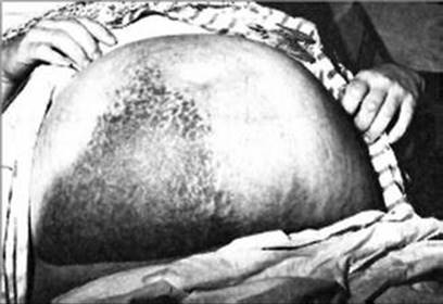
Fig. 11.12
Staining of the skin and ecchymosis are very common over the palpable mass (Cullen’s sign) not extending over the midline [296]
11.6.8.6 Diagnosis
The diagnosis of RSH should be suspected on clinical grounds and supported by additional imaging evidence. Abdominal ultrasonography, computed tomography (CT), radionucleotide imaging, and magnetic resonance imaging (MRI) have all been used to establish the diagnosis. Ultrasonography often shows a heterogeneous hypoechoic mass in the abdominal wall and is most useful for detecting RSH [287, 297]. Type III hematomas are large and could mimic intraperitoneal emergency such as torsion of the adnexa or placenta percreta with bleeding into the extrauterine compartment (Fig. 11.13) [298]. In unequivocal cases Doppler can aid the diagnosis showing nonvascularized structure (Fig. 11.14). During pregnancy, exposure of the fetus to radiation has limited the use of CT and radionucleotide imaging [287, 297, 299]. On the other hand, MRI appears to be a safe option (Fig. 11.15) [297].
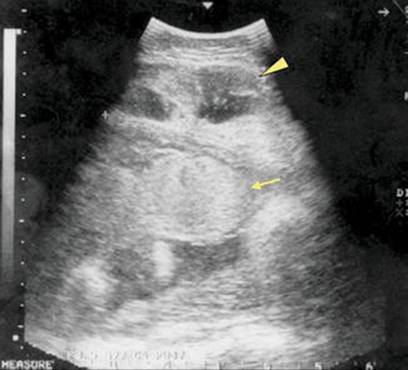
Fig. 11.13
Abdominal ultrasonography shows a heterogeneous hypoechoic mass (arrowhead) adjoining the placenta (arrow) [298]
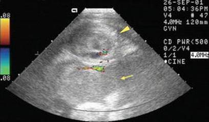
Fig. 11.14
Doppler sonography shows vascularity with blood flowing around the mass (arrowhead) and between the mass and the placenta (arrow) [298]

Fig. 11.15
A coronal T2-weighted magnetic resonance image showing a large left-sided rectus sheath hematoma measuring 160 × 70 × 55 mm (arrow). Note the subcutaneous fat stranding and a small hemoperitoneum coexisting with a fetus of 33 weeks of gestation [272]
11.6.8.7 Treatment
Conservative Treatment
Management is guided by the size of the hematoma and hemodynamic stability of the patient. Where the hematoma was small, it usually spontaneously resolves uneventfully within 1–2 weeks, although complete resolution of the hematoma may take as long as 2–3 months. Treatment generally consists of rest, peroral analgesia, and discontinuation of anticoagulation (if present). After resolution, RSHs usually do not recur and typically do not cause long-term sequels.
Embolization
Identification of a bleeding point is useful in guiding management; a massive hematoma that extends into the retroperitoneum may originate from a bleeding inferior epigastric vessel or may be coming from a retroperitoneal structure such as a leaking iliac or abdominal aortic aneurysm [300]. Selective percutaneous transcatheter arterial embolization is considered an effective hemostatic in the treatment of a patient with a large hematoma [272]. With regard to the safety of embolization during pregnancy, the general late onset of this pathology reduces the risks associated with fetal irradiation. The large hematoma displaces the uterus and the fetus to contralateral side reducing the dose of X-rays delivered to the fetus [272]. Selective epigastric embolization in severe RSH during the third trimester of pregnancy should be considered as the potential primary and alternative management path to classical laparotomy. However, this technique is time consuming, expensive, and not always available. This procedure is also associated with complications such as contrast-induced nephropathy [301] or bleeding from the puncture site [302]. Another disadvantage is that the bleeding vessel cannot always be identified. Further studies are required to confirm that the maternal and fetal benefits outweigh the fetal risks associated with embolization.
Surgical Treatment
In cases with rupture into the peritoneum, infection, or active bleeding with unstable hemodynamics, prompt surgical intervention is indicated. If the hematoma is very extensive and when shock is controlled, a paramedian incision with section of the rectus sheath and retraction of the muscle laterally will facilitate visualization of the bleeding vessel. Suture of the ruptured vessel or muscle should be performed. If the rupture is intraperitoneally, laparotomy is indicated to evacuate blood and clots and to control bleeding. If infection is present, evacuation of purulent material is mandatory with bacterial swabs and drains left in place.
11.6.8.8 Prognosis
In Maxwell series of 11 patients, eight were during pregnancy with 0 % maternal mortality and 37.5 % fetal mortality (additional two of eight cases do not mention fetal outcome). Three spontaneous RSHs during labor resulted in 0 % maternal and fetal mortality. In Torpin’s series there was a 15 % maternal mortality and a 50 % fetal mortality [269]. Reports from the 1950s show a maternal mortality of 12 % and a fetal mortality of 25 %. In 1997, maternal mortality rate of 11 % and a perinatal morbidity rate of 34 % were reported [286].
11.7 Omental Infarction
11.7.1 Incidence
Omental infarction is a rare clinical event that affects predominantly young and middle-aged women [303]. Omental infarction was first reported in 1882 by Oberst [304], and, since then, only a few hundred cases have been published in the English literature [305].
There are three published cases describing omental infarction in the postpartum period – one after Cesarean section and two after vaginal deliveries [306–308].
11.7.2 Etiology
Torsion of the omentum in general population is the main reason for infarction, and two different forms have been described: primary torsions (without other pathologic intra-abdominal findings) and secondary torsions (tumors, cysts, inflammatory changes, adhesions, hernias). Predisposing factors for torsion are anomalies of the omentum, such as a small root, irregular vascular anatomy, abdominal trauma, cough, and physical strain [304].
The etiology of omental infarction without torsion remains uncertain, but several mechanisms have been proposed, such as an anomaly of the venous vessels [309]. Other possible causes for primary infarctions could be disorders of hemostasis or vascular diseases. It is known that hematological changes occur during pregnancy and the puerperium and that hypercoagulability leads to an increased risk of thromboembolic events [310]. Depending on the duration and the degree of torsion, omental necrosis ensues (Fig. 11.16).

Fig. 11.16
Histological findings of major omentum show fresh hemorrhagic circulation disorders (arrows), partial necrosis of fatty tissue with acute inflammatory cell infiltrate (hematoxylin, 100×) [307]
The exact mechanism leading to infarction in puerperium remains unclear. Possible changes during the return of the mother’s body to the prepregnancy physiological condition may have provoked the infarction.
11.7.3 Clinical Presentation
Usually the clinical symptoms of an omental infarction are localized peritoneal irritation on the right side of the abdomen, sometimes associated with low-grade fever.
11.7.4 Differential Diagnosis
The clinical picture often misleads physicians to assume an incorrect preoperative diagnosis such as [304, 311, 312]:
· Acute cholecystitis
· Acute appendicitis
· Diverticulitis
· Appendagitis epiploica
· Umbilical hernia
11.7.5 Diagnosis
11.7.5.1 Laboratory Findings
The C-reactive protein and white blood count may be elevated.
11.7.5.2 Abdominal CT Scan
As most patients show symptoms of an acute abdomen, CT of the abdomen and pelvis should be the diagnostic imaging of choice [313]. If omental infarction is caused by torsion, characteristic CT findings might be detectable. The torsion leads to the presence of concentric linear strands in the fatty mass, a so-called fat spiral pattern (Fig. 11.17) [314].

Fig. 11.17
Computed tomography scan of the abdomen showing a hypoperfused mass in the anterior portion of the median epigastrium with fatty density (white arrows) and a thin layer of free fluid surrounding the liver [308]
Differentiating the omental infarction from other abdominal or omental diseases was challenging, and the radiological findings could be misinterpreted as a small incarcerated umbilical hernia as in case by Tachezy et al. [308].
11.7.5.3 Diagnostic Laparoscopy/Laparotomy
Diagnosis of an omental infarction has traditionally been made intraoperatively during an exploratory laparotomy or laparoscopy, and the treatment has been partial or total omentectomy. If there is other underlying pathology, it should be treated during the exploration.
11.7.6 Therapy
11.7.6.1 Conservative Therapy
Recent reports highlight cases of patients with CT-diagnosed omental torsions who have been successfully treated conservatively without any other complications (such as bacterial superinfections) [315–318]. Whenever conservative treatment fails or the clinical status of the patient worsens, a surgical intervention should be quickly implemented.
11.7.6.2 Surgical Therapy
Patients present symptoms of an acute abdomen. The clinical findings are very unspecific, and, therefore, in most cases surgical exploration leads to the diagnosis. All parts of the omentum that are macroscopically changed should be resected to eliminate the possibility of recurrent torsion and infection that can supervene omental necrosis.
11.8 Gastrointestinal: Genital Communications
11.8.1 Introduction
The high mortality rate associated with these cases indicates the seriousness of perforation of the vascular gravid genital system into the intestinal tract, the need for suspicion of the condition, and the knowledge of the principles essential for successful management.
11.8.2 Incidence
Communication of the gastrointestinal tract with the genital system from any cause is an unusual occurrence. Franco and Clough, reporting a review of the world’s literature to 1956, found only 75 cases of entero-uterine fistula [319]. In a review of over 1,000 consecutive cases of ectopic pregnancy at the Cook County Hospital (1940–1956), Webster and Kerr found only one associated with bowel invasion [320]. Documentation of entero-amniotic fistulas resulting from complications of ectopic pregnancy is even more unusual, only occasional cases being found in the literature. Only four cases presenting with rectal hemorrhage from an unknown source, ultimately proved to have arisen from rupture of an ectopic pregnancy into the bowel, have been reported (Table 11.6). The first known case is by Armstrong, in 1835, of a woman in her sixth month of pregnancy who suddenly passed bloody stools containing fetal bones and died of hemorrhage [324].
Table 11.6
Obscure intestinal hemorrhage (up to 1964) [321]
|
Author |
Year |
Site of ectopic pregnancy |
Site of perforation |
Symptoms |
Treatment |
Outcome |
|
Edgar [322] |
1901 |
Unknown |
Sigmoid colon |
Rectal hemorrhage, abdominal pain |
Posterior colpotomy |
Expired |
|
Webster and Kerr [320] |
1956 |
Right interstitial |
Terminal ileum and appendix |
Rectal hemorrhage, abdominal pain |
Resection of ileum, ileoileostomy, appendectomy, right salpingectomy, wedge resection of the right cornu of the uterus |
Expired |
|
Engel [323] |
1961 |
Left interstitial |
Mid-ileum |
Rectal hemorrhage, hypotension |
None |
Expired |
|
Shirkey et al. [321] |
1963 |
Left interstitial |
Terminal ileum |
Rectal hemorrhage, abdominal pain |
Suture ileum, total abdominal hysterectomy, left salpingectomy, right salpingo-oophorectomy |
Recovered |
11.8.3 Etiology
Le Jemtel, in 1909, presented an etiological classification of entero-uterine fistulas which with addition by Hawkes [325, 326]. There are four major etiological categories:
· Cancer – by infiltration and invasion arising in either the bowel or uterus
· Peritonitis – from trauma, puerperal infections, appendicitis, or diverticulitis, and fistulas following abscess formation involving the inflamed adjacent walls of both the uterus and intestine
· Traumatic or spontaneous rupture of the gravid uterus with strangulation of a loop of bowel caught in the defect
· Perforation of the uterus and bowel at the time of curettage
Danforth and Case, reviewing the relative incidence of the various causes of 58 cases of communication between the bowel and uterus dating from the 200 years previous to 1933, found 64 % due to injury, 24 % due to inflammatory or congenital processes, and 12 % due to carcinoma [327]. Diverticulitis has been reported as the underlying cause for a small number of fistulas between the uterus and large bowel [328–330]. In 1929, Noecker was the first to report a colouterine fistula secondary to diverticulitis in general female population [329]. Chronic salpingitis [326], criminal abortion [331, 332], retained pregnancy [333], and curettage of a bicornuate uterus [334] have been reported as specific causes for fistulas between the intestinal and genital tracts. An additional cause for fistula formation between the intestinal and genital tracts is rupture of an ectopic pregnancy into the bowel, causing an entero-amniotic fistula.
11.8.4 Pathogenesis
Infection is of paramount importance in the pathogenesis of entero-amniotic fistulas resulting from complications of pregnancy. Stock has previously described the role of infection in a case of secondary abdominal pregnancy complicated by fistula formation and rupture through the umbilicus [335]. The approximation of a vascular placenta, with the potential of villous invasion of adjacent structures, to intestine creates a precarious set of circumstances. Following villous invasion of the bowel wall, on approximation of the gestational sac to the intestine, inflammatory reaction and infection may create fistula formation. The source of infection may vary according to the location of the ectopic sac. Intraperitoneal ectopic sacs are most commonly infected from the adjacent bowel, whereas intraligamentary sacs are most often contaminated by bacteria from the vagina or uterus [335]. The vascular gestational structures aggravated by infection and villous invasion of adjacent vascular structures provide a dangerous source of massive hemorrhage (Fig. 11.18).

Fig. 11.18
Drawing and illustrating terminal ileum firmly attached to the left cornu of uterus with fistula between ectopic gestation and lumen of the bowel as seen diagrammatically through the bowel wall [321]
11.8.5 Prevention
The importance of meticulous surgical technique in reperitonealization of the pelvic floor, uterus, and Fallopian tubes and in manipulation of the bowel is exemplified in the case reported by Engel [323]. This case, documented by careful postmortem examination of involved structures, illustrates how inadequate reperitonealization of the uterus during a previous ipsilateral salpingo-oophorectomy allowed a loop of ileum to become adherent to the uterus. This set of circumstances made perforation of the adjacent ileum by a subsequent interstitial pregnancy a more likely possibility, ultimately costing the patient her life due to profuse hemorrhage into the bowel from an intrauterine vessel.
11.8.6 Management
Knowledge of the possibility of fistula formation between an ectopic gestational sac and bowel and suspicion of its presence are essential in successful management. Intestinal hemorrhage in any potentially pregnant woman should stimulate consideration of this dangerous condition. Consideration of the role played by an inevitable presence of infection in these cases makes necessary complete excision of all involved structures when possible. The patient reported by Masterson and Baum that died of sepsis and peritonitis resulting from infected placental tissue left in situ after removal of only the fetus demonstrates this concept [336]. When technically feasible, all structures, including the uterus, should be excised when involved in the infected fistula and inflammatory process. One exception is the abdominal pregnancy, where extensive intimate invasion of the surrounding structures by the placenta makes removal dangerous and usually impossible. Even in this situation, consideration should be given to resection of as much involved tissue as possible with drainage in an attempt to prevent intra-abdominal abscess formation. In simple fistula formation with only a small opening into the intestine and absence of marked inflammatory reaction, simple closure of the bowel is acceptable. Otherwise, segmental resection with end-to-end anastomosis in the case of the small bowel and colon, or diverting procedure in involvement of the rectum, is indicated.
References
1.
Witz CA. Current concepts in the pathogenesis of endometriosis. Clin Obstet Gynecol. 1999;42:566–85.PubMed
2.
Schweitzer KJ, van Bekkum E, de Groot CJ. Endometriosis with intestinal perforation in term pregnancy. Int J Gynaecol Obstet. 2006;93:152–3.PubMed
3.
Decker D, König J, Wardelmann E, et al. Terminal ileitis with sealed perforation – a rare complication of intestinal endometriosis: case report and short review of the literature. Arch Gynecol Obstet. 2004;269:294–8.PubMed
4.
Nezhat CR, Berger GS, Nezhat FR, et al. Endometriosis: advanced management and surgical techniques. New York: Springer; 1995. p. 19–25.
5.
Garg NK, Bagul NB, Doughan S, Rowe PH. Intestinal endometriosis – a rare cause of colonic perforation. World J Gastroenterol. 2009;15:612–4.PubMed
6.
Yantiss RK, Clement PB, Young RH. Endometriosis of the intestinal tract: a study of 44 cases of a disease that may cause diverse challenges in clinical and pathologic evaluation. Am J Surg Pathol. 2001;25:445–54.PubMed
7.
Cameron IC, Rogers S, Collins MC, Reed MW. Intestinal endometriosis: presentation, investigation, and surgical management. Int J Colorectal Dis. 1995;10:83–6.PubMed
8.
Haufler F. Unusual complication of pregnancy due to heterotopic endometriosis in the small intestine. Virchows Arch. 1931;280:822–8.
9.
Gini PC, Chukudebelu WO, Onuigbo WI. Perforation of the appendix during pregnancy: a rare complication of endometriosis. Case report. Br J Obstet Gynaecol. 1981;88:456–8.PubMed
10.
Nakatani Y, Hara M, Misugi K, Korehisa H. Appendiceal endometriosis in pregnancy. Report of a case with perforation and review of the literature. Acta Pathol Jpn. 1987;37:1685–90.PubMed
11.
Faucheron JL, Pasquier D, Voirin D. Endometriosis of the vermiform appendix as an exceptional cause of acute perforated appendicitis during pregnancy. Colorectal Dis. 2008;10:518–9.PubMed
12.
Beamish RE, Aslam R, Gilbert JM. Postpartum caecal perforation due to endometriosis. JRSM Short Rep. 2010;1:61.PubMed
13.
Clement PB. Perforation of the sigmoid colon during pregnancy: a rare complication of endometriosis. Case report. Br J Obstet Gynaecol. 1977;84:548–50.PubMed
14.
Rud B. Colonic endometriosis with perforation during pregnancy. Ugeskr Laeger. 1979;141:2831–2.PubMed
15.
Floberg J, Bäckdahl M, Silferswärd C, Thomassen PA. Postpartum perforation of the colon due to endometriosis. Acta Obstet Gynecol Scand. 1984;63:183–4.PubMed
16.
Pisanu A, Deplano D, Angioni S, et al. Rectal perforation from endometriosis in pregnancy: case report and literature review. World J Gastroenterol. 2010;16:648–51.PubMed
17.
McArthur JW, Ulfelder H. The effect of pregnancy upon endometriosis. Obstet Gynaecol Surv. 1965;20:709–33.
18.
King A. Uterine leukocytes and decidualization. Hum Reprod Update. 2000;6:28–36.PubMed
19.
Morton JH, Hubbard LT. Unexpected intestinal rupture in association with pregnancy. Obstet Gynecol. 1959;14:214–20.PubMed
20.
Greenhaigh R. Particulars of a case of Cesarean operation. Lancet. 1866;2:203.
21.
Warren RP. Carcinoma of the rectum and pregnancy. Br J Surg. 1957;45:61–7.PubMed
22.
Girard RM, Lamarche J, Baillot R. Carcinoma of the colon associated with pregnancy: report of a case. Dis Colon Rectum. 1981;24:473–5.PubMed
23.
Bacon HE, Rowe RJ. Abdominoperineal proctosigmoidectomy for rectal cancer complicating pregnancy: report of four cases. South Med J. 1947;44:471–9.
24.
Sadugor MG. Carcinoma of the rectum complicating pregnancy. Calif Med. 1949;70:489.PubMed
25.
Cappell MS. Colon cancer during pregnancy: the gastroenterologist’s perspective. Gastroenterol Clin North Am. 1998;27:225–56.PubMed
26.
Bernstein MA, Madoff RD, Caushaj PF. Colon and rectal cancer in pregnancy. Dis Colon Rectum. 1993;36:172–8.PubMed
27.
Nash AG. Perforated large bowel carcinoma in late pregnancy (two cases). Proc R Soc Med. 1967;60:504.PubMed
28.
Walsh C, Fazio VW. Cancer of the colon, rectum, and anus during pregnancy: the surgeon’s perspective. Gastroenterol Clin North Am. 1998;27:257–67.PubMed
29.
Slattery ML, Samowitz WS, Holden JA. Estrogen and progesterone receptors in colon tumors. Am J Clin Pathol. 2000;113:364–8.PubMed
30.
Singh S, Sheppard MC, Langman MJS. Sex differences in the incidence of colorectal cancer: an exploration of oestrogen and progesterone receptors. Gut. 1993;34:611–5.PubMed
31.
Lamerz R, Ruider H. Significance of CEA determinations in patients with cancer of the colon-rectum and the mammary gland in comparison to physiological states in connection with pregnancy. Bull Cancer. 1976;63:575–86.PubMed
32.
Fletcher RH. Carcinoembryonic antigen. Ann Intern Med. 1986;104:66–73.PubMed
33.
Minter A, Malik R, Ledbetter L, et al. Colon cancer in pregnancy. Cancer Control. 2005;12:196–202.PubMed
34.
Covens AL, van der Putten HW, Fyles AW, et al. Laparoscopic ovarian transposition. Eur J Gynaecol Oncol. 1996;17:177–82.PubMed
35.
Farber LA, Ames JW, Rush S, Gal D. Laparoscopic ovarian transposition to preserve ovarian function before pelvic radiation and chemotherapy in a young patient with rectal cancer. MedGenMed. 2005;7:66.PubMed
36.
Morice P, Thiam-Ba R, Castaine D, et al. Fertility results after ovarian transposition for pelvic malignancies treated by external irradiation or brachytherapy. Hum Reprod. 1998;13:660–3.PubMed
37.
Treissman MJ, Miller D, McComb PF. Laparoscopic lateral ovarian transposition. Fertil Steril. 1996;65:1229–31.PubMed
38.
Tulandi T, Al-Took S. Laparoscopic ovarian suspension before irradiation. Fertil Steril. 1998;70:381–3.PubMed
39.
Tinga DJ, Dolsma WV, Tamminga RY, et al. Preservation of ovarian function in 2 young women with Hodgkin disease by laparoscopic transposition of the ovaries prior to abdominal irradiation. Ned Tijdschr Geneeskd. 1999;143:308–2.PubMed
40.
Morice P, Castaigne D, Haie-Meder C, et al. Laparoscopic ovarian transposition for pelvic malignancies: indications and functional outcomes. Fertil Steril. 1998;70:956–60.PubMed
41.
Chan YM, Ngai SW, Lao TT. Colon cancer in pregnancy: a case report. J Reprod Med. 1999;44:733–6.PubMed
42.
Woods JB, Martin Jr JN, Ingram FH, et al. Pregnancy complicated by carcinoma of the colon above the rectum. Am J Perinatol. 1992;9:102–10.PubMed
43.
Isbister WH, Fraser J. Large-bowel cancer in the young: a national survival study. Dis Colon Rectum. 1990;33:363–6.PubMed
44.
Berry J. Dilatation and rupture of the sigmoid flexure. Br Med J. 1894;1:301.
45.
Maurer CA, Renzulli P, Mazzucchelli L, et al. Use of accurate diagnostic criteria may increase incidence of stercoral perforation of the colon. Dis Colon Rectum. 2000;43:991–8.PubMed
46.
Russell WL. Stercoraceous ulcer. Am Surg. 1976;42:416–20.PubMed
47.
Sung JF, Salvay HB, Hansman MF, Taslimi MM. Stercoral perforation of the colon with favorable pregnancy outcome. Obstet Gynecol. 2009;113:491–2.PubMed
48.
Matsushita T, Yumoto Y, Fukushima K, et al. Stercoral perforation of the colon during pregnancy. J Obstet Gynaecol Res. 2011;37:1685–8.PubMed
49.
Serpell JW, Nicholls RJ. Stercoral perforation of the colon. Br J Surg. 1990;77:1325–9.PubMed
50.
Elliot MS, Jeffery PC. Stercoral perforation of the large bowel. J R Coll Surg Edinb. 1980;25:38–40.PubMed
51.
Kasahara Y, Matsumoto H, Umemura H, et al. Idiopathic perforation of the sigmoid colon in Japan. World J Surg. 1981;5:125–30.PubMed
52.
Charles A, Domingo S, Goldfadden A, et al. Small bowel ischemia after Roux-en-Y gastric bypass complicated by pregnancy: a case report. Am Surg. 2005;71:231–4.PubMed
53.
Jawahar D, Leo PJ, Anandarao N, Pachter BR. Cocaine-associated intestinal gangrene in a pregnant woman. Am J Emerg Med. 1997;15:510–2.PubMed
54.
Moen MD, Caliendo MJ, Marshall W, Uhler ML. Hepatic rupture in pregnancy associated with cocaine use. Obstet Gynecol. 1993;82:687–9.PubMed
55.
Friedman M, Zimmer EZ, Graubard ZG. Mesenteric vein thrombosis associated with oral contraceptive administration during pregnancy. Ann Chir Gynaecol. 1984;73:296–8.PubMed
56.
Quancard X, Melon B. Post-partum development of a mesenteric venous infarct and portal thrombosis in a young woman taking an oral contraceptive. J Mal Vasc. 1981;6:307–11.PubMed
57.
Laguardia KD, Rotholz MV, Belfort P. A 10-year review of maternal mortality in a municipal hospital in Rio de Janeiro: a cause for concern. Obstet Gynecol. 1990;75:27–32.PubMed
58.
Bhattacharya S, Mukherjee G, Mistri P, Pati S. Safe abortion – still a neglected scenario: a study of septic abortions in a tertiary hospital of Rural India. Online J Health Allied Scs. 2010;9:7.
59.
Sedgh G, Henshaw S, Singh S, et al. Induced abortion: estimated rates and trends worldwide. Lancet. 2007;370:1338–45.PubMed
60.
Imoedemhe DA, Ezimokhai M, Okpere EE, Aboh IF. Intestinal injuries following induced abortion. Int J Gynaecol Obstet. 1984;22:303–6.PubMed
61.
Mabula JB, Chalya PL, McHembe MD, et al. Bowel perforation secondary to illegally induced abortion: a tertiary hospital experience in Tanzania. World J Emerg Surg. 2012;7:29.PubMed
62.
Jain V. Unsafe abortion: a neglected tragedy. Review from a tertiary care hospital in India. J Obstet Gynaecol Res. 2004;30:197–201.PubMed
63.
Naib JM, Siddiqui MI, Afridi B. A review of septic induced abortion cases in one year at Khyber teaching hospital, Peshwar. J Ayub Med Coll Abbottabad. 2004;16:59–62.PubMed
64.
Rana A, Pradhan N, Gurung G, Singh M. Induced septic abortion: a major factor in maternal mortality and morbidity. J Obstet Gynaecol Res. 2004;30:3–8.PubMed
65.
Oludiran OO, Okonofua FE. Morbidity and mortality from bowel injury secondary to induced abortion. Afr J Reprod Health. 2003;7:65–8.PubMed
66.
Ogundiran OO, Aziken ME. Transmural migration of an intraperitoneal textiloma. Nig J Surg Sci. 2001;11:81–3.
67.
Coffman S. Bowel injury as a complication of induced abortion. Am Surg. 2001;67:924–6.PubMed
68.
Ntia IO, Ekele BA. Bowel prolapse through perforated uterus following induced abortion. West Afr J Med. 2000;19:209–11.PubMed
69.
Okobia MN, Osime U, Ehigiegba AE. Intestinal injuries from complicated abortion – a report of five cases. Nig J Clin Pract. 1999;2:61–4.
70.
Osime U. Intestinal injury following induced abortion. A report of 4 cases. Nig Med J. 1978;8:378–80.
71.
Chalya PL, Mabula JB, Koy M, et al. Typhoid intestinal perforations at a University teaching hospital in Northwestern Tanzania: a surgical experience of 104 cases in a resource-limited setting. World J Emerg Surg. 2012;7:4.PubMed
72.
Thapa S, Satyal I, Malla K. Safe abortion service and post abortion care: understanding complications. N J Obstet Gynaecol. 2007;2:44–9.
73.
Saleem S, Fikree FF. Induced abortions in low socio-economic settlements of Karachi, Pakistan: rates and women’s perspectives. J Pak Med Assoc. 2001;51:275–9.
74.
Rehman A, Fatima S, Gangat S, Ahmed A et al. Bowel injuries secondary to induced abortion: a dilemma. Pak J Surg. 2007;23:122–5.
75.
Bhutta SZ, Aziz S, Korejo R. Surgical Complications following Unsafe Abortion. J Pak Med Assoc 2003;53:286.
76.
Smith LG, Moise KJ, Dildy GA, Carpenter RJ. Spontaneous rupture of liver during pregnancy: current therapy. Obstet Gynecol. 1991;77:171–5.PubMed
77.
Schwartz ML, Lien JM. Spontaneous liver haematoma in pregnancy not clearly associated with pre-eclampsia: a case presentation and literature review. Am J Obstet Gynecol. 1997;176:1328–33.PubMed
78.
Abdi S, Cameron IC, Nakielny RA, Majed AW. Spontaneous hepatic rupture and maternal death following an uncomplicated pregnancy and delivery. Br J Obstet Gynaecol. 2000;108:431–3.
79.
Matheï J, Janssen A, Olivier F, et al. Spontaneous postpartum subcapsular liver rupture. Acta Chir Belg. 2007;107:713–5.PubMed
80.
Rademaker L. Spontanoeus rupture of liver complicating pregnancy. Ann Surg. 1943;118:396–401.PubMed
81.
Abercrombie J. Hemorrhage of the liver. Lond Med Gaz. 1844;34:92–5.
82.
Devic E, Beriel L. L’apoplexie hépatique dans la syphilis: considérations sur les ruptures spontanées du foie. Ann de dermat et syph. 1906;7:642–65.
83.
McEwan JS, McEwan D. Spontaneous rupture of the liver in malaria. Ann Surg. 1938;107:473–5.
84.
Corriden TF. Subcapsular rupture of the liver in a child. Surgery. 1940;8:446.
85.
Allessandri R. Surgery of the liver. In: Whipple A (ed). Nelson’s new loose-leaf surgery, vol 5. Thomas Nelson & Sons, New York, 1927. p. 535.
86.
Sciacca F. Le rotture quasi spontanee del fegator. Polichinico (sez chir). 1937;44:254.
87.
Burton-Brown JRC, Shepard JA. Rupture of the liver associated with parturition. Br Med J. 1949;1:941–3.PubMed
88.
Mazel M. Aupropos des ruptures spontanees de doie. Rev de Med Legale. 1914;20:238.
89.
Bis KA, Waxman B. Rupture of the liver in association with pregnancy: a review of the literature and report of two cases. Obstet Gynaecol Surv. 1976;31:763–73.
90.
Wolf JL. Liver disease in pregnancy. Med Clin North Am. 1996;80:1167–87.PubMed
91.
Henny CP, Lim AE, Brummelkamp WH, et al. A review of the importance of acute multidisciplinary treatment following spontaneous rupture of the liver capsule during pregnancy. Surg Gynecol Obstet. 1982;156:593–8.
92.
Fiona FA, Kaufmann SJ, Bhabra K. Surviving hepatic rupture in pregnancy-a literature review with an illustrative case report. J Obstet Gynaecol. 2003;23:109–13.
93.
Goodlin RC, Anderson JC, Hodgson PE. Conservative treatment of liver hematoma in the postpartum period. J Reprod Med. 1985;30:368–70.PubMed
94.
Neerhof MG, Felman W, Sullivan T. Hepatic rupture in pregnancy. Obstet Gynecol Surv. 1989;44:407–9.PubMed
95.
Weinstein L. Syndrome of hemolysis, elevated liver enzymes, and low platelet count: a severe consequence of hypertension in pregnancy. Am J Obstet Gynecol. 1982;142:159–67.PubMed
96.
Nelson EW, Archibald L, Albo D. Spontaneous hepatic rupture in pregnancy. Am J Surg. 1977;134:817–20.PubMed
97.
Hakim-Elahi E. Spontaneous rupture of the liver in pregnancy. Obstet Gynecol. 1965;26:435–40.PubMed
98.
Castaneda H, Garcia-Romero H, Canto M. Hepatic hemorrhage in toxemia of pregnancy. Am J Obstet Gynecol. 1970;107:578–84.PubMed
99.
Rolfes DB, Ishak KG. Liver disease in toxemia of pregnancy. Am J Gastroenterol. 1986;81:1138–44.PubMed
100.
Manas KJ, Welsh JD, Rankin RA, Miller DO. Hepatic hemorrhage without rupture in preeclampsia. N Engl J Med. 1985;312:424–6.PubMed
101.
Pollak EW, Walker TA. A therapeutic dilemma: non-ruptured subcapsular liver hematoma during pregnancy and puerperium. J Kans Med Soc. 1979;80:15–7.PubMed
102.
Severino U, Freedman WL, Maheshkumar AP. Spontaneous subcapsular hematoma of liver during pregnancy. N Y State J Med. 1970;70:2818–21.PubMed
103.
Terisaki KK, Quinn MF, Lundell CJ, et al. Spontaneous hepatic hemorrhage in preeclampsia: treatment with hepatic arterial embolization. Radiology. 1990;174:1039–41.
104.
Rinehart B, Terrone D, Maganne E, et al. Pre-eclampsia associated hepatic haemorrhage and rupture: mode of management related to maternal and perinatal outcome. Obstet Gynecol Surv. 1999;54:196–202.PubMed
105.
Hoveyda F, Mackenzie IZ. Secondary postpartum haemorrhage: incidence, morbidity and current management. BJOG. 2001;108:927–30.PubMed
106.
Alexander J, Thomas P, Sanghera J. Treatments for secondary postpartum haemorrhage. Cochrane Database Syst Rev. 2002;1, CD002867.PubMed
107.
Bryan PJ, Dinn WM, Grossman ZD, et al. Correlation of computed tomography, gray scale ultrasonography, and radionucleotide imaging of the liver in detecting space-occupying processes. Radiology. 1977;124:387–93.PubMed
108.
Furlan A, Fakhran S, Federle MP. Spontaneous abdominal hemorrhage: causes, CT findings, and clinical implications. Am J Roentgenol. 2009;193:1077–87.
109.
Lindheimer MD, Katz AI. Hypertension in pregnancy. N Engl J Med. 1985;313:675–80.PubMed
110.
Loevinger EH, Vujic I, Lee WM, Anderson ME. Hepatic rupture associated with pregnancy: treatment with transcatheter embole therapy. Obstet Gynecol. 1985;65:281–4.PubMed
111.
Mays ET, Conti S, Fallahzadeh H, Rosenblatt M. Hepatic artery ligation. Surgery. 1979;86:536–41.PubMed
112.
Stain SC, Yellin AE, Donovan AJ. Hepatic trauma. Arch Surg. 1988;123:1251–5.PubMed
113.
Flint LM, Polk HE. Selective hepatic artery ligation: limitations and failures. J Trauma. 1979;19:319–21.PubMed
114.
Mays ET, Wheeler CS. Demonstration of collateral arterial flow after interruption of hepatic arteries in man. N Engl J Med. 1974;290:993–6.PubMed
115.
Wagner WH, Lundell CJ, Donovan AJ. Percutaneous angiographic embolization for hepatic arterial hemorrhage. Arch Surg. 1985;120:1241–9.PubMed
116.
Lewis FR, Lim RC, Blaisdell FW. Hepatic artery ligation: adjunct in the management of massive hemorrhage from the liver. J Trauma. 1974;14:743–55.PubMed
117.
Stain SC, Woodburn DA, Stephens AL, et al. Spontaneous hepatic hemorrhage associated with pregnancy treatment by hepatic arterial interruption. Ann Surg. 1996;224:72–8.PubMed
118.
Feliciano OV, Mattox KL, Jordan GL. Intra-abdominal packing for control of the hepatic hemorrhage: a reappraisal. J Trauma. 1981;21:285–90.PubMed
119.
Herbert WN, Brenner WE. Improving survival with liver rupture complicating pregnancy. Am J Obstet Gynecol. 1982;142:530–4.PubMed
120.
Hibbard LT. Spontaneous rupture of the liver in pregnancy: a report of eight cases. Am J Obstet Gynecol. 1976;126:334–8.PubMed
121.
Golan A, White RG. Spontaneous rupture of the liver associated with pregnancy. S Afr Med J. 1979;56:133–6.PubMed
122.
Heller TO, Goldfarb JP. Spontaneous rupture of liver during pregnancy: a case report and a review of the literature. NY State J Med. 1986;84:314–5.
123.
Aziz S, Merrell RC, Collins JA. Spontaneous hepatic hemorrhage during pregnancy. Am J Surg. 1983;146:680–2.PubMed
124.
Gonzalez DO, Rubel HR, Giep NN, Bottsford JE. Spontaneous hepatic rupture in pregnancy: management with hepatic artery ligation. South Med J. 1984;77:242–5.PubMed
125.
Clark DH. Peptic ulcer in women. Br Med J. 1953;1:1254–7.PubMed
126.
Jones FA. Haematomesis and melaena with special reference to bleeding peptic ulcer. Br Med J. 1947;2:477–82.PubMed
127.
Becker-Andersen H, Husfeldt V. Peptic ulcer in pregnancy. Report of two cases of surgically treated bleeding duodenal ulcer. Acta Obstet Gynecol Scand. 1971;50:391–5.PubMed
128.
Crisp WE. Pregnancy complicating peptic ulcer. Postgrad Med. 1960;27:445–7.PubMed
129.
Durst JB, Klieger JA. A report of a fatal hemorrhage due to peptic ulcer in pregnancy. Am J Obstet Gynecol. 1955;70:448–51.PubMed
130.
Stevenson IH. Bleeding peptic ulcer in pregnancy. Br Med J. 1962;1:54.
131.
Strange SL. Massive haemorrhage from duodenal ulcer in pregnancy. Proc R Soc Med. 1962;55:798–9.PubMed
132.
Vasicka A, Lin TJ, Bright RH. Peptic ulcer and pregnancy. Review of hormonal relationships and a report of one case of massive gastrointestinal hemorrhage. Obstet Gynecol Surv. 1957;12:1–13.PubMed
133.
Bernstine JB, Friedman MHF. Peptic ulcer in pregnancy. Am J Obstet Gynecol. 1948;56:973–6.PubMed
134.
Carangelo J, Efstation TD. Massive gastric hemorrhage in pregnancy. Am J Obstet Gynecol. 1948;56:191–4.PubMed
135.
Johnston JL. Peptic ulcer and pregnancy. Deaths from perforation and hemorrhage of peptic ulcer during pregnancy. Obstet Gynecol. 1953;2:290–6.PubMed
136.
King CG, Steedman R, Berk JE. Duodenal ulcer with massive hemorrhage complicating pregnancy. Calif Med. 1967;106:401–4.PubMed
137.
MacNalty AS. Ministry of Health report in an investigation into maternal mortality in Great Britain and Wales. Presented by the Minister of Health to Parliament, Apr 1937. Printed and Published by His Majesty’s Stationery Office.
138.
Hooker R. Maternal mortality in New York City. A study of all puerperal deaths. Commonwealth Fund. New York Academy of Medicine. Committee on Public Health Relations. 1930–1932. p. 234.
139.
Sandweiss DJ, Saltzstein HC, Farbman AA. The relation of sex hormones to peptic ulcer. Am J Dig Dis. 1939;6:6–12.
140.
Sandweiss DJ, Podolsky HM, Saltzstein HC, Farbman AA. Deaths from perforation and hemorrhage of gastroduodenal ulcer during pregnancy and puerperium. Am J Obstet Gynecol. 1943;45:131–6.
141.
Elliot JW. The operative relief of gangrene of intestine due to occlusion of the mesenteric vessels. Ann Surg. 1895;21:9–23.PubMed
142.
Köster v H. Deutsch Med Wchnsch. 1898;21:325.
143.
Fagge CH. Mesenteric venous thrombosis. Trans Path Soc London. 1876;27:124–8.
144.
Warren S, Eberhard TP. Mesenteric vein thrombosis. Surg Gynecol Obstet. 1935;61:102–21.
145.
Skandalakis JE, Acosta FV, Veatch Jr JW. Venous mesenteric thrombosis. J Med Assoc Ga. 1958;47:222–8.PubMed
146.
Ottinger LW, Austen WG. A study of 136 patients with mesenteric infarction. Surg Gynecol Obstet. 1967;124:251–61.PubMed
147.
Hansen HJB, Christoffersen JK. Occlusive mesenteric infarction: a retrospective study of 83 cases. Acta Chir Scand Suppl. 1976;472:103–8.PubMed
148.
Abdu RA, Zakhour BJ, Dallis DJ. Mesenteric venous thrombosis – 1911 to 1984. Surgery. 1987;101:383–8.PubMed
149.
Rhee RY, Gloviczki P, Mendonca CT, et al. Mesenteric venous thrombosis: still a lethal disease in the 1990s. J Vasc Surg. 1994;20:688–97.PubMed
150.
Morasch MD, Ebaugh JL, Chiou AC, et al. Mesenteric venous thrombosis: a changing clinical entity. J Vasc Surg. 2001;34:680–4.PubMed
151.
Kumar S, Kamath PS. Acute mesenteric venous thrombosis: one disease or two? Am J Gastroenterol. 2003;98:1299–304.PubMed
152.
Acosta S, Alhadad A, Svensson P, Ekberg O. Epidemiology, risk and prognostic factors in mesenteric venous thrombosis. Br J Surg. 2008;95:1245–51.PubMed
153.
Abu-Daff S, Abu-Daff N, Al-Shahed M. Mesenteric venous thrombosis and factors associated with mortality: a statistical analysis with five-year follow-up. J Gastrointest Surg. 2009;13:1245–50.PubMed
154.
Acosta-Merida MA, Marchena-Gomez J, Hemmersbach-Miller M, et al. Mesenteric venous thrombosis. Associated systemic disorders and hypercoagulability status of 21 surgical patients. Hepatogastroenterology. 2007;54:1080–4.PubMed
155.
Rhee RY, Gloviczki P. Mesenteric venous thrombosis. Surg Clin North Am. 1997;77:327–38.PubMed
156.
Chambers JSW, Goodbody RA. Portal phlebothrombosis in the puerperium: a report of three cases. Br Med J. 1963;2:1104–6.PubMed
157.
Cardozo JB, Duncan T. Portal Phlebothrombosis in the puerperium. Br Med J. 1963;2:1409–10.PubMed
158.
Bucura C. Puerperaler Mesenterialgefäßverschluß nebst einem Beitrag zur allgemeinen Thrombosenätiologie. Arch Gynäk. 1923;119:275.
159.
Heimrath T. Pregnancy complicated by thrombosis of portal, mesenteric and splenic veins. Ginekol Pol. 1961;32:311–6.PubMed
160.
Atakan Al R, Borekci B, Ozturk G, et al. Acute mesenteric venous thrombosis due to protein S deficiency in a pregnant woman. J Obstet Gynaecol Res. 2009;35:804–7.PubMed
161.
Sönmezer M, Aytaç R, Demirel LC, Kurtay G. Mesenteric vein thrombosis in a pregnant patient heterozygous for the factor V (1691 G → A) Leiden mutation. Eur J Obstet Gynecol Reprod Biol. 2004;114:234–5.PubMed
162.
McGurgan P, Holohan M, McKenna P, Gorey TF. Idiopathic mesenteric thrombosis following caesarean section. Ir J Med Sci. 1999;168:164–6.PubMed
163.
Zamani F, Amiri A, Mohit M, et al. CMV infection in a pregnant woman complicated by toxic megacolon and mesenteric vein thrombosis. Turk J Gastroenterol. 2009;20:234–5.PubMed
164.
Chan CM, Chen WL, Chen JH, et al. Pregnancy-induced acute intestinal infarction in a woman with chronic idiopathic mesenteric vein thrombosis under regular anticoagulation treatment. Med Princ Pract. 2009;18:422–4.PubMed
165.
Terzhumanov R, Uchikova E, Paskaleva V, et al. Mesenteric venous thrombosis and pregnancy – a case report and a short review of the problem. Akush Ginekol (Sofiia). 2005;44:47–9.
166.
Engelhardt TC, Kerstein MD. Pregnancy and mesenteric venous thrombosis. South Med J. 1989;82:1441–3.PubMed
167.
Van Way CW, Brockman SK, Rosenfeld L. Spontaneous thrombosis of the mesenteric veins. Ann Surg. 1970;173:561–8.
168.
Staszewicz W, Christodoulou M, Marty F, Bettschart V. Damage control surgery by keeping the abdomen open during pregnancy: favorable outcome, a case report. World J Emerg Surg. 2009;4:33.PubMed
169.
Milch E, Masotti G. Am J Dig Nutr. 1936;3:536.
170.
Jackson JM, Porter CA, Quinby WC. Mesenteric embolism and thrombosis. A study of two hundred and fourteen cases. J Am Med Assoc. 1904;42:1469–75.
171.
Bermúdez RR, Rodríguez Pumarejo PG. Mesenteric venous thrombosis (MVT) in the puerperium. A report of 2 cases. Ginecol Obstet Mex. 1993;61:323–5.
172.
Efthimiadis C, Kosmidis C, Anthimidis G, et al. Enterectomy during pregnancy for mesenteric vein thrombosis factor V Leiden homozygosity. J Gynecol Surg. 2011;27:111–3.
173.
Muris C, Refahi N, Roche Y, Dreyfus M. Atypical abdominal pain in the first trimester of pregnancy. J Gynecol Obstet Biol Reprod (Paris). 2008;37:204–6.
174.
Lockwood CJ. Heritable coagulopathies in pregnancy. Obstet Gynecol Surv. 1999;54:754–65.PubMed
175.
Hallak M, Senderowicz J, Cassel A, et al. Activated protein C resistance (factor V Leiden) associated with thrombosis in pregnancy. Am J Obstet Gynecol. 1997;176:889–93.PubMed
176.
Fouad MA, Pathania AG, Marouf R. Primary mesenteric venous thrombosis in a 28-week pregnant woman. Med Princ Pract. 2001;10:204–6.
177.
Salati SA, Rather AA. Primary mesenteric vein thrombosis in pregnancy. J Symptoms Signs. 2012;1:1–3.
178.
Wright HP. Changes in the adhesiveness of blood platelets following parturition and surgical operations. J Path Bact. 1942;54:461–8.
179.
Lin H, Lin CC, Huang WT. Idiopathic superior mesenteric vein thrombosis resulting in small bowel ischemia in a pregnant woman. Case Rep Obstet Gynecol. 2011;2011:687250.PubMed
180.
Unuigbe JA, Nouri S. Postpartum collapse caused by extensive mesenteric venous thrombosis. Ann Saudi Med. 1988;18:47–8.
181.
Graubard ZG, Friedman M. Mesenteric venous thrombosis associated with pregnancy and oral contraception. A case report. S Afr Med J. 1987;71:453.PubMed
182.
Hassan HA, Raufman JP. Mesenteric venous thrombosis. South Med J. 1999;92:558–62.PubMed
183.
Klugewitz K, Rehermann B, Seifert U, et al. A rare case of bloody diarrhea: thrombosis of the V. mesenterica inferior following laparoscopic cholecystectomy. Z Gastroenterol. 1998;36:35–9.PubMed
184.
Aurousseau MH, Samama MM, Belhassen A, et al. Risk of thromboembolism in relation to an in-vitro fertilization programme: three case reports. Human Reprod. 1995;10:94–7.
185.
Dorais J, Jones K, Hammoud A, et al. A superior mesenteric vein thrombosis associated with in vitro fertilization. Fertil Steril. 2011;95:804.e11–13.
186.
Maylard AE. Two cases of post-operative thrombosis of the mesenteric vessels followed by death. Br Med J. 1901;2:1454–6.PubMed
187.
Reed DL, Coon WW. Thromboembolism in patients receiving progesterone drugs. N Engl J Med. 1963;269:622–4.PubMed
188.
Hoyle M, Kennedy A, Prior AL, Thomas GE. Small bowel ischaemia and infarction in young women taking oral contraceptives and progestational agents. Br J Surg. 1977;18:533–7.
189.
Kilpatrick ZM, Silverman JF, Betancoun E. Vascular occlusion of the colon and oral contraceptives. N Engl J Med. 1968;278:438–40.PubMed
190.
Lescher TJ, Bombeck T. Mesenteric vascular occlusion associated with oral contraceptive use. Arch Surg. 1977;112:1231–2.PubMed
191.
Chan WS. The “ART” of thrombosis: a review of arterial and venous thrombosis in assisted reproductive technology. Curr Opin Obstet Gynecol. 2009;21:207–18.PubMed
192.
Chan WS, Ginsberg JS. A review of upper extremity deep vein thrombosis in pregnancy: unmasking the “ART” behind the clot. J Thromb Haemost. 2006;4:1673–7.PubMed
193.
Berry FB, Bougas JA. Agnogenic venous mesenteric thrombosis. Ann Surg. 1950;132:450–74.PubMed
194.
Donaldson JK, Stout BF. Mesenteric thrombosis. Am J Surg. 1935;29:208–15.
195.
Cabot C. Miscellany. New Engl J Med. 1934;210:1125.
196.
Goldstone J, Moore WS, Hall AD. Chronic occlusion of the superior mesenteric veins: report of a case. Am Surg. 1970;36:235–7.PubMed
197.
Thomas ML, Cotton PB. Ischaemic colitis and the contraceptive pill. Br Med J. 1971;3:27–8.
198.
Gelfand MD. Ischemic colitis associated with a depot synthetic progesterone. Am J Dig Dis. 1972;17:275–7.PubMed
199.
Trotter W. Embolism and thrombosis of mesenteric vessels. London: Cambridge University Press; 1919.
200.
Divino CM, Park IS, Angel LP, et al. A retrospective study of diagnosis and management of mesenteric vein thrombosis. Am J Surg. 2001;181:20–3.PubMed
201.
Grieshop RJ, Dalsing MC, Cikrit DF, et al. Acute mesenteric venous thrombosis: revisited in a time of diagnostic clarity. Am Surg. 1991;57:573–7.PubMed
202.
Boley SJ, Kaleya RN, Brandt LJ. Mesenteric venous thrombosis. Surg Clin North Am. 1992;72:183–201.PubMed
203.
Kaplan JL, Weintraub SL, Hunt JP, et al. Treatment of superior mesenteric and portal vein thrombosis with direct thrombolytic infusion via an operatively placed mesenteric catheter. Am Surg. 2004;70:600–4.PubMed
204.
Ozdogan M, Gurer A, Gokakin AK, et al. Thrombolysis via an operatively placed mesenteric catheter for portal and superior mesenteric vein thrombosis: report of a case. Surg Today. 2006;36:846–8.PubMed
205.
Gecelter G, Fahoum B, Gardezi S, Schein M. Abdominal compartment syndrome in severe acute pancreatitis: an indication for a decompressing laparotomy? Dig Surg. 2002;19:402–4.PubMed
206.
Shapiro SB, Mumme DE. Use of negative pressure wound therapy in the management of wound dehiscence in a pregnant patient. Wounds. 2008;20:46–8.
207.
Miller PR, Meredith JW, Johnson JC, Chang MC. Prospective evaluation of vacuum- assisted fascial closure after open abdomen: planned ventral hernia rate is substantially reduced. Ann Surg. 2004;239:608–14.PubMed
208.
Boele van Hensbroek P, Wind J, Dijkgraaf MG, et al. Temporary closure of the open abdomen: a systematic review on delayed primary fascial closure in patients with an open abdomen. World J Surg. 2009;33:199–207.PubMed
209.
Yapar EG, Bilge U, Dumanli H, et al. Portal vein thrombosis concomitant with thrombophilia during pregnancy. Eur J Obstet Gynecol Reprod Biol. 1996;68:213–7.PubMed
210.
Demertzis S, Ringe B, Gulba D, et al. Treatment of portal vein thrombosis by thrombectomy and regional thrombolysis. Surgery. 1994;115:389–93.PubMed
211.
Yankes JR, Uglietta JP, Grant J, Braun SD. Percutaneous transhepatic recanalization and thrombolysis of the superior mesenteric vein. Am J Roentgenol. 1988;151:289–90.
212.
Rivitz SM, Geller SC, Hahn C, Waltman AC. Treatment of acute mesenteric venous thrombosis with transjugular intramesenteric urokinase infusion. J Vasc Interv Radiol. 1995;6:219–28.PubMed
213.
Olah A, Decurtins M, Largiader F. Die chirurgische Therapie der frischen Pfortaderthrombose. Helv Chir Acta. 1990;57:51–5.PubMed
214.
Kispert JF, Kazmers A. Acute intestinal ischemia caused by mesenteric venous thrombosis. Semin Vasc Surg. 1990;3:157–71.
215.
Harward TRS, Seeger JM. Mesenteric venous thrombosis. In: Ernst CB, Stanley JC, editors. Current therapy in vascular surgery. St. Louis: Mosby; 1995. p. 710–3.
216.
Murray G. Heparin in surgical treatment of blood vessels. Arch Surg. 1940;40:307–25.
217.
Strohl EL, Lasner J. Mesenteric venous occlusion. Arch Surg. 1948;60:339–42.
218.
Naitove A, Weisman RE. Primary mesenteric venous thrombosis. Ann Surg. 1965;161:516–23.PubMed
219.
Amitrano L, Guardascione MA, Scaglione M, et al. Prognostic factors in noncirrhotic patients with splanchnic vein thromboses. Am J Gastroenterol. 2007;101:2464–70.
220.
Condat B, Pessione F, Denninger MH, et al. Recent portal and mesenteric venous thrombosis: increased recognition and frequent recanalization on anticoagulant therapy. Hepatology. 2000;32:466–70.PubMed
221.
Kumar S, Sarr MG, Kamath PS. Mesenteric venous thrombosis. New Engl J Med. 2001;345:1683–8.PubMed
222.
Nelson SM. Prophylaxis of VTE in women – during assisted reproductive techniques. Thromb Res. 2009;123 Suppl 3:S8–15.PubMed
223.
Foo E, Sim R, Ng BK. Case report of acute splenic and superior mesenteric vein thrombosis and its successful medical management. Ann Acad Med Singapore. 1996;25:755–8.PubMed
224.
Donaldson LB, Plant RK. Pregnancy complicated by extrahepatic portal hypertension: review of the literature and report of two cases. Am J Obstet Gynecol. 1971;10:255–64.
225.
Wilson C, Walker ID, Davidson JF, Imrie CW. Mesenteric venous thrombosis and antithrombin III deficiency. J Clin Pathol. 1987;40:906–8.PubMed
226.
Montany PF, Finley Jr RK. Mesenteric venous thrombosis. Am Surg. 1988;54:161–6.PubMed
227.
Gertsch P, Matthews J, Lerut J, et al. Acute thrombosis of the splanchnic veins. Arch Surg. 1993;128:341–5.PubMed
228.
Goodrich MA, James EM, Baldus WP, et al. Portal vein thrombosis associated with pregnancy: a case report. J Reprod Med. 1993;38:969–72.PubMed
229.
Jouini S, Fakunle YM, Azazy A. Portal vein thrombosis in pregnancy: case report and review of the literature. Ann Saudi Med. 2002;22:227–9.PubMed
230.
Dasari P, Balusamy S. Portal vein thrombosis during pregnancy. BMJ Case Rep. 2013;27:2013.
231.
Denninger MH, Chait Y, Casadevall N, et al. Cause of portal or hepatic venous thrombosis in adults: the role of multiple concurrent factors. Hepatology. 2000;31:587–91.PubMed
232.
Schmutz GR, Benko A, Billiard JS, et al. Computed tomography of superior mesenteric vein thrombosis following appendicectomy. Abdom Imaging. 1998;23:563–7.PubMed
233.
Novick SL, Fishman EK. Portal vein thrombosis: spectrum of helical CT and CT angiographics findings. Abdom Imaging. 1998;23:505–10.PubMed
234.
Kawamoto S, Soyer PA, Fishman EK, Blueke DA. Nonneoplastic liver disease: evaluation with CT and MR imaging. Radiographics. 1998;18:827–48.PubMed
235.
Okumura A, Watanabe Y, Dohke M, et al. Contrast-enhanced three dimensional MR portography. Radiographics. 1999;19:973–87.PubMed
236.
Jouini S, Lestrat JP, Koning E, et al. Portal vein thrombosis: diagnosis in the emergency situation by duplex Doppler ultrasound. Rev Im Med. 1991;3:633–7.
237.
Kim DI, Lee BB, Noh SI, et al. Conservative management of superior mesenteric and portal vein thrombosis associated with protein C and S deficiency. Case report. Int Angiol. 1997;16:235–8.PubMed
238.
Kemppainen E, Kokkoka A, Siren J, Kiviluoto J. Superior mesenteric and portal vein thrombosis following laparoscopic Nissen fundoplication. Dig Surg. 2000;17:279–81.PubMed
239.
Yang YY, Chan CC, Wang SS, et al. Case report: portal vein thrombosis associated with hereditary protein C deficiency: a report of two cases. J Gastroenterol Hepatol. 1999;14:1119–23.PubMed
240.
Distefano G, Rodono A, Cilauro S, et al. Fibrinolytic treatment of portal vein thrombosis after umbilical catheterization using systemic urokinase. Pediatr Int. 2000;42:82–4.PubMed
241.
Bilbao JI, Vivas I, Elduayen B, et al. Limitations of percutaneous techniques in the treatment of portal vein thrombosis. Cardiovasc Intervent Radiol. 1999;22:417–22.PubMed
242.
Ludwig DJ, Hauptmann E, Rosoff Jr L, Neuzil D. Mesenteric and portal vein thrombosis in a young patient with protein S deficiency treated with urokinase via the superior mesenteric artery. J Vasc Surg. 1999;30:551–4.PubMed
243.
Neidhardt JPH. Muscles of the abdomen. In: Chevrel JP, editor. Clinical anatomy – trunk. Paris: Springer; 1996. p. 306–33.
244.
Yalamanchili S, Harvey SM, Friedman A, et al. Transarterial embolization for inferior epigastric artery injury. Vasc Endovascular Surg. 2008;42:489–93.PubMed
245.
Haddad FS, Cobiella CE, Wilson L. Inferior epigastric artery avulsion: a fracture table complication. J Orthop Trauma. 1998;12:587–8.PubMed
246.
Park YJ, Lee SY, Kim SH, et al. Transcatheter coil embolization of the inferior epigastric artery in a huge abdominal wall hematoma caused by paracentesis in a patient with liver cirrhosis. Korean J Hepatol. 2011;17:233–7.PubMed
247.
Lavery S, Porter S, Trew G, et al. Use of inferior epigastric artery embolization to arrest bleeding at operative laparoscopy. Fertil Steril. 2006;86:719.e13–4.
248.
Hurd WW, Pearl ML, DeLancey JO, et al. Laparoscopic injury of abdominal wall blood vessels: a report of three cases. Obstet Gynecol. 1993;82:673–6.PubMed
249.
Samandouras G, Wadley J, Afshar F. Life-threatening intraabdominal haemorrhage following insertion of a lumboperitoneal shunt. Br J Neurosurg. 2002;16:192–3.PubMed
250.
Dumortier J, Poncet G, Belbouab S, et al. Severe hemorrhage from the epigastric after ascites paracentesis. Gastroenterol Clin Biol. 2002;26:180–1.PubMed
251.
Seal SL, Kamilya G, Bhattacharyya SK, et al. Relaparotomy after cesarean delivery: experience from an Indian teaching hospital. J Obstet Gynaecol Res. 2007;33:804–9.PubMed
252.
Shyamal D, Pratim SP, Aradhana K, et al. Early re-operations after gynecological and obstetrical surgery – a five years. J Obstet Gynecol India. 2010;60:507–10.
253.
Miyauchi T, Ishikawa M, Miki H. Rectus sheath hematoma in an elderly woman under anti-coagulant therapy. J Med Invest. 2001;48:216–20.PubMed
254.
Basile A, Medina JG, Mundo E, et al. Transcatheter arterial embolization of concurrent spontaneous hematomas of the rectus sheath and psoas muscle in patients undergoing anticoagulation. Cardiovasc Intervent Radiol. 2004;27:659–62.PubMed
255.
Brühwiler H, Krause M, Szoenyi A, Ulrich R. Spontaneous hematoma of the abdominal wall: diagnostic error of abruptio placentae. Z Geburtshilfe Neonatol. 1999;203:126–7.PubMed
256.
Meyers TJ, Smith WR, Ferrari JD, et al. Avulsion of the pubic branch of the inferior epigastric artery: a cause of hemodynamic instability in minimally displaced fractures of the pubic rami. J Trauma. 2000;49:750–3.PubMed
257.
Wong TC, Chan WL, Wu WC. Life threatening stable pubic rami fracture. Injury Extra. 2005;36:300–2.
258.
Mcdonald DJ, Tollan CJ, Robertson I, et al. Massive haemorrhage after a low-energy pubic ramus fracture in a 71-year-old woman. Postgrad Med J. 2006;82:e25.
259.
Loffroy R, Yeguiayan JM, Guiu B, et al. Stable fracture of the pubic rami: a rare cause of life-threatening bleeding from the inferior epigastric artery managed with transcatheter embolization. CJEM. 2008;10:392–5.PubMed
260.
Ko SF, Lin H, Ng SH, et al. Postpartum hemorrhage with concurrent massive inferior epigastric artery bleeding after cesarean delivery. Am J Obstet Gynecol. 2002;187:243–4.PubMed
261.
Randall IM, Costello J, Carvalho JC. Transversus abdominis plane block in a patient with debilitating pain from an abdominal wall hematoma following cesarean delivery. Anesth Analg. 2008;106:1928.PubMed
262.
Ko SY, Park SW, Sohn IS, et al. Interventional management for complications following caesarean section. Br J Radiol. 2011;84:204–9.PubMed
263.
Herrman C. Rupture of the deep epigastric vessels. Am J Surg. 1946;91:553–5.
264.
Maydl K. Ueber subcutane Muskel- und Sehnenzerreissungen, sowie Rissfracturen. Deutsche Zeitschrift Chir. 1882;17:306–61.
265.
Culbertson C. Hematoma occurring spontaneously in sheath of rectus abdominis muscle: consideration of its gynecologic and obstetric significance: Report of two cases. J Am Med Assoc. 1925;85:1955–58.
266.
Cullen TS. A new sign in ruptured extrauterine pregnancy. Am J Obstet Gynecol. 1918;78:457.
267.
Cullen TS, Brödel M. Lesions of the rectus abdominis muscle simulating an acute intra-abdominal condition. Bull Johns Hopkins Hosp. 1937;61:295–348.
268.
Maxwell AF. Spontaneous hematoma of the abdominal wall in women – report of cases. Cal West Med. 1929;30:407–10.PubMed
269.
Torpin R. Hematoma of the rectus abdominis muscle in pregnancy. Am J Obstet Gynecol. 1943;46:557–66.
270.
Teske JM. Hematoma of the rectus abdominis muscle: report of a case and analysis of 100 cases from the literature. Am J Surg. 1946;71:689–95.PubMed
271.
Aird I. Companion in surgical studies. Edinburgh: Livingstone; 1949. p. 523.
272.
Riera C, Deroover Y, Marechal M. Embolization of a rectus sheath hematoma in pregnancy. Int J Gynaecol Obstet. 2009;104:145–6.PubMed
273.
Rose D. Spontaneous hematoma of the abdominal wall in pregnancy; report of a case. N Engl J Med. 1946;234:582.PubMed
274.
Sheehan V. Spontaneous haematoma of the rectus abdominis muscle in pregnancy. Br Med J. 1951;2:1131–2.PubMed
275.
Badelon, Razoux, Rautureau. Hematoma of the rectus abdominis and pregnancy. Mars Chir. 1951;3:499–501.PubMed
276.
Chalmers JA. Haematoma of the rectus abdominis muscle in association with pregnancy. Edinb Med J. 1953;60:223–31.PubMed
277.
Dennis MS. Hematoma of the rectus abdominis muscle during pregnancy. J Mich State Med Soc. 1952;51:211–2.PubMed
278.
Toulouse R, Pelle A, Grenier AJ. Spontaneous hematoma of the large rectus abdominis muscle during pregnancy. Rev Fr Gynecol Obstet. 1956;51:243–7.PubMed
279.
Lucas WE, Baker Jr WS. Hematoma of the rectus abdominis muscle simulating rupture of the pregnant uterus; a diagnostic problem. Am J Obstet Gynecol. 1958;76:1302–5.PubMed
280.
Rodriguez F. Hematoma of the anterior rectus muscle of the abdomen and pregnancy. Bol Soc Chil Obstet Ginecol. 1959;24:177–9.PubMed
281.
Ragucci N. Hematoma of the rectus abdominis muscle in pregnancy. Clinical contribution. Arch Obstet Gynecol. 1960;65:346–57.
282.
Singh U, Mehrotra S, Gupta HP. Massive rectus sheath haematoma mimicking abruptio placenta. J Obstet Gynaecol. 2008;28:796–7.PubMed
283.
Cusack JD, Lawler FC. Hematoma of the rectus abdominis muscle simulating an acute abdomen in pregnancy. IMJ III Med J. 1964;126:668–71.
284.
Berna JD, Garcia-Medina V, Guirao J, Garcia-Medina J. Rectus sheath haematoma: diagnostic classification by CT. Abdom Imaging. 1996;21:62–4.PubMed
285.
Zainea GG, Jordan F. Rectus sheath hematomas: their pathogenesis, diagnosis, and management. Am Surg. 1988;54:630–3.PubMed
286.
Ramirez MM, Burkhead 3rd JM, Turrentine MA. Spontaneous rectus sheath hematoma during pregnancy mimicking abruptio placenta. Am J Perinatol. 1997;14:321–3.PubMed
287.
Humphrey R, Carlan SJ, Greenbaum L. Rectus sheath hematoma in pregnancy. J Clin Ultrasound. 2001;29:306–1.PubMed
288.
Ducatman BS, Ludwig J, Hurt RD. Fatal rectus sheath hematoma. J Am Med Assoc. 1983;249:924–5.
289.
Manier JW. Rectus sheath hematoma. Am J Gastroenterol. 1972;57:443–52.PubMed
290.
Jackson PP, Gray EJ. Abdominal-wall hematomas. Arch Surg. 1966;92:194–7.PubMed
291.
Kaftori JK, Rosenberger A, Pollack S, Fish JH. Rectus sheath hematoma: ultrasonographic diagnosis. Am J Roentgenol. 1977;128:283–5.
292.
Jones TW, Merendino KA. The deep epigastric artery: rectus muscle syndrome. Am J Surg. 1962;103:159–69.PubMed
293.
Carnett JB. Intercostal neuralgia as a cause of abdominal pain and tenderness. Surg Gynecol Obstet. 1926;42:625–32.
294.
Edlow JA, Juang P, Margulies S, Burstein J. Rectus sheath hematoma. Ann Emerg Med. 1999;34:671–5.PubMed
295.
Guthrie CM, Stanfey HA. Rectus sheath hematoma presenting with Cullen’s sign and Grey-Turner’s sign. Scott Med J. 1996;41:54–5.PubMed
296.
Amini SA, Haghighi L. Spontaneous hematoma of rectus sheath during pregnancy: a case report. Razi J Med Sci. 1995;2:97–101.
297.
Fukuda T, Sakamoto I, Kohzaki S, et al. Spontaneous rectus sheath hematomas: clinical and radiological features. Abdom Imaging. 1996;21:58–61.PubMed
298.
Lin YH, Hsieh ST, Yu HT, TWang TH. Rectus sheath hematoma during pregnancy – A severe but easily overlooked condition: a case report. Taiwan J Obstet Gynecol. 2004;43:168–71.
299.
Monsein LH, Davis M. Radionuclide imaging of a rectus sheath hematoma caused by insulin injections. Clin Nucl Med. 1990;15:539–41.PubMed
300.
de Donato G, Neri E, Baldi I, Setacci C. Rupture of internal iliac aneurysm presenting as rectus sheath hematoma: case report. J Vasc Surg. 2004;39:250–3.PubMed
301.
Paraskevas KI, Giannoukas AD, Kotsikoris I, Mikhailidis DP. Contrast-induced nephropathy and the vascular patient. Angiology. 2010;61:721–3.PubMed
302.
Zack JR, Ferral H, Postoak D, Wholey M. Coil embolization of rectus sheath hemorrhage. J Trauma. 2001;51:793–5.PubMed
303.
Kimber CP, Westmore P, Hutson JM, Kelly JH. Primary omental torsion in children. J Paediatr Child Health. 1996;32:22–4.PubMed
304.
Knoop M, Vorwerk T. Inflammatory alterations of the greater omentum – a difficult preoperative diagnosis. Zentralbl Chir. 2002;127:626–8.PubMed
305.
Leung R, Kreis Jr DJ. Infarction of the omentum in pregnancy. South Med J. 1986;79:1597.PubMed
306.
Guerquin B, Pannequin L, Gregoire J, Legoulme C. Tumor syndrome of omental origin in the post-partum period. J Gynecol Obstet Biol Reprod (Paris). 1994;23:96–8.
307.
Phillips RW, Peterson CM. Infarction of the omentum after cesarean section. A case report. J Reprod Med. 1988;33:382–4.PubMed
308.
Tachezy M, Grotelüschen R, Gebauer F, et al. Omental infarction in the postpartum period: a case report and a review of the literature. J Med Case Reports. 2010;4:368.
309.
Maternini M, Pezzetta E, Martinet O. Laparoscopic approach for idiopathic segmental infarction of the greater omentum. Minerva Chir. 2009;64:225–7.PubMed
310.
James AH. Pregnancy-associated thrombosis. Hematology Am Soc Hematol Educ Program. 2009;1:277–85.
311.
Basson SE, Jones PA. Primary torsion of the omentum. Ann R Coll Surg Engl. 1981;63:132–4.PubMed
312.
Tompkins RK, Sparks FC. Primary torsion of the omentum – mimic of appendicitis: review of six cases. Am Surg. 1966;32:399–402.PubMed
313.
Naffaa LN, Shabb NS, Haddad MC. CT findings of omental torsion and infarction: case report and review of the literature. Clin Imaging. 2003;27:116–8.PubMed
314.
Ceuterick L, Baert AL, Marchal G, et al. CT diagnosis of primary torsion of greater omentum. J Comput Assist Tomogr. 1987;11:1083–4.PubMed
315.
Puylaert JB. Right-sided segmental infarction of the omentum: clinical, US and CT findings. Radiology. 1992;185:169–72.PubMed
316.
Coulier B, Pringot J. Pictorial essay. Infarction of the greater omentum: can US and CT findings help to avoid surgery? JBR-BTR. 2002;85:193–9.PubMed
317.
van Breda Vriesman AC, Lohle PN, Coerkamp EG, Puylaert JB. Infarction of omentum and epiploic appendage: diagnosis, epidemiology and natural history. Eur Radiol. 1999;9:1886–92.PubMed
318.
Balthazar EJ, Lefkowitz RA. Left-sided omental infarction with associated omental abscess: CT diagnosis. J Comput Assist Tomogr. 1993;17:379–81.PubMed
319.
Franco FO, Clough DM. Parietal entero-uterine fistula. Am J Surg. 1956;91:377–80.PubMed
320.
Webster A, Kerr CH. A case of interstitial tubal pregnancy with rupture into the bowel. Am J Obstet Gynecol. 1956;72:430–2.PubMed
321.
Shirkey AL, Wukasch DC, Matthews GB, et al. Profuse intestinal hemorrhage from ruptured ectopic pregnancy: report of a successfully treated case and review of the literature. Ann Surg. 1964;160:839–43.PubMed
322.
Edgar J. Ectopic gestation with formation of large hematocele and secondary rupture into upper third of sigmoid flexure. Glasgow Med J. 1901;56:143–4.
323.
Engel G. Death from hemorrhage caused by perforation of an intramural pregnancy into the small intestine. Munchen Med Wsch. 1961;103:1762–3.
324.
Armstrong J. Anomalous case of ectopic pregnancy in a syphilitic patient; discharge of fetal bones by the rectum. Lond Med Gaz. 1835;16:51.
325.
Jemtel L. Des fistules intestino-uterines. Arch Prov de Chir (Paris). 1909;18:628–54.
326.
Hawkes SZ. Enterouterine fistula; with a review of the literature and report of an unusual case. Am J Obstet Gynecol. 1946;52:150–3.PubMed
327.
Danforth WC, Case JT. Enterouterine fistula with a review of the literature and report of a case studied radiologically. Am J Obstet Gynecol. 1933;25:300.
328.
Johnston MH, Stubbs GM. Sigmoidouterine fistula complicating diverticulitis. Report of a case. Ann Surg. 1955;141:138–40.PubMed
329.
Noecker CB. Perforation of sigmoid and small bowel into uterus secondary to diverticulitis of the sigmoid. Pennsylvania Med J. 1929;32:496.
330.
Pickles BG. A case of utero-colic fistula due to diverticulitis. J Obstet Gyneacol Br Emp. 1957;64:252–4.
331.
Holden FC. Traumatic uterointestinal fistula. Am J Obstet Gynecol. 1934;27:770.
332.
Martin DH, Hixson CH, Wilson Jr EC. Enterouterine fistula; review; report of an unusual case. Obstet Gynecol. 1956;7:466–9.PubMed
333.
Dwyer WA, Paterson NJ. Enterouterine fistulas. Am J Obstet Gynecol. 1939;38:169.
334.
Rose BT. Fistula between the small intestine and one horn of a uterus bicornis. Br Med J. 1945;1:630.PubMed
335.
Stock FE. Secondary abdominal pregnancy. Br Med J. 1944;2:661–2.PubMed
336.
Masterson JG, Baum H. Spontaneous perforation of the intestine by an abdominal pregnancy. Am J Obstet Gynecol. 1956;72:1143–5.PubMed