
Maya Basu1
(1)
Obstetrics and Gynaecology, Medway NHS Foundation Trust, Windmill Road, Gillingham, Kent, ME7 5NY, UK
Maya Basu
Email: mayabasu@aol.com
Abstract
Perineal trauma is the most common form of morbidity experienced during childbirth. Accurate assessment by an appropriately qualified professional and adequate analgesia are essential before undertaking repair. Repair should be undertaken as soon as possible after delivery, with good lighting, and an aseptic technique. The principles of repair of second, third and fourth degree tears are outlined in this chapter. There appears to be insufficient evidence to recommend either overlapping or end to end repair of the external anal sphincter in terms of continence outcomes. Symptoms of anal incontinence are seen in up to 43 % of women following obstetric anal sphincter injury, but the aetiology of this is likely to be multifactorial. Careful debriefing and assessment post-natally is of importance, and this is ideally done within a specialized clinic such that persisting symptoms can be managed effectively in a standardized manner. Advice for management in subsequent pregnancies will depend on factors such as symptoms of anal incontinence, endoanal scan and anorectal manometry findings, as well as patient choice.
Keywords
Perineal traumaObstetric anal sphincter injuryEndoanal ultrasound scanPerineal pain
Introduction
Lower genital tract trauma is the most common form of morbidity seen in postnatal women. All practitioners caring for postnatal women therefore require a good knowledge of the management of women with all types of trauma, from first degree to fourth degree tears. In addition, women sustaining obstetric anal sphincter injuries will require assessment for, and management of sequelae including faecal incontinence. Planning for delivery in future pregnancies is also an essential component in such women. Complications such as perineal wound breakdown can be hugely distressing for women and so require careful management. In this chapter, an overview of pertinent management issues in women sustaining childbirth trauma will be discussed.
Management of Perineal Trauma in the Acute Setting
The first stage of managing any childbirth injury in the acute setting will be a comprehensive assessment of the structures involved in the injury by a suitably qualified practitioner. There is evidence that hands-on training workshops improve knowledge of perineal anatomy and recognition of anal sphincter injury [1], and training in recognition and repair of obstetric anal sphincter injury is now a mandatory component of obstetrics training in the UK. A prospective study reported sonographic evidence of persistent sphincter defects in 10 % of women following repair by doctors who had undergone structured training in repair of anal sphincter injuries, with no deterioration in symptoms at 1 year postnatal; this compares very favourably with previous reports of sphincter defects of up to 92 % [2].
Certain basic surgical principles should be adhered to when approaching repair of lower genital tract trauma [3]:
· Repair should be undertaken by an appropriately qualified practitioner.
· Repair of perineal tears should be undertaken as soon as possible after delivery to reduce the risk of bleeding and tissue oedema, which may make repair technically more difficult.
· Adequate analgesia making the woman comfortable enough to allow for visualization and good approximation of the tear is an essential step. Local anaesthesia with 1 % lidocaine, or an epidural top up are suitable options.
· Basic surgical principles of asepsis, good lighting and ensuring swab counts are correct should be followed.
To Suture or Not to Suture?
Whilst the general approach of most practitioners is to suture vaginal and perineal tears, the question of whether leaving the skin unsutured has been asked by some researchers. A trial of 80 women with first- or second-degree tears were randomized to suturing or non-suturing (40 in each arm) and followed up at up to 6 months for pain and healing. There were no differences between the groups in terms of healing defects or pain scores, although more women in the sutured group visited the midwife in the early postnatal period because of discomfort [4]. This study should be viewed with caution however as non-standardised instruments were used for data collection. A later randomized trial of suturing versus non-suturing of first and second degree tears reported a significant difference in healing between the two groups, with women in the non-sutured group having a significantly higher incidence of poor wound approximation [5]. There was no difference in pain scores and depressive symptoms between the two groups; however the findings of this trial should also be interpreted with caution since the eventual sample size led to it being underpowered. A non-randomised observational study of pain and pelvic floor function in 172 women with sutured second degree tears, non-sutured perineal tears and intact perineums reported an increase in analgesic use in women with sutured second degree tears in the early postnatal period, but no differences in pain, resumption of sexual activity and bladder and bowel function at 12 weeks’ postpartum [6].
Other authors have focused on whether the presence of sutures in the skin causes excess pain. A randomized trial of 1780 women with a first- or second-degree tear or an episiotomy compared leaving the skin unsutured with skin closure using interrupted or subcuticular sutures. At up to 10 days postpartum there was no difference in the incidence of perineal pain between the two groups, although at 3 months there was a higher incidence of pain in the sutured group [7]. There was also a higher incidence of dyspareunia in the sutured group, with fewer patients having resumed sexual activity in this group. Conversely a trial of 400 women randomized to skin suturing or not suturing reported no differences in pain scores, wound gaping, dyspareunia and patient satisfaction between the two groups [8].
Overall, there does not seem to be enough consistent evidence to support a change in practice of leaving perineal trauma unsutured. More randomized studies allowing for meta-analysis are necessary to answer this question.
What Type of Suture Should Be Used?
There have been a relatively large number of studies on this issue, meaning that the level of evidence for this question is high. The question of whether chromic catgut or the now more commonly used multifilament polyglactin 910 should be used has been addressed in several randomized trials, which have reported less short term pain in the polyglactin 910 group with a trend towards a lower requirement for resuturing [9]. A head to head trial of chromic catgut versus rapidly absorbed polyglactin found significantly less short term pain, wound dehiscence, wound infection and discomfort from the sutures in the rapidly absorbed polyglactin group [10]. A systematic review of the available trials has confirmed a lower incidence of short term pain with absorbable synthetic sutures [11]. Other trials have evaluated standard polyglactin 910 versus rapidly absorbed polyglactin 910. In one large trial there was no difference in pain at 10 days or dypareunia at 3 months between the two groups, but there was a higher requirement for suture removal in the standard polyglactin 910 group [12].
Technique for Repair of First- or Second-Degree Tears
· After obtaining informed consent and ensuring adequate pain relief, the woman should be positioned such that the vaginal and perineal components of the tear can be clearly and easily visualised; this may require the lithotomy position.
· A digital rectal examination should be carried out prior to repair to check for anal sphincter trauma or buttonhole tears between the vagina and rectum.
· The first suture should be inserted and tied above the apex of the vaginal component of the tear to ensure haemostasis.
· The vaginal part of the wound should then be sutured with a continuous, non-locking technique; this has been found to be associated with less pain and dyspareunia than interrupted sutures [13].
· The perineal muscles should be apposed and sutured with the same continuous suture, aiming to approximate the muscle such that the skin edges can be closed without tension; if the defect in the muscle layer is deep, this may require two layers of continuous sutures.
· The perineal skin should be closed with a continuous subcuticular suture.
· Following repair, a vaginal and rectal examination should be carried out to ensure the repair is complete, and that there is no other trauma.
Technique for the Repair of Third- and Fourth-Degree Tears [14]
· In the case of third- and fourth-degree tears, repair should be undertaken by a clinician who has undergone formal training and attained competence in repair of obstetric anal sphincter injury.
· A trial of immediate versus delayed repair (8–12 h) concluded that there is no difference in functional outcome if the repair is delayed [15], e.g., because of lack of trained staff; however, it is good practice to repair perineal trauma as soon as possible after the delivery.
· Repair should be carried out in an operating theatre environment under regional or general anaesthesia; this permits muscle relaxation, which allows for easier identification of the torn ends of the anal sphincter.
· A pre-repair rectal examination may identify a buttonhole tear between the rectum and vagina; this should be repaired using two layers of interrupted polyglactin sutures to minimise the risk of a fistula. In the case of gross faecal contamination of the wound, an opinion should be sought from a colorectal surgeon.
· In the case of a fourth-degree tear, trauma to the anal epithelium should be repaired with interrupted 3/0 polyglactin sutures with the knots tied in the anal lumen [14].
· Any trauma to the internal anal sphincter should be repaired separately with interrupted sutures using a fine suture such as 3/0 polydioxanone (PDS) or polyglactin. Separate identification and repair of the internal anal sphincter is associated with better continence outcomes [16].
· The torn ends of the external anal sphincter are held with Allis tissue forceps and sutured using either an overlap (if the muscle is completely torn, i.e., 3B/3C) or end-to end approximation (see below); a systematic review found no difference in perineal pain, dyspareunia, faecal incontinence or flatal incontinence between the two techniques, although there is some evidence of a lower incidence of faecal urgency and lower anal incontinence symptom scores in the overlap group [17].
· Following repair of the sphincter, it is important to perform a robust reconstruction of the perineal body to provide support to the repaired sphincter muscles and to minimise the risk of the perineum being deficient and more vulnerable to subsequent trauma.
· Repair of the vagina and perineum should proceed as for a second degree tear.
· A rectal examination should be carried out to ensure that the repair is complete and that no sutures have been placed inadvertently through the rectal mucosa.
· An indwelling catheter should be left in the bladder for 12–24 h.
· Patients should be given an intravenous dose of broad spectrum antibiotics (including cover for anaerobic organisms) at the time of the repair, plus oral antibiotics for 5–7 days after, since the development of infection and breakdown around the sphincter repair will result in a high risk of fistula formation or anal incontinence [18].
· Comprehensive documentation of the extent of the tear and the type of repair undertaken, including diagrams, is useful for debriefing the woman at a later date, and also in the case of potential litigation.
· Laxatives are recommended in the post-natal period to avoid passage of a hard stool, which could disrupt the repair. The use of a laxative will lead to earlier and less painful bowel opening when compared with a constipating agent. Clinical guidelines recommend the use of a stool softener plus a bulking agent for 10 days [14]; however, there is some evidence that there is a higher incidence of anal incontinence in the early postnatal period with this regime compared to stool softeners alone [19]. There are no long-term differences in long-term symptoms or pain between the two regimes.
· Good pain relief should be prescribed for the post-operative period, avoiding the use of constipating agents if possible. Evidence supports the use of diclofenac suppositories following perineal repair [20], but there are no trials evaluating its use in third- and fourth-degree tears specifically.
Techniques for Repairing the External Anal Sphincter: Overlap or End to End?
If the external sphincter is completely torn, reconstruction may proceed by either overlapping one end over the other or by approximating the two torn edges together. As mentioned above, there have been a number of studies evaluating whether either one of these techniques is associated with superior outcomes.
A randomised trial of primiparous women who had sustained an obstetric anal sphincter injury with complete disruption to the external sphincter used validated questionnaires to assess rates of faecal and flatal incontinence in women undergoing an end to end repair versus an overlapping repair at 1, 2 and 3 years following delivery. At one year, there was a significantly lower risk of flatal incontinence in women who had undergone an end to end repair (31 % vs. 56 %, p = 0.01), with a trend towards a lower risk of faceal incontinence that did not reach significance (7 % versus 16 %, p = 0.1) [21]. However there were no long term differences in symptoms between the two groups. An earlier study with a similar design evaluated symptom outcomes at 3 months postnatally, and identified no significant differences in faecal or flatal incontinence or urgency between the two methods of repair [22]. This study also evaluated the endoanal ultrasonographic appearance of the anal sphincter. Although there was no significant difference in the proportion of women with a significant (more than one quadrant) defect, it is interesting to note that more that two thirds of the cohort had a residual full thickness defect on imaging. Another smaller study of 64 women randomised to overlap or end to end repair again reported differing results. For the primary outcome measure of faecal incontinence, there was a significant difference favouring overlap repair (0 % vs. 24 %) [23]. Faecal urgency was also significantly more likely in the end to end group.
Overall, the available evidence is somewhat conflicting. A recent meta-analysis of the data available at the time reported that overlap repair was associated with a lower risk of anal incontinence [17]. However, the trials included did not take operator experience into account, and the authors therefore concluded that there is insufficient evidence to recommend one method over the other.
Continence Outcomes
There have been a number of cohort studies reporting outcomes after repair of obstetric anal sphincter injury. The reported incidence of anal incontinence following OASIS seems to be lower in more recent studies than in past studies, and this is likely to be due to improvements in training for recognition and repair.
In the first 12 months after delivery, symptoms of anal incontinence have been reported in approximately 20–43 % of women with a previous OASIS [24, 25]. Sonographic evidence of internal anal sphincter injury in the early postnatal period has been identified as a significant risk factor for the development of anal incontinence [24], underlining the importance of carefully identifying and repairing trauma to the internal anal sphincter. Other independent factors associated with a higher risk of anal incontinence include fourth-degree tears [25] and evidence of persistent sphincter defects. Interestingly, a prospective study evaluating risk factors for postnatal anal incontinence in a cohort of unselected women (i.e., including all modes of delivery, with and without perineal trauma) found that anal sphincter defects account for only 45 % of cases of anal incontinence overall [26]. This implies that the aetiology of anal incontinence in postnatal women is multifactorial. Other factors such as pre-existing anal incontinence and intrapartum injury to the pudendal nerve may be relevant factors.
Follow-Up Care and Future Pregnancies
It is recommended that all women who sustain a third- or fourth-degree tear be seen by a senior obstetrician at between 6 and 12 weeks following delivery [14]. A more recent concept is that of a dedicated Perineal Clinic, where women can be seen by a professional with training in perineal trauma, with access to endoanal imaging and anal manometry; this allows for follow-up assessment and management of postnatal women as well as counselling of women antenatally with regards to mode of delivery [27]. Although there is no direct evidence to support mandatory assessment with anal manometry and ultrasonography, this has been suggested based on expert opinion, since endoanal sonography has been found to be more accurate than purely clinical assessment at diagnosis of sphincter defects [28]. There is a significant association between sonographic sphincter defects, anal incontinence symptom scores and low rest and squeeze pressure differentials [29, 30]. A multivariate analysis of risk factors for faecal incontinence following sphincter injury in 500 women showed significant internal anal sphincter defects specifically to be associated with the development of symptoms at 3 months [31]. Sonographic sphincter defects are also predictive of the development of faecal incontinence in later life [32].
Women with anal incontinence symptoms are managed according to the severity of their symptoms. Those with mild symptoms such as faecal urgency may be successfully managed by dietary modification, constipating agents and physiotherapy with bowel retraining and biofeedback. Women with more severe incontinence symptoms should be referred to a colorectal surgeon for further management. Secondary sphincter repair has been the surgical option for refractory symptoms for many years. Although this will usually lead to an improvement in symptoms, the rate of complete resolution of symptoms is poor [33]. This is likely to be because the aetiology of anal incontinence in post birth injury patients is likely to be multifactorial. A newer treatment for faecal incontinence is sacral nerve stimulation. An evaluation of medium term results from sacral nerve stimulation in faecally incontinent patients reported a decrease in median number of incontinence episodes from 11 per week to 0, with significant improvements seen in urgency [34]. A small study specifically evaluating the use of sacral nerve stimulation in 8 patients with faecal incontinence following obstetric anal sphincter injury reported similarly good outcomes at a median follow up of 26 months [35]. Sacral nerve stimulation has been found to be an effective treatment in the presence of pudendal neuropathy or after a previous sphincter repair [36].
The benefit of incorporating endoanal ultrasonography into the follow up assessment lies in the ability to use the information gained to advise on mode of delivery in subsequent pregnancies. A suggested pathway for the management of future pregnancies is given in Fig. 9.1 [38].
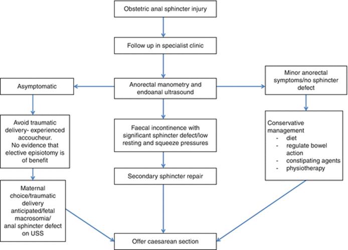
Fig. 9.1
Suggested algorithm for management of subsequent pregnancies after obstetric anal sphincter injury
Generally speaking, women with symptoms of anal incontinence may be offered delivery by caesarean section, since a second vaginal delivery may be associated with a deterioration in symptoms [37]. A study using validated questionnaires and endoanal ultrasonography to evaluate the effect of a second vaginal delivery found that significant sphincter defects were present in 53 % of women after their first delivery and were associated with higher anal incontinence scores. The risk of incontinence was highest in women with a significant sphincter defect who had undergone a second vaginal delivery (39 %) [39]. A further study of women undergoing a second vaginal delivery following a previous forceps delivery found a 26 % risk of developing new or worsening anal incontinence symptoms in women with a significant anal sphincter defect identified prior to the second delivery, although none developed severe symptoms [40]. This implies that women with an asymptomatic defect following obstetric anal sphincter injury may be offered delivery by caesarean section in subsequent pregnancy; however, there is currently no evidence that this will alter outcomes.
Management of Complications of Perineal Trauma
Although the healing process and complications are presented in detail in Chapter 13, we will outline here the salient points on the management of the common complications for ease of reference.
Perineal Pain and Dyspareunia
Perineal pain is common after any degree of perineal trauma and has been reported to affect 92 % of women, resolving by 2 months’ postnatally in the majority of cases [41]. Whilst most perineal pain is self-limiting and manageable with simple analgesia, a small proportion of women will develop longer term symptoms. In the short term, women should be reassured and treated with analgesia as necessary. Perineal trauma is known to be associated with a decrease in sexual function at 6 months postpartum, with second degree tears leading to an 80 % increased risk of dyspareunia and third/fourth degree tears leading to a 270 % increased risk of dyspareunia [42]. Pain, refractory to conservative measures may be addressed with a variety of interventions, but there is no extensive evidence base to support these measures. Local perineal injections with hydrocortisone, marcaine and hyaluronidase are well tolerated and have been reported to lead to a significant fall in pain visual analogue scale scores (from 6.1 to 4.1) and resolution of dyspareunia in 89 % of sexually active women [43], but there is currently no randomised control trial evidence to support these data. There are very little other data in the literature on strategies for long term perineal pain in the obstetric population. Dyspareunia secondary to scarring or tightness at the fourchette following suturing is generally initially treated with dilators and topical oestrogens, but there are no data on outcomes of this intervention. In the presence of obvious scarring and anatomical constriction of the fourchette, women with refractory symptoms may be considered for a surgical revision of the perineum. This will generally involve a longitudinal incision of the scarred area with mobilisation of the underlying tissues and suturing transversely to ensure that a persistent skin bridge does not form. As with other interventions in this patient group, there is little in the literature on outcomes. A prospective study of 9 women who underwent a perineal revision for dyspareunia following perineal trauma reported a significant decrease in pain scores from 6.1 to 0.5 and an increase in coital frequency and satisfaction in 89 % of the cohort [44]. In the presence of associated spasm of the levator muscles, botulinum toxin injections to the levator muscles may also be an effective treatment [45].
Perineal Wound Infection and Breakdown
It is estimated that 11 % of women having had a perineal tear will have a wound infection [46], with prolonged rupture of membranes and instrumental delivery being significant risk factors. Administration of prophylactic antibiotics at the time of third/fourth-degree tear repair has been found to lead to a significantly lower risk of wound infection [47], which is important after a sphincter repair; however, women with second-degree tears are not routinely given antibiotics since there is no proven benefit. Wound infections should be treated with broad spectrum antibiotics including anaerobic cover, unless sensitivities based on wound swabs suggest otherwise. The majority of perineal infections will resolve with a course of antibiotics and good perineal hygiene. Perineal wound breakdown is less common, but can lead to considerable distress and impact on quality of life for affected women. As with other postnatal perineal complications, there is a poor evidence base to guide management. By convention, most practitioners manage the wound expectantly, treating any infection, and awaiting healing by secondary intention. Women should be seen at intervals by an experienced professional to keep the wound under review and provide reassurance. A recent systematic review of suturing versus expectant management for perineal wound breakdown identified only two small randomised trials of poor quality [48]. Only one of these trials evaluated wound healing as a primary outcome measure, and although there was a trend towards better healing in the resuturing group, this did not reach statistical significance and the authors also did not specify how this outcome was quantified. The authors concluded that there is currently insufficient evidence to favour either treatment. An adequately powered randomised trial of these two interventions is underway to evaluate for the primary outcome measure of wound healing, in order to effectively evaluate these management strategies [49].
References
1.
Andrews V, Thakar R, Sultan AH. Structured hands-on training in repair of obstetric anal sphincter injuries (OASIS): an audit of clinical practice. Int Urogynecol J Pelvic Floor Dysfunct. 2009;20(2):193–9.PubMed
2.
Andrews V, Thakar R, Sultan AH. Outcome of obstetric anal sphincter injuries (OASIS): role of structured management. Int Urogynecol J Pelvic Floor Dysfunct. 2009;20(8):973–8.PubMed
3.
National Institute of Health and Care Excellence. CG190: Intrapartum care of healthy women and babies NICE, London, 2014.
4.
Lundquist M, Olsson A, Nissen E, Norman M. Is it necessary to suture all lacerations after a vaginal delivery? Birth. 2000;27(2):79–85.PubMed
5.
Fleming V, Hagen S, Niven C. Does perineal suturing make a difference? The SUNS trial. BJOG. 2003;110(7):684–9.PubMed
6.
Leeman L, Rogers R, Greulich B, Albers L. Do unsutured second-degree perineal lacerations affect postpartum functional outcomes? J Am Board Fam Med. 2007;20(5):451–7.PubMed
7.
Gordon B, Mackrodt C, Fern E, Truesdale A, Ayers S, Grant A. The Ipswich Childbirth Study 1: a randomised evaluation of two stage postpartum perineal repair leaving the skin unsutured. Br J Obstet Gynaecol. 1998;105(4):435–40.PubMed
8.
Kindberg S, Stehouwer S, Hvidman L, Henriksen TB. Postpartum perineal repair performed by midwives: a randomised trial comparing two suture techniques leaving the skin unsutured. BJOG. 2008;115(4):472–9.PubMed
9.
Mackrodt C, Gordon B, Fern E, Ayers S, Truesdale A, Grant A. The Ipswich Childbirth Study 2: a randomised comparison of ployglactin 910 with chromic catgut for postpartum perineal repair. Br J Obstet Gynaecol. 1998;105(4):441–5.PubMed
10.
Bharathi A, Reddy D, Kote G. A prospective randomized comparative study of vicryl rapide verus chromic catgut for episiotomy repair. J Clin Diagn Res. 2013;7(2):326–30.PubMed
11.
Kettle C, Dowswell T, Ismail K. Absorbable suture materials for primary repair of episiotomy and second degree tears. Cochrane Database Syst Rev. 2010;(6):CD000006.
12.
Kettle C, Hills R, Jones P, Darby L, Gray R, Johanson R. Continuous versus interrupted perineal repair with standard or rapidly absorbed sutures after spontaneous vaginal birth: a randomised controlled trial. Lancet. 2002;359(9325):2217–23.PubMed
13.
Kettle C, Dowswell T, Ishmail K. Continuous and interrupted suturing techniques for repair of episiotomy or second-degree tears. Cochrane Database Syst Rev. 2012;(11):CD000947.
14.
Royal College of Obstetricians and Gynaecologists. The Management of Third- and Fourth-Degree Perineal Tears, Green-top Guideline No. 29. RCOG, London, June 2015.
15.
Nordenstam J, Mellgren A, Altman D, Lopez A, Johansson C, Anzen B, Li Z, et al. Immediate or delayed repair of obstetric anal sphincter tears- a randomised controlled trial. BJOG. 2008;115(7):857–65.PubMed
16.
Norderval S, Oian P, Revhaug A, Vonen B. Anal incontinence after obstetric sphincter tears: outcome of anatomic primary repairs. Dis Colon Rectum. 2005;48(5):1055–61.PubMed
17.
Fernando R, Sultan A, Kettle C, Thakar R, Radley S. Methods of repair for obstetric anal sphincter injury. Cochrane Database Syst Rev. 2013;(12):CD002866.
18.
Sultan A, Monga A, Kumar D, Stanton S. Primary repair of obstetric anal sphincter rupture using the overlap technique. BJOG. 1999;106:318–23.
19.
Eogan M, Daly L, Behan M, O’Connell P, O’Herlihy C. Randomised clinical trial of a laxative alone versus a laxative and a bulking agent after primary repair of obstetric anal sphincter injury. BJOG. 2007;114(6):736–40.PubMed
20.
Dodd J, Heydayati H, Pearce E, Hotham M, Crowther C. Rectal analgesia for the relief of perineal pain after childbirth: a randomised controlled trial of diclofenac suppositories. BJOG. 2004;111(10):1059–64.PubMed
21.
Farrell S, Flowerdew G, Gilmour D, Turnbull G, Schmidt M, Baskett T, Fanning C. Overlapping compared with end-to-end repair of complete third-degree or fourth-degree obstetric tears: three year follow up of a randomised controlled trial. Obstet Gynecol. 2012;120(4):803–8.PubMed
22.
Fitzpatrick M, Behan M, O’Connell P, O’Herlihy C. A randomised clinical trial comparing primary overlap with approximation repair of third-degree obstetric tears. Am J Obstet Gynecol. 2000;183(5):1220–4.PubMed
23.
Fernando R, Sultan A, Kettle C, Radley S, Jones P, O’Brian P. Repair techniques for obstetric anal sphincter injuries: a randomized controlled trial. Obstet Gynecol. 2006;107(6):1261–8.PubMed
24.
Vaccaro C, Clemons J. Anal sphincter defects and anal incontinence symptoms after repair of anal sphincter lacerations in primiparous women. Int Urogynecol J Pelvic Floor Dysfunc. 2008;19(11):1503–8.
25.
Laine K, Skjeldestad F, Sanda B, Horne H, Spydslaug A, Staff A. Prevalence and risk factors for anal incontinence after obstetric anal sphincter rupture. Acta Obstet Gynecol Scand. 2011;90(4):319–24.PubMed
26.
Abramowitz L, Sobhani I, Ganansia R, Vuagnat A, Benifla J, Darai E, et al. Are sphincter defects the cause of anal incontinence after vaginal delivery? Results of a prospective study. Dis Colon Rectum. 2000;43(5):590–6.PubMed
27.
Fitzpatrick M, Cassidy M, O’Connell P, O’Herlihy P. Experience with an obstetric perineal clinic. Eur J Obstet Gynecol Reprod Biol. 2002;100(2):199–203.PubMed
28.
Sultan A, Kamm M, Talbot I, Nicholls R, Bartram C. Anal endosonography for identifying external sphincter defects confirmed histologically. Br J Surg. 1994;81(3):463–5.PubMed
29.
Faltin D, Boulvain M, Irion O, Bretones S, Stan C, Weil A. Diagnosis of anal sphincter tears by postpartum endosonography to predict fecal incontinence. Obstet Gynecol. 2000;95(5):643–7.PubMed
30.
Starck M, Bohe M, Valentin L. The effect of endosonographic anal sphincter defects after primary repair of obstetric sphincter tears increases over time and is related to anal incontinence. Ultrasound Obstet Gynecol. 2006;27(2):188–97.PubMed
31.
Mahony R, Behan M, Daly L, Kirwan C, O’Herlihy C, O’Connell P. Internal anal sphincter defect influences continence outcome following obstetric anal sphincter injury. Am J Obstet Gynecol. 2007;196(3):217.e1–5.
32.
Oberwalder M, Dinnewitzer A, Baig M, Thaler K, Cotman K, Nogueras J, et al. The association between late onset fecal incontinence and obstetric anal sphincter defects. Arch Surg. 2004;139(4):429–32.PubMed
33.
Johnson E, Carlsen E, Steen T, Backer Hjorthaug J, Eriksen M, et al. Short- and long-term results of secondary anterior sphincteroplasty in 33 patients with obstetric injury. Acta Obstet Gynecol Scand. 2010;89(11):1466–72.PubMed
34.
Kenefick N, Vaizey C, Nicholls R, Cohen R, Kamm M. Medium term results of permanent sacral nerve stimulation for faecal incontinence. Br J Surg. 2002;89(7):896–901.PubMed
35.
Jarrett M, Dudding T, Nicholls R, Vaizey C, Cohen R, Kamm M. Sacral nerve stimulation for faecal incontinence related to obstetric anal sphincter damage. Dis Colon Rectum. 2008;51(5):531–7.PubMed
36.
Brouwer R, Duthie G. Sacral nerve neuromodulation is effective treatment for fecal incontinence in the presence of a sphincter defect, pudendal neuropathy, or a previous sphincter repair. Dis Colon Rectum. 2010;53(3):273–8.PubMed
37.
Fynes M, Donnelly V, Behan M, O’Connell P, O’Herlihy C. Effect of second vaginal delivery on anorectal physiology and faecal continence. Lancet. 1999;354(9183):983–6.PubMed
38.
Sultan A, Thakar R. Third and fourth degree tears. In: Sultan A, Thakar R, Fenner D, editors. Perineal and anal sphincter trauma. London: Springer; 2007. p. 33–51.
39.
Faltin D, Sangalli M, Roche B, Floris L, Boulvain M, Weil A. Does a second delivery increase the risk of anal incontinence? BJOG. 2001;108(7):684–8.PubMed
40.
Mahony R, Behan M, O’Connell P, O’Herlihy C. Effect of second vaginal delivery on anal function in patients at risk of occult anal sphincter injury after first forceps delivery. Dis Colon Rectum. 2008;51(9):1361–6.PubMed
41.
Andrews V, Thakar R, Sultan A, Jones P. Evaluation of postpartum perineal pain and dyspareunia: a prospective study. Eur J Obstet Gynecol Reprod Biol. 2008;137(2):152–6.PubMed
42.
Signorello L, Harlow B, Chekos A, Repke J. Postpartum sexual functioning and its relationship to perineal trauma: a retrospective cohort study of primiparous women. Am J Obstet Gynecol. 2001;184(5):881–8.PubMed
43.
Doumouchtsis S, Boama V, Gorti M, Tosson S, Fynes M. Prospective evaluation of combined local bupivacaine and steroid injections for the management of chronic vaginal and perineal pain. Arch Gynecol Obstet. 2011;284(3):681–5.PubMed
44.
Woodward A, Matthews C. Outcomes of revision perineoplasty for persistent postpartum dyspareunia. Female Pelvic Med Reconstr Surg. 2010;16(2):135–9.PubMed
45.
Romito S, Bottanelli M, Pellegrini M, Vicentini S, Rizzuto N, Bertolasi L. Botulinum toxin for the treatment of genital pain syndromes. Gynecol Obstet Invest. 2004;58(3):164–7.PubMed
46.
Johnson A, Thakar R, Sultan A. Obstetric perineal wound infection: is there under-reporting? Br J Nursing. 2012;21(5):S28. S30, S32-5.
47.
Duggal N, Mercado C, Daniels K, Bujor A, Caughey A, El-Sayed Y. Antibiotic prophylaxis for prevention of postpartum perineal wound complications: a randomized controlled trial. Obstet Gynecol. 2008;111(6):1268–73.PubMed
48.
Dudley L, Kettle C, Ismail K. Secondary suturing compared to non-suturing for broken down perineal wounds following childbirth. Cochrane Database Syst Rev. 2013;(9):CD008977.
49.
Dudley L, Kettle C, Carter P, Thomas P, Ismail K. Perineal re-suturing versus expectant management following vaginal delivery complicated by a dehisced wound (PREVIEW): protocol for a feasibility and pilot randomised controlled trial. BMJ Open. 2012;2(4). pii: e001458.