
From the Old to the New
Oded Langer, MD, PhD
Doctors pour drugs of which they know little, To cure diseases of which they know less, Into patients of whom they know nothing.
—Voltaire
Key Points
• Insulin therapy should mimic as much as possible the physiological pattern of insulin secretion.
• There are different patterns of insulin requirements for gestational diabetes mellitus (GDM; bi-phasic) and pre-existing diabetes (tri-phasic).
• Regular human insulin, despite improved purity and stability, does not successfully imitate physiological insulin secretion.
• Regular human insulin should be administered 30 to 45 minutes prior to eating; its peak effect occurs 2 to 4 hours after the injection and its duration of action lasts 6 to 8 hours.
• Short-acting insulin analogs successfully imitate physiological insulin secretion. They are absorbed quickly, achieving peak plasma concentrations about twice as high and within approximately half the time compared to structurally unchanged insulin.
• In GDM women, fasting plasma glucose of >95 mg/dL is the threshold for insulin initiation; insulin dose is unlimited.
• Self-monitoring blood glucose is a prerequisite for insulin therapy.
INTRODUCTION
In 1889, von Mehring and Minkowski identified the pancreas as the origin of diabetes mellitus. Thirty-two years later, Banting, Best, Collip, and Macleod successfully extracted and introduced the active factor, insulin, and soon followed with its administration to a young patient in Canada. The stage was set for a new era of diabetes care and research with the ultimate goal of disease prevention and/or discovery of a cure. Since the breakthrough of the source of this complex metabolic disorder, there have been major developments in insulin production, refinement, pharmaceutical formulation, and methods of delivery. However, the persistent complications that lead to morbidity and mortality in patients with GDM and type 1 and type 2 diabetes are an unrelenting reminder that we may have won many of the major therapeutic and management battles but are still far from winning the war.1-3
There are approximately 120 to 150 million people worldwide who suffer from diabetes. According to the World Health Organization (WHO), in some parts of Africa, the mortality rate from insulin-dependent diabetes is estimated at 50% in the first five years of the illness. In many African countries, insulin-dependent diabetes is a death sentence, since the insulin may be unavailable or beyond the purchasing power of the patient's family. In Russia, access to insulin is restricted. Under Communism, the Russian medical services supplied medicine including insulin to people who needed it. When Communism collapsed and free enterprise moved in, the situation in Russia changed, that is, only people with money could afford insulin. According to a 1997 International Diabetes Federation Task Force survey, “15 countries (out of 73 countries surveyed) have severe problems with access to insulin,” www.diabetesinterview.com (April 2000). The main barriers to insulin access appear to be affordability, distribution, and transportation. Another major barrier includes the luxury tax laws and war. Although the WHO has listed insulin as an “essential drug,” and, therefore, should not be taxed as a luxury item, several countries still tax insulin, for example, the Philippines. Obtaining insulin and proper diabetes care in a poor country is a major challenge for diabetic sufferers. However, most authoritative bodies and researchers neglect to address the drain that this disease causes on family resources, www.diabetesinterview.com (April 2000).
THE PHYSIOLOGY OF INSULIN
Insulin is produced in the beta cells of the islets of Langerhans as a single polypeptide precursor, preproinsulin, which is then converted to proinsulin and 86-amino acid polypeptide. Proinsulin forms equimolar amounts of insulin and C peptide (i.e., connecting peptide) through the removal of 4-amino acid residues. The resulting insulin consists of a 20-amino acid A chain and a 31-amino acid B chain connected by two disulfide bridges, with the addition of a third disulfide bridge within the A chain. The final product that is released from the beta cells into the portal venous system is 90% to 97% insulin with an equimolar amount of C peptide. Proinsulin and the intermediates of change comprise the remainder.4
The liver removes about 50% of the insulin that is released into the portal system during its first passage.5 However, since the amount the liver extracts is variable, estimating the rates of insulin secretion based on peripheral venous concentrations is inexact. C-peptide is primarily cleared by the kidney, and this has, under various conditions, provided the precise quantification of insulin secretion rates. Insulin secretion into the portal system occurs in the basal state at a rate of about 1 U/h in normal adults. A 5- to 10-fold increased rate of insulin secretion occurs with the intake of food. The total daily secretion of insulin is approximately 40 U.5
Insulin secretion rates in either the fasting or the postprandial state decrease rapidly to prevent hypoglycemia with moderate exercise. With strenuous exercise, hyperglycemia may occur and result in enhanced secretion of insulin in the postexercise period. Therefore, insulin's secretory responses to physiologic stimuli are multifaceted and present a challenge to care providers and researchers trying to duplicate them with available therapeutic regimens.6-8
In general, exercise helps diabetic patients improve their ability to metabolize glucose. But for those people who are not inclined to use exercise machines such as the treadmill or Stairmaster, the exercise involved in walking downhill leads to even bigger improvements than hiking up! A study conducted in Austria found that both forms of exercise improved glucose tolerance but that the downhill hike had a greater effect—a 25% change in tolerance compared with 9% after the stint of uphill climbing. This finding may be explained by the fact that eccentric exercise (muscle cells are lengthened as they resist a force) may increase blood flow more than concentric (muscle cells shorten to exert force on an object) exercise.9
A Finnish study found that physical activity on the job was as effective as leisure-time workouts in reducing deaths from heart disease among patients with type 2 diabetes. When the survey data were adjusted to account for factors such as age and weight, researchers found that having an active job or participating for more than three hours a week of a demanding leisure-time activity cut the risk of death from heart disease by approximately 33%. However, since more and more jobs are sedentary, the researchers recommended exercising during work breaks, walking more on the job, or walking to work.10
INSULIN ANALOGS
The development of drugs is driven by the goal to correct pathophysiology to approximate normal physiology. Normal secretion of insulin includes a basal stage that prevents excessive hepatic production and mobilization of free fatty acids from the adipose tissue stores. This basal insulin is needed regardless of meal pattern and patient physical activity. The second component in insulin physiology is related to insulin secretion in association with meals. Ninety percent of nutrients are absorbed within 90 minutes after a meal. After two hours, the plasma glucose and insulin levels return to normal premeal values.11
Several generations of insulin have been developed since its original discovery. Today the most commonly used are human and insulin analogs. Regular human insulin, despite improved purity and stability, is not entirely successful in imitating physiological insulin secretion. It should be administered 30 to 45 minutes prior to ingestion of a meal; its peak effect occurs two to four hours after the injection, and its duration of action lasts for six to eight hours. The pharmacokinetics following subcutaneous injection of regular or soluble human insulin preparations makes it difficult to achieve day-long normoglycemia. This low rise to peak insulin concentration is likely to account for much of the observed hyperglycemia following meals in people with diabetes. The delay in the absorption of subcutaneously administered structurally unchanged insulin is due to the fact that in this preparation, insulin tends to associate in “clusters” of six molecules (hexamers), and time is needed after injection for these clusters to dissociate to single molecules that can be used by the body.12 There is often nonadherence to the insulin protocol due to the inconvenience and limitations of injecting approximately a half hour prior to a meal so the patient ultimately takes the insulin with the meal (Table 16-1).13
Considerable research in the past two decades has been devoted to the development and improvement of insulin analogs with pharmacokinetic profiles that differ from those of existing insulin preparations. Insulin lispro is an analog of human insulin (HumalogP) in which the amino acids praline and lysine, which occupy B28 and B29 positions, respectively, are interchanged. Although insulin lispro, like soluble insulin, forms hexamers, they dissociate more rapidly following subcutaneous injection.14
In the short-acting insulin analog aspart (NovoRapidR), praline at position 29 of the B-region is replaced by aspartic acid. The change reduces the stability of the interactions within the hex- amer, thereby increasing absorption of the insulin after injection.15 Aspart can be administered shortly before a meal but with the addition of basal insulin to preclude late postprandial and fasting hyperglycemia. Studies of aspart involving nonpregnant type 1 diabetic patients reported decreased postprandial glucose concentrations, a reduction in nighttime hypoglycemia, and overall improvement in glycemic control and enhanced patient satisfaction. In addition, hemoglobin A1C values were lower with comparable frequency of adverse events as with the use of regular insulin.16,17
Short-acting insulin analogs are, therefore, absorbed more quickly, achieving peak plasma concentrations about twice as high and within approximately half the time compared to structurally unchanged insulin.18,19 These analogs facilitate lower glucose levels after meals18,20 and should enhance overall glycemic control. Haffner proposed that lowering postprandial glucose levels may be associated with a decreased risk for developing cardiovascular complications in diabetes.21 One suggested advantage of short-acting insulin analogs is the possibility to inject insulin immediately before meals, even if in daily life, most diabetic patients seem to use short- or even no injection-meal interval.20 Further proposed advantages in terms of quality of life are changes in injection modes with the possibility of injecting short-acting insulin analogs after meals.22
TABLE 16-1 Action Profile of Commonly Used Insulins
|
Type |
Onset of Action |
Peak of Action (h) |
Duration of Action (h) |
FDA Pregnancy Classification |
Cross Placenta |
Excreted in Breast Milk |
|
Insulin Lispro |
1-15 min |
1-2 |
4-5 |
B |
Minimal |
No |
|
Insulin aspart |
1-15 min |
1-2 |
4-5 |
B |
Minimal |
No |
|
Regular insulin |
30-60 min |
2-4 |
6-8 |
B |
Yes |
Yes |
|
Isophane insulin Suspension |
1-3 h |
5-7 |
13-18 |
B |
Unknown |
No |
|
Insulin zinc Suspension |
1-3 h |
4-8 |
13-20 |
B |
Unknown |
No |
|
Extended insulin Zinc suspension |
2-4 h |
8-14 |
18-30 |
B |
Unknown |
No |
|
Insulin glargine |
1 h |
No peak |
24 |
C |
Minimal |
Unknown |
|
Insulin detamir |
1 h |
No peak |
14 |
C |
Unknown |
Unknown |
Treatment with the two short-acting insulin analogs (Lispro— HumalogR; Aspart-Novo RapidR) currently available on the market is promoted with purported advantages with respect to metabolic control and reduced incidence of hypoglycemic episodes for patients with type 1 or type 2 diabetes, diabetic children, and diabetic pregnant women.23-25 On the other hand, another study failed to show a positive effect on overall blood glucose levels when short-acting insulin analogs were compared with structurally unchanged insulin.26 In the case of hypoglycemic episodes, two other studies reported contradictory results with respect to hypoglycemic episodes.27,28 Insulin treatment strategies where short-acting insulin analogs can be used include intensified insulin therapy (short-acting insulin before meals, basal insulin at bedtime or twice daily, including adjustment of insulin dose based on carbohydrate intake) or conventional insulin therapy (basal or premixed insulin up to three times daily with or without oral hypoglycemic agents). Only patients treated with continuous subcutaneous insulin infusion (CSII) performing intensified insulin therapy showed a significant decrease in HbA1C when short-acting insulin analogs were used in the nonpregnant state.29,30 Insulin analogs are more expensive than structurally unchanged insulin, and in the year 2000, insulin lispro and insulin aspart had a 30% share of the market for short-acting insulin in most developed countries.
Insulin lispro is 1.6-fold more potent than human insulin in binding to human placental insulin-like growth factor I (IGF-I) receptors. Both have 0.2% of the binding capacity of IGF-I itself.31 Structural homology of insulin analogs to IGF-I has caused concern regarding the progression of late complications and potential mitogenic (induction of cell division) effects, especially with long-term use of insulin analogs. IGF-I may affect the progression of retinopathy,32,33 and certain modified insulin analogs have shown a carcinogenic effect in the mammary glands in female rats34 or mitogenic potency in osteosarcoma cells.31 Despite the potentially adverse properties of insulin analogs, very limited data on long-term safety are currently available, mainly because patients with clinically relevant microvascular complications have been excluded from most clinical trials. In summary, to prevent adverse outcomes such as congenital malformations and macro- somia, it is recommended that patients be placed on a regimen of daily multiple injections of rapid- and long-acting insulin, or an external insulin pump, together with intensive blood glucose self-monitoring. Rapid-acting insulin analogs such as lispro and aspart are now widely used during pregnancy in women with type 1 diabetes with demonstrated efficacy and safety.35,36
INTERMEDIATE- AND LONG-ACTING INSULIN
Evaluation of the teratogenicity of a medicinal product in humans requires a sample size large enough to show an increase in the occurrence of rare events. If the risk of malformation in a given population is only 3%, then at least 220 to 240 pregnancies need to be analyzed to detect a two- to threefold increase with a power of 80%.37 The use of long-acting insulin analogs (glargine and leve- mir) is currently not recommended during pregnancy. However, since treatment with glargine can facilitate good glycemic control with a reduced risk of hypoglycemia, it may be a valuable alternative in the management of pregnant women with type 1 diabetes. Moreover, since many patients with type 1 diabetes are usually already being treated with short- and long-acting insulin analogs, they may be reluctant to change their insulin regimen when planning a pregnancy if their diabetes is already well controlled.
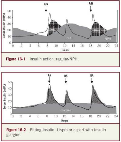
Intermediate- and long-acting insulin are components of the insulin algorithm in the care of patients. The neutral protamine hage- dorn insulin (NPH) is more commonly used than the lente and the ultra-lente insulin in pregnancy mainly because its absorption pattern and duration are more accurate. However, the 24-hour NPH concentration pattern is not ideal and resembles a bimodal distribution; therefore, it cannot create a stable monotonous basal level throughout the day (Figures 16-1 and 16-2).
Insulin glargine and detemir are long-acting insulin analogs that were developed to mirror the basal pancreatic insulin secretion. However, neither has been tested in pregnancy for placental transfer and effect on the fetus; they are not currently recommended in pregnancy. Glargine has high IGF qualities. It has been reported to have 6.5 times more potency than human insulin in binding the IGF receptors.31 In a malignant cell line, increased mitogenicity was found with glargine versus regular insulin. Finally, a case report suggested that prolonged use of the drug (more than a year) may be associated with progression in retinopathy.38 Insulin glargine is an analog in which glycine is substituted for aspartic acid at position 21 on the alpha chain and two basic argenines are added to the C-terminus of the beta chain. The addition of zinc to the molecule results in stabilization of the hexamer (prolongation of molecule action), a decrease in absorption, and increase in association rates. In contrast to NPH, the glargine has a stable monotonous basal profile that minimizes the peaks and valleys in the former insulin. Thus, it has been suggested that the use of glargine results in decreased hypoglycemic episodes when used as part of the insulin administration algorithm in conjunction with lispro or aspart insulin. In the nonpregnant state, the use of glargine demonstrated a decrease in fasting glucose levels, hemoglobin A1C, and nocturnal hypoglycemia.39-40 Use of glargine in pregnancy may be promising since it has the potential to decrease nocturnal hypoglycemic episodes common with tight glycemic control in type 1 pregnant diabetics. Further studies are needed to test the safety for mother and fetus before recommending its use in pregnancy. This is especially significant since the alteration of the insulin molecule for the creation of the insulin analog results, at least in the case of lispro, in placental transfer that is dose dependent.38,41
Reports on animal studies have demonstrated the safety of insulin glargine during pregnancy.42-45 Studies46 have reported the perinatal outcomes in 102women with type 1 diabetes treated with insulin glargine before conception and throughout pregnancy. The observed rate of large-for-gestational-age (LGA) infants (30%) compares favorably with the rate of macrosomia seen in infants of women treated with human insulin and is in accordance with a recent pilot study showing that the use of insulin glargine is not associated with an increased risk of fetal macrosomia. However, the current study has certain important limitations that need to be taken into account when interpreting the results. It has the inherent weaknesses of all observational studies, that is, recall bias and the absence of matching. There is also a potential for selection bias, as it involved self-selected rather than population-based centers. Furthermore, data on maternal age, body mass index, duration of diabetes, socioeconomic status, and occurrence of severe hypoglycemia were not included in the data collection process.47 Some concerns, however, have been raised over the use of glargine during pregnancy since the analog exhibits an increased affinity for the IGF-1 receptor, a tendency that is not seen with human insulin.48
Insulin detemir is modified by the addition of 14-carbon fatty acid side chain at position 29 of the beta chain that enhances aggregation and delays absorption.49 The action profile of detemir is close to the physiological profile of insulin regarding its baseline characteristics. The drug does neither peak like NPH nor is it flat like glargine. In addition, it has been demonstrated that there is a fivefold decrease in potency in binding the IGF receptor in comparison to regular insulin. Many consider it unwise to administer glargine or detemir in the management of diabetes in pregnancy.31,49 In a randomized study,50 the authors sought to compare the efficacy and safety of insulin detemir (IDet: n = 139) with neutral protamine Hagedorn (NPH: n = 145), both with insulin aspart, in pregnant women with type 1 diabetes. They concluded that IDet is as well tolerated as NPH as regards perinatal outcomes in pregnant women with type 1diabetes and no safety issues were identified. But the study also reported 20 cases of early fetal losses and three perinatal deaths. Sixteen children had malformations (IDet: n = 8/142, 5.6%; NPH: n = 8/145, 5.5%).
In the nonpregnant state, short- and long-acting insulin analogs have been shown to result in better glycemic control with less hypoglycemia than human insulin in subjects with diabetes. Recently, a randomized trial showed similar benefits with short-acting analogs in pregnancy complicated by type 1 diabetes.36 In contrast, few studies have examined the efficacy and safety of long-acting analogs in women with type 1 diabetes during pregnancy, despite their increasing use in this subject group. Consequently, basal insulin analogs have been used off-label.51,52
Insulin detemir is an insulin analog that has a consistent phar- macokinetic/pharmacodynamic profile with lower intrasubject variability in terms of glucose-lowering effect compared with either NPH or insulin glargine in subjects with type 1 or type 2 diabetes. Studies have shown that IDet provides similar glycemic control, but with lower rates of hypoglycemia and less weight gain than NPH insulin in nonpregnant subjects with type 1 or type 2 diabetes. The aim of the study was to compare the efficacy and safety of IDet with NPH in pregnant women with type 1 diabetes. The report presented primary data on perinatal and obstetric pregnancy outcomes. Data on glycemic control, maternal hypoglycemia, and maternal safety were reported separately.53 The drug has been shown in previous studies to have a consistent glucose-lowering effect and one that was similar in magnitude to that seen with NPH insulin in nonpregnant subjects with type 1 or type 2 diabetes.54
In a recent study,55 the objective was to compare glycemic control and pregnancy outcome in women with type 1 diabetes treated with long-acting insulin analogs detemir or glargine. One offspring in each group was born with a major congenital malformation. However, in the majority of women on detemir, this treatment was initiated after the organogenesis, whereas only a small group was on detemir before conception. The authors concluded that pregnancy outcomes were comparable in women using insulin detemir or glargine. The incidence of severe hypoglycemia was comparable 23% versus 23% (P = .98). Preeclampsia 14% versus 18%, P = .52; lower prevalence of LGA infants in women on glargine 49% versus 30% (P = .046); hemoglobin A1C was comparable at eight weeks (median 6.6% [range 5.6-9.8] vs. 6.8% [5.4-10.1] and at 33 weeks gestation (6.1% [5.1-7.6] vs. 6.2% [4.8-7.2]). Of note, there was slight improvement in small changes (8%) in the level of glycemic control; however, the threshold used was higher than the recommended threshold for prevention of fetal macrosomia (<100 mg/dL, HbA1c 5.5%). The overall results of current studies appear reassuring for the continued use of insulin analogs.56,57 Another concern has been the increased affinity of insulin glargine for the IGF-1 receptor and, therefore, potential for increasing the risk of excessive fetal growth; this is in contrast to insulin detemir. Surprisingly, results showed a decreased risk of LGA infants in the glargine group compared with the detemir group. The prevalence of LGA infants in the detemir group of 49% is comparable with that generally reported for infants of mothers using human intermediate NPH insulin during pregnancy.58-60 The prospective randomized study comparing insulin detemir to human intermediate NPH insulin demonstrated no significant differences in the prevalence of LGA infants between the insulin detemir and the human intermediate NPH insulin groups. The incidence of LGA infants reported in the majority of former studies on the use of insulin glargine during pregnancy ranged from 30% to 47%. A single study of 37 pregestational diabetics treated with insulin glargine during pregnancy reported a decreased risk of LGA infants compared with women treated with human intermediate NPH insulin (19% vs. 50%).43,45,47,61,62
PREGNANCY AND THE USE OF INSULIN ANALOGS
In pregnancy, data on insulin lispro are limited and abstracted from studies with relatively small sample sizes. Most of these reports demonstrated an improvement in glycemic control and enhanced quality of patient lifestyle. Human insulin became widely available in the 1980s when the preferred method of production was recombinant DNA technology. This led to the availability of mutant insulin (insulin analogs) during the mid- 1990s that were designed primarily to have improved pharmacokinetic features for subcutaneous administration.63 Human insulin is recommended when insulin is prescribed in pregnancy since the use of insulin analogs has not been adequately tested in GDM.64
The comparison between lispro and human insulin needs to address not only placental transfer but also the efficacy of one drug over the other as well as the cost/benefit ratio. There is little or no difference between insulin lispro, insulin aspart, and human insulin in receptor binding and metabolic and mitogenic potency with a slightly increased binding of insulin lispro to the receptor for IGF-1.31 Mounting evidence of the beneficial effects of insulin lispro in type 1 and type 2 nonpregnant diabetic subjects includes decreased frequency of severe hypoglycemic episodes, limited postprandial glucose excursions, and a possible decrease in glycosylated hemoglobin when the drug is administered by continuous subcutaneous infusion.31 In addition, insulin lispro provides greater convenience in the timing of administration (analogs administered up to 15 minutes after start of a meal compared to soluble insulin taken 30 minutes before the meal), patient satisfaction, and reduction in hypoglycemic episodes. The omission of maternal/fetal outcome data in the majority of these studies limits the ability to draw any firm conclusions about the efficacy of insulin lispro in comparison to human insulin.
Controversy persists on the association of proliferative retinopathy with the insulin lispro. In a cohort of 10 women, with type 1 or type 2 diabetes who had been retinopathy free prior to pregnancy, 3 women developed proliferative retinopathy that required laser therapy during the third trimester. Several authors have debated these findings.38,63-76 In one study, 16 women were treated with insulin lispro and the remainder with regular insulin. None of the lispro patients had any ophthalmo- logical changes during pregnancy.65 In another study of pregnant type 1 diabetic women, 12 were treated with insulin lispro and 42 with regular insulin. None of the lispro-treated patients showed ophthalmological changes.64 The development of diabetic retinopathy with the use of insulin lispro remains questionable. However, the likelihood is that patients’ level of glycemia and not the drug is responsible for the proliferative retinopathy. A randomized trial with a substantial sample size may provide a definitive answer.
DOES INSULIN CROSS THE PLACENTA?
Academic scientists acknowledge that they often get things wrong. However, they also recognize that these errors sometimes get corrected over time since there are other researchers pursuing the same issues. However, the literature will confirm that this assumption for self-correction is often false. There are errors and misinterpretations of data in a lot more of the scientific papers being published and the guidelines emanating from them than anyone would normally suppose. For example, The Practice Bulletin no 137, August 2013, declared that insulin does not cross the placenta. The results of the above declarative statement give practitioners false confidence that they can freely administer insulin with potentially no harm to the fetus.
Various factors contribute to the problem of prolific distribution of questionable study results. There is a lot of scientific research that is poorly thought through, or executed, or both. Statistical mistakes are widespread, and most scientists are not statisticians. Some scientists use inappropriate techniques because those are the ones they are comfortable with; others experiment with new ones without understanding their refinements. Some just rely on the methods built into their software, even if they don’t understand them. The peer reviewers who evaluate papers before journals commit to publishing them are much worse at spotting mistakes than they or others appreciate. Professional pressure, competition, and ambition pressure scientists to publish more quickly than is prudent. The medical academic career structure that lays great pressure on publishing numerous papers aggravates all these problems. There is an academic price to pay for not getting published. The pervasive bias favors publication of claims to have found something new. By and large, scientists want surprising results especially when they are able to use these new data to reinforce current opinions and dogma.
Bauman and Yalow77 demonstrated that beef and pork insulin unilaterally do not cross the placenta; they will, however, cross when complexed to insulin antibodies. Anecdotal cases have reported the presence of congenital anomalies with the use of insulin analogs. However, this unlikely finding still queries if insulin lispro and other analogs cross the placenta. Others found that in 51 infants of type 1 diabetic mothers, both animal and human, insulin crossed the placenta. The transfer was directly related to the level of anti-insulin antibodies in the mother.78 In another study of 19 GDM women treated with insulin lispro, 4 of the subjects received intravenous infusion of the drug in labor; insulin lispro was not detected in the umbilical cord blood of their infants.79 Holcberg et al.80 using a perfusion model, reported that insulin lispro does not cross the human placental membranes at low concentrations. The maternal steady-state concentration reached 48 ± pU in the maternal artery and 28 ± 1 pU in the maternal vein, while in the fetal site insulin lispro was not detected. However, the concentration of insulin lispro in placental tissue was 1836 ± 220 pU. It has been shown that human insulin concentration in the fetus corresponds to peak serum insulin levels after doses of 14, 24, 104, and 278 units. This maternal dose is relatively common in the majority of GDM and type 2 women.78 Insulin antibodies were detected in the cord blood of 95% of the offspring at birth. Others concluded that there was no appreciable transplacental transfer of either aspart or actarapid.81,82 Boskovic et al.41 evaluated 11 term human placentas from uncomplicated pregnancies immediately after delivery. Insulin lispro, at concentrations ranging from 100 to 1000 micro U/mL, was introduced into the maternal reservoir. The maternal side of the placenta was perfused with a constant concentration of lispro insulin; the fetal circulation was closed. Samples were drawn from both the maternal and the fetal circulations at regular intervals. The appearance of insulin lispro in the fetal circulation was analyzed by a specific radioimmunoassay. No placental transfer of lispro could be detected during perfusion with 100 and 200 pU/mL. In contrast, there was a concentration-dependent transfer to the fetus at >580 pU/mL, which corresponds to a maternal dose of approximately 75 units. This dose of insulin lispro is quite common in type 2 and obese GDM. Finally, they compared serum level and administered doses of lispro. Mothers treated with 50 units of insulin lispro achieved serum concentrations >200 µU/mL with an apparent linear correlation between dose and levels. The rate of placental transfer was 0.019 pU x min (-1) x g tissue(-1) at maternal levels of 580 µU/mL and 0.045 pU x min(-1) x g(-1) tissue at maternal levels of 1000 pU/mL. Moreover, a dose of 50 units may achieve serum concentrations >200 µU/mL with apparent linear correlation between dose and levels.41 The author concluded that insulin lispro is not likely to cross the placenta at a single-standard dose and unlikely to reach or harm the unborn baby. However, they never took into consideration that the average type 2 GDM or type 2 diabetic women receive much higher doses of insulin than the authors assumed and, therefore, the fetus is exposed to insulin lispro in the majority of cases. Pollex et al.83 reported that glargine in therapeutic concentration levels is not likely to cross the placenta based on a relatively low maternal dose. These studies clearly demonstrate that insulin crosses the placenta in a dose-dependent pattern.
Finally, the Cochrane Reviewers summarized their findings84:
Our analysis suggests only a minor benefit of short acting insulin analogs in the majority of diabetic patients treated with insulin. Until long-term efficacy and safety data are available, we suggest a cautious response to the vigorous promotion of insulin analogs. Due to fears of potentially carcinogenic and proliferative effects, most studies to date have excluded patients with advanced diabetic complications. For safety purposes, we need a long-term follow-up of large numbers of patients who use short acting insulin analogs. Furthermore, we need well-designed studies in pregnant women to determine the safety profiles for both the mother and the unborn child.
We need to refrain from using the expression “unlikely to cross and/or harm the unborn fetus” because we are fostering misinformation. On the other hand, we need to remember that the majority of drugs cross the placenta. Thus, the question is not so much which drug crosses but which drug crosses and causes harm to the fetus?
THERAPEUTIC INSULIN REQUIREMENTS IN PREGESTATIONAL DIABETES
Type 1 diabetes is characterized as an inability to secrete insulin and type 2 diabetes and GDM have impaired insulin secretion and resistance, in addition to the physiological insulin resistance of pregnancy. The insulin requirements are not evidence of “physiological” need in diabetic patients but rather a reflection of the therapeutic dose of insulin required to achieve the established levels of glucose targeted by the physician.
Several prerequisites need to be addressed on the subject of therapeutic insulin requirements in pregnancy are as follows:
1. What level of glycemia does the care provider want to target? Since the normal glycemic profile in the nondiabetic pregnant woman is different than that of clinical thresholds targeted to optimize pregnancy outcome, different thresholds will require varying doses of insulin.
2. Is the method used to assess the level of glycemia accurate enough to reflect the true glycemic profile in the diabetic patient? Weekly blood glucose and HbA1C are not sensitive enough to demonstrate this association and only multiple glucose determinations throughout the day can provide the answer. Normal daily conditions rather than the hospital environment will provide the true level of glycemia that will mandate the insulin dose.85,86
Several studies have evaluated insulin requirements in pre-GDM.87-89 The sensitivity to insulin changes throughout pregnancy. In general, during the first trimester and even in the first half of the second trimester, there is a state of increased insulin sensitivity mainly due to the increased level of estrogen. Thereafter, due to the increased level of progesterone, human placental lactogen, prolactin, and other factors, there is a state of increased insulin resistance that, in turn, results in the need for increased insulin throughout pregnancy.
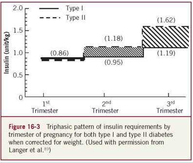
Jovanovic and Peterson90 reported that there are increased insulin needs throughout pregnancy, 0.7 units/kg/d in the first trimester (weeks 5-12); 0.8 units/kg/d at week 18, 0.9 units/kg/d at week 26, and 1.0 units/kg/d at 36 weeks gestation. We89 analyzed insulin requirements of type 1 and type 2 diabetic women using memory-based reflectance meters (SMBG) that reassured the accuracy of the glycemic control in all study participants. In both type 1 and type 2 diabetic subjects, we reported a triphasic insulin pattern. In comparison with type 1 patients whose daily insulin requirements were first trimester: 0.86 units/kg/d; second trimester: 0.95 units/kg/d; and third trimester: 1.19 units/kg/day, type 2 women required higher insulin doses during each trimester (first trimester: 0.86 units/kg/d, second trimester: 1.18 units/kg/d, and third trimester: 1.62 units/kg/d) (Figure 16-3). Our data were comparable to reports by other investigators.87,88,90
Although many insulin algorithms have been suggested for the management of the pregnant diabetic patient, the majority can only provide a basic guideline. It goes without saying that no algorithm or cook book approach can address the physiological/ psychological needs of the human body. In addressing the needs of pregestational diabetic women, the care provider needs to assess, adjust, and customize the insulin dosage throughout pregnancy. In general, when insulin therapy is initiated, the total calculated insulin dose is based on the insulin requirements during a stage of pregnancy. For example, a type 1 diabetic patient during the second trimester who weighs 60 kg will require 57 units of insulin daily, 2/3s in the morning in a ratio of 2:1 (intermediate/rapid acting) and 1/3 in late afternoon/evening divided in a ratio of 1:1 (rapid acting with dinner and intermediate at bedtime). Further adjustment and the addition of insulin at lunchtime customize per patient needs.
GESTATIONAL DIABETES MELLITUS
The majority of women (over 90%), whose pregnancies are compromised by diabetes, are diagnosed with GDM. Approximately 9% of these women are undiagnosed type 2 diabetics.92 Women who develop GDM often have higher insulin resistance already evident prior to conception, frequently in association with obesity. The beta cells are unable to increase insulin secretion as an adaptation to the decreased sensitivity. This may be a universal response to the insulin resistance since it is found in many ethnic groups.93,94
Women with a history of GDM have an increased risk to develop type 2 diabetes later in life. Therefore, many investigators refer to GDM and type 2 diabetes as the same disease with different names.92 The reported risk ranges from 6.8% to 92% when impaired glucose tolerance test is combined with overt diabetes and 3% to 50% for overt diabetes alone.95,96 Approximately 56% of GDM women will require pharmacological therapy. The options are either oral antidiabetic drugs or insulin. To identify the GDM women who need pharmacological therapy, the following questions need to be addressed.
Who Needs to Get Insulin Therapy?
When diet therapy fails to achieve established levels of glycemic control, insulin and antidiabetic agents are the validated treatment options. In GDM, opinions of authoritative bodies differ regarding the threshold of fasting plasma glucose for the initiation of pharmacological therapy (glyburide or insulin). Several surveys have been performed in the United States to evaluate physician practice.97-99 Landon et al.97 reported that 11% of the participants initiated insulin treatment at fasting plasma values of 90 to 104 mg/dL; 54% at fasting of 105 mg/dL; 23% at a threshold fasting of 110 to 119 mg/dL; and 9% at fasting levels in the range of 120 to 150 mg/dL. Only 22% of the survey respondents used 120 mg/dL as the postprandial threshold for insulin initiation; the remaining 78% used values ranging from 121 to 160 mg/dL. Subspecialists reported mean fasting and postprandial thresholds of 105.1 ± 7.0 and 138.7 ± 15.3, respectively. A review of the literature of the past two decades86 reveals that the majority of authors used a fasting plasma threshold of 95 to 105 mg/dL and a postprandial threshold of >120 to 130 mg/dL. Despite these criteria, the majority of the studies reported a relatively high rate of macrosomia and LGA infants.
Since the goal of therapy for type 1 diabetes is to mimic physiological insulin replacement, keeping in mind that there is a 24-hour requirement for basal insulin secretion, many advocate the use of insulin pumps (CSII) to approximate the insulin needs. The pump provides the basal insulin requirements and is especially effective during exercise and overcoming “the dawn phenomenon.” However, the clinical experience with the insulin pump for type 1 diabetes in pregnancy to date is limited. Pump therapy requires that the patient be highly motivated, compliant and capable of calculating insulin dose; patient satisfaction for this subgroup appears to be high. Therefore, the only major limiting factor is the relatively high cost of the pump, which usually does not qualify for third-party reimbursement. Published data have reported that this therapy is as safe as multiple-injection therapy in achieving glucose control and perinatal outcome.91
The majority of fasting and postprandial plasma glucose thresholds in use are not based on peer-reviewed studies but rather on clinical opinions. Additional controversy addresses the use of the 1- and 2-hour postprandial determinations. Therefore, it is surprising that only one set of criteria is recommended as the glucose target to optimize pregnancy outcome in light of the fact that there are so many reported thresholds. The Fourth International Workshop on Gestational Diabetes, the North American Diabetes in Pregnancy Study Group, and the American College of Obstetrics and Gynecology all recommended a threshold of >95 mg/dL,100-103,105,106 whereas others recommended >105 mg/dL.100,104
It is the author’s opinion that using a fasting plasma glucose threshold >95 mL/dL will decrease rates of macrosomia and LGA infants. This is based on a large prospective study105,106 demonstrating that the rate of LGA was similar with either diet or insulin therapies when fasting plasma from the oral glucose tolerance test (OGTT) was <95 mg/dL. In contrast, in patients with fasting plasma between 95 and 105 mg/dL, the rate of LGA was threefold higher (27%) in diet versus insulin-treated subjects (Figure 16-4).
Most authorities agree on initiation of drug therapy with elevated postprandial values >120 mg/dL for 2 hours or >130 to 140 mg/dL for 1 hour. Using the above fasting plasma standards and the postprandial criteria, approximately 30% to 50% of women with GDM will require pharmacological therapy when diet therapy alone fails to reduce glycemic levels. These thresholds are probably too high; it will not be surprising if in the future lower thresholds are used as the cutoff. In a recent study using continuous blood glucose measurements, we found that the maximum excursion of the postprandial glucose level occurred after 90 minutes and reached glycemic levels of 110 mg/dL in nondiabetic pregnant women.107 However, this glycemic profile in nondiabetic subjects still needs to be validated in association with pregnancy outcome.
Using the Bergman Minimal Model, patients who qualified for diet therapy were evaluated; only those who achieved established levels of glycemic control improved insulin secretion and sensitivity. Patients who failed to achieve glycemic control, although exhibiting slightly improved sensitivity, did not achieve the same level of insulin response and sensitivity as nondiabetic women. Furthermore, using the same methodology, stratifying the diabetic women into two groups of above and below 95 mg/dL, we found that the lower fasting group improved their primary lesion significantly compared to the other group.108 In another study, we demonstrated that women with fasting plasma <95 significantly improved glycemic levels, whereas those with fasting >95 failed. Thus, the data suggest that pharmacological therapy should be initiated at fasting levels <95 order to maximize pregnancy outcome.109

Can the Fetus Itself Provide a Marker for Pharmacological Initiation?
The main therapy in GDM is to prevent fetal complications. Therefore, it would be beneficial if the fetus could be used as the marker for initiation of pharmacological therapy rather than treating a large group of mothers of whom 30% will demonstrate neonatal morbidity. Weiss et al.110,111 suggested the use of amniotic fluid insulin at 29 weeks gestation as a marker for insulin initiation. Measurement of the fetal abdominal circumference (>70-75th percentile) early in the third trimester has also been suggested as a marker for insulin initiation to prevent macrosomia. A few studies primarily in pregnancies with maternal fasting glucose levels of <105 mg/dL have evaluated this approach. The measurement of both maternal and fetal factors may eventually enhance fetal outcome in a subset of GDM patients.112
An opposite approach was proposed by Coustan and Imarah113 who suggested treatment of all GDM women with insulin as a prophylactic approach, which may decrease fetal morbidity. Using this approach will commit all GDM women to insulin therapy and will increase the rate of women treated unnecessarily with pharmacological therapy.
Although the methods of using the fetus as a marker in his/ her own therapy is attractive, a few limitations are apparent. Fetal insulin is metabolized mainly in the liver and, therefore, the insulin level found in the amniotic fluid will not identify all hyper- insulinemic fetuses but rather only the severely ill ones. We114 compared 38 pregnant diabetic subjects for insulin levels in the umbilical cord at delivery and amniotic fluid within 1 day of delivery. By utilizing the Hollister U-Bag, first void neonatal urine was collected to validate the amniotic fluid insulin values. Insulin levels were measured using the radioimmune assay double antibody technique (coefficient of variation = 4.408). The fetal cord blood insulin level was twofold higher than neonatal urine insulin level (24.5 ± 9 vs. 11.8 ± 5, P < .05) and fourfold higher than amniotic insulin levels (23.4 ± 4 vs. 6.8 ± 0.7, P < .01, respectively). A positive association was found between amniotic fluid insulin levels and neonatal urine insulin (r = 0.55) and fetal serum insulin (r = 0.42). Our data suggest that amniotic fluid insulin is of fetal origin but represents only the most severe hyperinsuline- mic fetuses. The use of amniotic fluid as a diagnostic tool for an abnormal fetus will result in a high false negative rate.
Regarding abdominal circumference, it is well known that most fetal growth occurs in the third trimester and the growth, especially in diabetic patients, is influenced by environmental factors (nutrition and glucose). Furthermore, studies have failed to demonstrate that a given gestational age, early in the third trimester, can predict fetal weight at delivery. Therefore, a snapshot approach at 28 to 29 weeks gestation may result in under estimation of the potential fetal disease. A combination of maternal criteria at entry (fasting and postprandial) and throughout pregnancy, and a blood glucose profile in association with ultrasound examination will provide a more customized approach. For example, a fetus with an abdominal circumference of 68 at 29 weeks gestation but with an elevated fasting plasma and/or postprandial glucose whose mother cannot achieve targeted levels of glycemic control will benefit from pharmacological therapy. On the other hand, a fetus with an abdominal circumference of 25, even with an abnormal glycemic profile, may not weigh above the 90th percentile at delivery. The question that still needs to be answered is the effect of excessive fetal growth within the norm (11th-89th birth percentiles). For example, a fetus that is expected to match growth for the 15th percentile surpasses that growth to the 80th percentile because of the effect of the diabetes. This situation would most likely be associated with adverse outcome.
How Long Should a Patient Remain on Diet Therapy Before the Introduction of Pharmacological Treatment?
Although opinions and regimens proliferate, there is a lack of consensus and hard data on how long a pregnant diabetic woman should remain on diet before initiation of pharmacological therapy. To date only a single study evaluated the time required to achieve desired levels of glycemic control with diet alone during a four-week study period. Seventy percent of the subjects with initial fasting plasma glucose <95 mg/dL achieved targeted levels of glycemia within a two-week period with no significant improvement thereafter.109 The failure to initiate a timely introduction of insulin therapy may lead to fetal hyperinsulinemia and associated complications. However, premature initiation of insulin therapy to the patient who could have achieved glycemic control with diet alone may cause unnecessary drug treatment. In cases of GDM in which a diagnosis is made after 30 to 33 weeks of gestation and there is scant time available to affect the desired level of control, initiation of pharmacological therapy is recommended. There is greater flexibility in treatment modalities when GDM is diagnosed early in the third trimester.
INSULIN REQUIREMENTS IN GDM
What is the required dose to achieve the established level of glycemic control? The paucity of information regarding insulin requirements in GDM stems from the fact that most studies of diabetes in pregnancy have been conducted on subjects with pre-existing diabetes. We115 in a prospective study evaluated GDM women who underwent an OGTT six to eight weeks postpartum to exclude all undiagnosed type 2 patients. To ascertain the correct insulin dose needed to achieve targeted levels of glycemic control, we used memory reflectance meters to obtain accurate and reliable verified glucose data. Fifty-seven patients with a normal OGTT postpartum were included in the study. Insulin requirements demonstrated a biphasic pattern. The first phase, 24 to 30 weeks gestation, was characterized by a significant weekly increase in insulin dose to maintain the targeted levels of glycemic control. In the second phase, from 31 to 39 weeks, the level of glycemic control remained constant without the necessity to alter the insulin dose (Figures 16-5 and 16-6). Insulin requirements for obese patients were 0.9 units/kg and 0.8 units/kg for nonobese subjects.

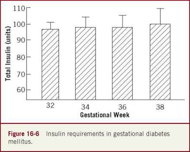

There was a significant difference in variability measured by the coefficient of variation (45% vs. 25%, P < .01), respectively.
The total insulin dose required to reach the established level of glycemic control for the majority of patients range from 40 to 90 units (body-weight dependent). For the GDM patient during the first phase, it is beneficial to schedule frequent visits to adjust insulin dose to maximize glycemic control. During the second phase, the care provider is vigilant about fetal growth and surveillance in addition to blood glucose testing although a significant increase in insulin dose is not anticipated.
The insulin to be administered should be calculated in a similar pattern as that recommended for type 1 diabetic patients. The insulin is divided into three doses: regular and intermediate-acting insulin in the morning; regular at dinner; and intermediate acting at bedtime. The standard formula for the amount of insulin prescribed should be 2/3s of all insulin in the morning (2:1, intermediate acting: regular) and 1/3 in the evening (1:1 regular [dinner]: intermediate acting [bedtime]). If after three days of self-monitoring blood glucose levels with insulin administration the overall glycemic profile fails to reach targeted levels, insulin should be increased at the rate of 15% to 20% for each dose. The procedure is repeated with a 10% to 15% increase in overall insulin dose. Thereafter, alteration in insulin dose is based on achievement of glucose target ranges for overall, preprandial, and postprandial levels (Figures 16-6, and 16-7).
In summary, insulin therapy should mimic as much as possible the physiological pattern of insulin secretion. Although different kinds of insulin are on the market today, no specific type (from human to analog) has demonstrated a better overall effect. When a pregnant woman is treated with insulin, the care provider needs to take into account the presence of insulin resistance that requires a relatively high insulin dose to overcome the abnormal glycemic profile. Overall, insulin requirements after two weeks of insulin therapy increase by 26% for type 1 diabetes; 34% for type 2 diabetes; and more than 40% for GDM from the calculated dose at entry to the actual dose that maintains targeted levels of glycemic control. Pregnancy provides a relatively narrow window of opportunity to prevent fetal complications. Therefore, an intensified approach in insulin management should be the goal.
REFERENCES
1. Banting F, Best C, Collip J, et al. Pancreatic extracts in the treatment of diabetes mellitus. Can Med Assoc J. 1922;12:141-146.
2. Owens D, Zinman B, Bolli G, et al. Insulins today and beyond. Lancet. 2001;358:739-746.
3. Zinman B. Diabetes research: are we getting anywhere? Can Med Assoc. 1996;155(9):1271-1274.
4. Robbins D, Tager H, Rubenstein A. Biologic and clinical importance of proinsulin. N Engl J Med. 1984;310:1165-11675.
5. Polonsky K, Rubenstein A. C-peptide as a measure of the secretion and hepatic extraction of insulin: pitfalls and limitations. Diabetes. 1984;33:486-494.
6. Zinman B, Murray F, Vranic M, et al. Glucoregulation during moderate exercise in insulin treated diabetes. J Clin Endocrinol Metab. 1977;45:641-652.
7. Vranic M, Kawamori R, Pek S, et al. The essentiality of insulin and the role of glucagons in regulating glucose utilization and production during strenuous exercise indogs. J Clin Invest. 1976;57:245-255.
8. Mitchell T, Abraham G, Schiffrin A, et al. Hyperglycemia after intense exercise in IDDM subjects during continuous subcutaneous insulin infusion. Diabetes Care. 1988;11:311-317.
9. Physical Exercise Must Be “Made-to-Order.” Medical Congress. com. Updated November 2004.
10. Occupational Exercise Lowers Heart Disease Risk for Diabetics. patienthealthinternational.com. Updated July 2004.
11. Malherbe C, deGaspro M, deHertogh R, et al. Circadian variations of blood sugar and plasma insulin levels in man. Diabetologia. 1969;5:397-404.
12. Mosekilde E, Skovbo J, Binder C, et al. Modeling absorption kinetics of subcutaneous injected soluble insulin. J Pharmacokinet Biopharm. 1989;17(1):67-87.
13. Zinman B. The physiological replacement of insulin: an elusive goal. N Engl J Med. 1989;321(6):363-370.
14. Humalog—A new insulin analogue. Drug Ther Bull. 1997;35:57-58.
15. Hermansen L, Colombo M, Storgaard H, et al. Improved postprandial glycemic control with biphasic insulin aspart relative to biphasic insulin lispro and biphasic human insulin in patients with type 2 diabetes. Diabetes Care. 2002;25:883-888.
16. Lindholm A, Jensen L, Home P, et al. Immune responses to insulin aspart and biphasic insulin aspart in people with type 1 and type 2 diabetes. Diabetes Care. 2002;25:876-882.
17. Preece R, Jovanovic L. New and future diabetes therapies: are they safe during pregnancy? J Matern Fetal Neonatal Med. 2002;12:365-375.
18. Howey D, Bowsher R, Brunelle R, et al. [(Lys (B28), Pro (B29)]- human insulin: a rapidly absorbed analog of human insulin. Diabetes. 1994;43:396-402.
19. Torlone E, Fanelli C, Rambotti A, et al. Pharmacokinetics, pharmacodynamics and glucose counterregulation following a subcutaneous injection of the monomeric insulin analogue [Lys (B28), Pro (B29)] in IDDM. Diabetologia. 1994;37(7):713-720.
20. Heinemann L, Kapitza C, Starka A, et al. [Time-action profile of the insulin analogue B28Asp]. Diab Med. 1996;13:683-684.
21. Haffner S. The importance of hyperglycemia in the non-fasting state to the development of cardiovascular disease. Endocr Rev. 1998;19(5):583-592.
22. Schernthaner G, Wein W, Sandholzer K, et al. Postprandial insulin lispro: a new therapeutic option for type 1 diabetic patients. Diabetes Care. 1998;21(4):570-573.
23. Martin J, Anderson D; UK Lispro Study Group. A short-acting insulin analog in clinical practice. Diabet Med. 1994;11(3):35.
24. Garg S, Carmain J, Braddy K, et al. Pre-meal insulin analog insulin lispro vs. humulin insulin treatment in young subjects with type 1 diabetes. Diabet Med. 1996;13(1):47-52.
25. Pfutzner A, Kuestner E, Forst T, et al. Intensive insulin therapy with insulin lispro in patients with type 1 diabetes reduces the frequency of hypoglycemic episodes. Exp Clin Endocrinol Diabetes. 1996;104(1):25-30.
26. Gale E. A randomized controlled trial comparing insulin lispro with human soluble insulin in patients with type 1 diabetes on intensified insulin therapy. Diabet Med. 2000;17(3):209-214.
27. Davey P, Grainger D, MacMillan J, et al. Clinical outcomes with insulin lispro compared with human regular insulin: a meta-analysis. Clin Ther. 1997;19(4):656-674.
28. Brunelle R, Llewelyn J, Anderson J, et al. Meta-analysis of the effect of insulin lispro on severe hypoglycemia in patients with type 1 diabetes. Diabetes Care. 1998;21(10):1726-1731.
29. Zinman B, Tildesley H, Chiasson J, et al. Insulin lispro in CSII: results of a double-blind crossover study. Diabetes. 1997;46(3):440-443.
30. Melki V, Renard E, Lassmann-Vaue V, et al. Improvement of HbA1c and blood glucose stability in IDDM patients treated with lispro insulin-analog in external pumps. Diabetes Care. 1998;21(6):977-982.
31. Kurthals P, Schaffer L, Sorensen A, et al. Correlations of receptor binding and metabolic and mitogenic potencies of insulin analogs designed for clinical use. Diabetes. 2000;49(6):999-1005.
32. King G, Goodman A, Buzney S, et al. Receptors and growth-promoting effects of insulin and insulin-like growth factors on cells from bovine retinal capillaries and aorta. J Clin Invest. 1985;75:1028-1036.
33. Grant M, Mames R, Fitzgerald C, et al. Insulin-like growth factor I acts as an angiogenic agent in rabbit cornea and retina: comparative studies with basic fibroblast growth factor. Diabetologia. 1993;36:282-291.
34. Jorgensen L, Didriksen L, Drejer K. Carcinogen effect of the human insulin analog B10 Asp in female rats [Abstract]. Diabetologia. 1992;35:A3.
35. Wyatt J, Frias J, Hoyme H, et al. Congenital anomaly rate in offspring of mothers with diabetes treated with insulin lispro during pregnancy. Diabet Med. 2005;22:803-807.
36. Mathiesen E, Kinsley B, Amiel S, et al. Maternal glycemic control and hypoglycemia in type 1 diabetic pregnancy: a randomized trial of insulin aspart versus human insulin in 322 pregnant women. Diabetes Care. 2007;30:771-776.
37. Koren G, Pastuszack A, Ito S. Drugs in pregnancy. N Engl J Med. 1998;338:1128-1137.
38. Devlin J, Hothersall L, Wilkis J. Use of insulin glargine during pregnancy in a type 1 diabetic woman. Diabetes Care. 2002;25:1095-1096.
39. Leslie C. New insulin replacement technologies: over-coming barriers to tight glycemic control. Cleve Clin J Med. 1999;66:293-302.
40. Yki-Jarvinen H, Dressler A, Ziemen M. Less nocturnal hypoglycemia and better post-dinner glucose control with bedtime insulin glargine compared with bedtime NPH insulin during insulin combination therapy in type 2 diabetes. Diabetes Care. 2000;23:1130-1136.
41. Boskovic R, Feig D, Derewlany L, et al. Transfer of insulin lispro across the human placenta. Diabetes Care. 2003;26(5):1390-1394.
42. Hofmann T, Horstmann G, Stammberger I. Evaluation of the reproductive toxicity and embryotoxicity of insulin glargine (LANTUS) in rats and rabbits. Int J Toxicol. 2002;21:181-189.
43. Poyhonen-Alho M, Ronnemaa T, Saltevo J, et al. Use of insulin glargine during pregnancy. Acta Obstet Gynecol Scand. 2007;29:1-4.
44. Gallen I, Jaap A, Roland J, et al. Survey of glargine use in 115 pregnant women with type 1 diabetes. Diabet Med. 2008;25:165-169.
45. Di Cianni G, Torlone E, Lencioni C, et al. Perinatal outcomes associated with the use of glargine during pregnancy. Diabet Med. 2008;25:993-996.
46. Lepercq J, Jacqueminet S, Hieronimus D, et al. Use of insulin glargine throughout pregnancy in 102 women with type 1 diabetes. Diabetes Metab. 2010;36:209-212.
47. Price N, Bartlett C, Gillmer M. Use of insulin glargine during pregnancy: a case-control pilot study. BJOG. 2007;114:453-457.
48. Kurtzhals P, Schaffer L, Sorensen A, et al. Potencies of insulin analogs designed for clinical use. Diabetes. 2000;49:999-1005.
49. Hermansen K, Madsbad S, Perrid H, et al. Comparison of the soluble basal insulin analog detemir with NPH insulin. Diabetes Care. 2001;4:296-301.
50. Hod M, Mathiesen E, Jovanovic L, et al. A randomized trial comparing perinatal outcomes using insulin detemir or neutral protamine Hagedorn in type 1 diabetes. J Matern Fetal Neonatal Med. 2014;27:7-13.
51. Mathiesen E, Damm P, Jovanovic L, et al. Basal insulin analogues in diabetic pregnancy: a literature review and baseline results of a randomized, controlled trial in type 1 diabetes. Diabetes Metab Res Rev. 2011;27:543-551.
52. Heise T, Nosek L, R0nn B, et al. Lower within-subject variability of insulin detemir in comparison to NPH insulin and insulin glargine in people with type 1diabetes. Diabetes. 2004;53:1614-1620.
53. Mathiesen E, Hod M, Ivanisevic M, et al. Maternal efficacy and safety outcomes in a randomized controlled trial comparing insulin detemir with NPH insulin in 310 pregnant women with type 1 diabetes mellitus. Diabetes Care. 2012;35:2012-2017.
54. Bartley P, Bogoev M, Larsen J, et al. Long-term efficacy and safety of insulin detemir compared to neutral protamine Hagedorn insulin in patients with type 1 diabetes using a treat-to-target basal-bolus regimen with insulin aspart at meals: a 2-year, randomized, controlled trial. Diabet Med. 2008;25:442^49.
55. Nicoline F, Callesen, Damm J, et al. Treatment with the long-acting insulin analogs detemir or glargine during pregnancy in women with type 1 diabetes: comparison of glycemic control and pregnancy outcome. J Matern Fetal Neonatal Med. 2013;26(6):588-592.
56. Hod M, McCance D, Ivanisevic M, et al. Perinatal outcomes in pregnancy: a randomized trial comparing insulin detemir with NPH insulin in 310 subjects with type 1 diabetes. Diabetologia. 2011;54 (suppl 1):S487.
57. Mathiesen E, Damm P, Hod M, et al. Maternal glycemic control and hypoglycemia in pregnancy: a randomized trial comparing insulin detemir with NPH insulin in 310 subjects with type 1 diabetes. Diabetologia. 2011;54 (suppl 1):S486-S487.
58. Jensen D, Damm P, Moelsted-Pedersen L, et al. Outcomes in type 1 diabetic pregnancies: a nationwide, population-based study. Diabetes Care. 2004;27:2819-2823.
59. Nielsen L, Pedersen-Bjergaard U, Thorsteinsson B, et al. Hypoglycemia in pregnant women with type1 diabetes: predictors and role of metabolic control. Diabetes Care. 2008;31:9-14.
60. Hod M, Damm P, Kaaja R, et al.; Insulin Aspart Pregnancy Study Group. Fetal and perinatal outcomes in type 1 diabetes pregnancy: a randomized study comparing insulin aspart with human insulin in 322 subjects. Am J Obstet Gynecol. 2008;198:186.el-186.e7.
61. Bruttomesso D, Bonomo M, Costa S, et al.; Italian Group for Continuous Subcutaneous Insulin Infusion in Pregnancy. Type 1 diabetes control and pregnancy outcomes in women treated with continuous subcutaneous insulin infusion (CSII) or with insulin glargine and multiple daily injections of rapid-acting insulin analogues (glargine-MDI). Diabetes Metab. 2011;37:426-431.
62. Fang Y, MacKeen D, Egan JF, et al. Insulin glargine compared with neutral protamine Hagedorn insulin in the treatment of pregnant diabetics. J Matern Fetal Neonatal Med. 2009;22:249-253.
63. Bhattacharyya A, Vice P. Insulin lispro, pregnancy, and retinopathy. Diabetes Care. 1999;22:2101-2102.
64. Buchbinder A, Miodovnik M, McElvy S, et al. Is insulin lispro associated with the development or progression of diabetic retinopathy during pregnancy? Am J Obstet Gynecol 2000;183:1162-1165.
65. Bhattacharyya A, Brown S, Hughes S, et al. Insulin lispro and regular insulin in pregnancy. QJM. 2001;94:255-260.
66. Persson B, Swahn M, Hjertberg R, et al. Insulin lispro therapy in pregnancies complicated by type 1 diabetes mellitus. Diabetes Res Clin Pract. 2002;58:115-121.
67. Loukovaara S, Immonen I, Teramo K, et al. Progression of retinopathy during pregnancy in type 1 diabetic women treated with insulin lispro. Diabetes Care. 2003;26(4):1193-1198.
68. Durand-Gonzalez K, Guillausseau N, Anciaux M, et al. Allergy to insulin in a woman with gestational diabetes mellitus: transient efficiency of continuous subcutaneous insulin lispro infusion. Diabetes Metab. 2003;29:432-434.
69. Garg S, Frias J, Anil S, et al. Insulin lispro therapy in pregnancies complicated by type 1 diabetes: glycemic control and maternal and fetal outcomes. Endocr Pract. 2003;9(3):187-193.
70. Mecacci F, Carignani L, Cioni R, et al. Maternal metabolic control and perinatal outcome in women with gestational diabetes treated with regular or lispro insulin: comparison with non-diabetic pregnant women. Eur J Obstet Gynecol Reprod Biol. 2003;111:19-24.
71. Anderson J Jr, Brunelle R, Koivisto V, et al. Reduction of postprandial hyperglycaemia and frequency of hypoglycaemia in IDDM patients on insulin analog treatment. Diabetes. 1997;46:265-270.
72. Carr K, Idama T, Masson E, et al. A randomized controlled trial of insulin lispro given before or after meals in pregnant women with type 1 diabetes—the effect on glycemic excursion. J Obstet Gynaecol. 2004;24(4):382-386.
73. Idama T, Lindow S, French M, et al. Preliminary experience with the use of insulin lispro in pregnant diabetic women. J Obstet Gynecol. 2001;21(4):350-351.
74. Cypryk K, Sobczak M, Pertynska-Marczewska M, et al. Pregnancy complications and perinatal outcome in diabetic women treated with Humalog (insulin lispro) or regular human insulin during pregnancy. Med Sci Monit. 2004;10(2):129-132.
75. Masson E, Patmore J, Brash P, et al. Pregnancy outcome in type 1 diabetes mellitus treated with insulin lispro (Humalog). Diabet Med. 2003;20:46-50.
76. Pettitt D, Opsina P, Kolaczynski J, et al. Comparison of an insulin analog, insulin aspart, and regular human insulin with no insulin gestational diabetes mellitus. Diabetes Care. 2003;26:183-186.
77. Bauman W, Yalow R. Transplacental passage of insulin complexed to antibody. Proc Natl Acad Sci USA. 1981;78(7):4588-4590.
78. Menon R, Cohen R, Sperling M, et al. Transplacental passage of insulin in pregnant women with insulin-dependent diabetes melli- tus; its role in fetal macrosomia. N Engl J Med. 1990;323:309-315.
79. Jovanovic L, Ilic S, Pettitt, D, et al. Metabolic and immunologic effects of insulin lispro in gestational diabetes. Diabetes Care. 1999;22:1422-1427.
80. Holcberg G, Tsadkin-Tamir M, Sapir O, et al. Transfer of insulin lispro across the human placenta. Eur J Obstet Gynecol Reprod Biol. 2004;115(1):117-118.
81. McCance D, Damm P, Mathiesen, et al. Evaluation of insulin antibodies and placental transfer of insulin aspart in pregnant women with type 1 diabetes mellitus. Diabetologia. 2008;51(11):2141-2143.
82. Lindsay R, Ziegler A, Hamilton B, et al.; Scottish Multicenter Study of Diabetes in Pregnancy. Type 1 diabetes-related antibodies in the fetal circulation: prevalence and influence on cord insulin and birth weight in offspring of mothers with type 1 diabetes. J Clin Endocrinol Metab. 2004;89:3436-3439.
83. Pollex E, Feig D, Lubetsky A, et al. Insulin glargine safety in pregnancy. Diabetes Care. 2010;33(1):29-33.
84. Siebenhofer A, Plank J, Berghold A, et al. Short acting insulin analogs versus regular human insulin in patients with diabetes mellitus [Cochrane Review]. In: The Cochrane Collaboration. The Cochrane Review Library. Chichester, UK: John Wiley & Sons, Ltd; (2) 2004.
85. Langer O, Mazze R. Diabetes in pregnancy: evaluating self-monitoring performance and glycemic control with memory-based technology. Am J Obstet Gynecol. 1986;155:635-638.
86. Langer O, Rodriguez D, Xenakis E, et al. Intensified versus conventional management of gestational diabetes. Am J Obstet Gynecol. 1994;170(4):1036-1047.
87. Rayburn W. Changes in insulin therapy during pregnancy. Am J Perinatol. 1985;2:271-277.
88. Weiss P, Hofmann H. Intensified conventional insulin therapy for the pregnant diabetic patient. Obstet Gynecol. 1984;64:629-633.
89. Langer O, Anyaegbunam A, Brustman L, et al. Pregestational diabetes: insulin requirements throughout pregnancy. Am J Obstet Gynecol. 1988;159(3):616-620.
90. Jovanovic L, Peterson M. Optimal insulin delivery for the pregnant diabetic patients. Diabetes Care. 1982;5:24-31.
91. Gabbe S. New concepts and applications in the use of the insulin pump during pregnancy. J Maternal-Fetal Med. 2000;9:42^5.
92. Pendergrass M, Fazioni E, DeFronzo R. Non-insulin dependent diabetes mellitus and gestational diabetes mellitus: same disease, another name? Diabetes Rev. 1995;3:584-601.
93. Buchanan T, Xiang A, Peters R. Response of pancreatic B-cells to improved insulin sensitivity in women at high risk for type 2 diabetes. Diabetes. 2000;49:782-788.
94. Catalano P, Kirwan J, Mouzon S-de, et al. Gestational diabetes and insulin resistance: role in short- and long-term implications for mother and fetus. JNutr. 2003;133:1674S-1683S.
95. Bevier W, Jovanovic-Peterson L, Peterson C. Endocrine disorders of pregnancy: pancreatic disorders of pregnancy. Endocrinol Metab Clin North America. 1995;24:103-138.
96. Ciampelli M, Lanzone A, Caruso A. Insulin in obstetrics: a main parameter in the management of pregnancy. Hum Reprod Update. 1998;4(6):904-914.
97. Landon M, Gabbe S, Sachs L. Management of diabetes mellitus and pregnancy: a survey of obstetricians and maternal-fetal specialists. Obstet Gynecol. 1990;75:635-640.
98. Marrero D, Moore P, Langerfeld C, et al. Care of diabetic pregnant women by primary care physicians. Diabetes Care. 1992;15:101-107.
99. Owen J, Phelan S, Landon M, et al. Gestational diabetes survey. Am J Obstet Gynecol. 1995;1722:615-620.
100. Metzger B, Coustan D. Organizing committee. Summary and recommendations of the fourth international workshop-conference on gestational diabetes. Diabetes Care. 1998;21(supp 2):B161-B167.
101. Reece E, Homko C, Miodovnik M, et al. A consensus report of the diabetes in pregnancy study group of North America. J Matern Fetal Neonatal Med. 2002;12:362-364.
102. ACOG Educational and Technical Bulletin #200; 1994.
103. American College of Obstetricians and Gynecologists. Clinical Management Guidelines for Obstetrician-Gynecologists. No.30 Gestational diabetes. Washington, DC: American College of Obstetricians and Gynecologists; 2001.
104. American Diabetes Association. Position statement on gestational diabetes mellitus. Diabetes Care. 2004;27(supp 1):S88-S90.
105. Langer O. Maternal glycemic criteria for insulin therapy in gestational diabetes mellitus. Diabetes Care. 1998;21(suppl 2):B91-B98.
106. Langer O, Berkus M, Brustman L, et al. Rationale for insulin management in gestational diabetes mellitus. Diabetes. 1991;40(suppl 2):186-190.
107. Ben-Haroush A, Yogev Y, Chen R, et al. The postprandial glucose profile in the diabetic pregnancy. Am J Obstet Gynecol. 2004;191(2):576-581.
108. Langer O. Management of gestational diabetes. Clin Obstet Gynecol. 2000;43(1):106-115.
109. McFarland M, Langer O, Conway D, et al. Dietary therapy for gestational diabetes: how long is long enough? Obstet Gynecol. 1999;93:978-982.
110. Weiss P, Hofman H, Kainer F, et al. Fetal outcome in gestational diabetes with elevated amniotic fluid insulin levels: dietary vs. insulin treatment. Diabetes Red Clin Pract. 1988;5:1-7.
111. Weiss P, Hofman H, Winter R, et al. Diagnosis and treatment of gestational diabetes according to amniotic fluid insulin levels. Arch Gynecol. 1986;239:81-91.
112. Buchanan T, Kjos S, Schaefer U, et al. Utility of fetal measurements in the management of gestational diabetes mellitus. Diabetes Care. 1998;21(suppl 2):B99-B106.
113. Coustan D, Imarah J. Prophylactic insulin treatment of gestational diabetes reduces the incidence of macrosomia, operative delivery, and birth trauma. Am J Obstet Gynecol. 1984;15:836-842.
114. Langer O, Davidson L. What is the source of insulin in amniotic fluid? American Diabetes Association. 4th International Workshop Conference on Gestational Diabetes Mellitus, Chicago, IL; 1997; Abstract 28:page 41.
115. Langer O, Anyaegbunam A, Brustman L, et al. Gestational diabetes: insulin requirements in pregnancy. Am J Obstet Gynecol. 1987;157(3):669-675.