
Paul Park
Ali F. Mallat
Presentation
A 75-year-old woman presents with possible small bowel obstruction. She is demented and resides at an extended care nursing facility. Over the past 6 days, she has had intermittent abdominal pain, confusion, and emesis. She was evaluated by the physician at her nursing facility 4 days ago, and she was treated empirically for a urinary tract infection. She continued to have episodic bouts of emesis, abdominal pain, and confusion over the ensuing days and was sent to the emergency room for further evaluation.
By her chart history, she has chronic obstructive pulmonary disease, diabetes mellitus, hyperlipidemia, Alzheimer’s dementia, hypothyroidism, and cholelithiasis. She has had no prior abdominal surgeries.
On initial evaluation, she is found to be confused and somnolent. She is afebrile, mildly tachycardic at 102 beats per minute, and with a blood pressure of 116/78. On examination of her abdomen, she has no prior surgical scars. Her abdomen is moderately distended and tympanitic. She has diffuse, nonlocalized tenderness over her abdomen without involuntary guarding or signs of peritonitis. There are no palpable masses. She has no costovertebral angle tenderness. Her rectal exam reveals no masses or fissures, normal tone, and no fecal impaction.
Differential Diagnosis
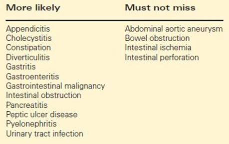
This is a common presentation suspicious for bowel obstruction. The time course is not acute but rather one of progressive decline. Her differential will need to be broad, and her initial studies directed toward narrowing down her differential diagnosis rapidly (Table 1).
TABLE 1. Differential Diagnosis
Workup
The initial workup should be directed toward narrowing the list of potential diagnoses quickly. In this case her history, clinical course, and examination are suspicious for potential obstruction, either mechanical or from an ileus. Her initial workup should include laboratory studies and imaging. A complete blood count, comprehensive metabolic panel, amylase and lipase, and urinalysis, along with multiview chest and abdominal radiographs would be an appropriate initial workup.
Her initial lab work reveals a mild leukocytosis at 11,000. Her creatinine is elevated to 2.4, and her chloride is low at 92. Her liver panel is within normal limits, and her amylase and lipase are normal. Urinalysis is normal. Her chest x-ray is unremarkable. On abdominal x-ray, she has diffusely dilated small bowel loops throughout her abdomen, with a paucity of gas in the colon (Figure 1). Careful inspection reveals a radiopaque density in the lower abdomen, consistent with an ectopic gallstone. Depending on the patient’s clinical condition, further imaging may not be necessary before making the decision to intervene surgically. However, in cases where the physical exam and plain radiographs do not prompt immediate surgical intervention, computed tomography (CT) with enteric contrast can be helpful in establishing a diagnosis of bowel obstruction and in delineating other pathology in the abdomen.
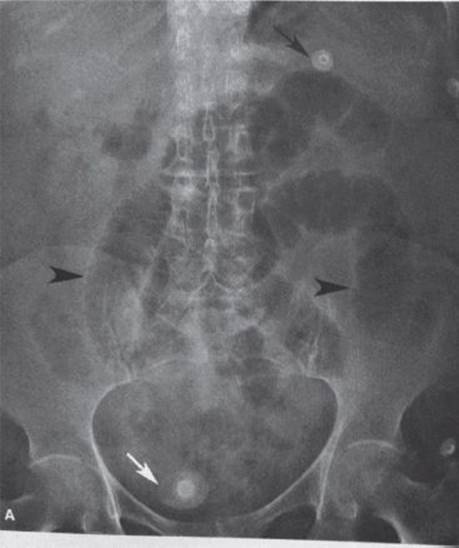
FIGURE 1 • Evidence of ileus with diffuse small bowel dilation (arrowheads), accompanied by an ectopic gallstone (white arrow) and pneumobilia (black arrow), is suggestive of gallstone ileus.
In this case, CT showed evidence of a cholecystoduodenal fistula (Figure 2), with contrast from the duodenum filling the gall bladder. Images of the lower abdomen confirmed the presence of the calculus at a transition point in the small intestine (Figure 3).
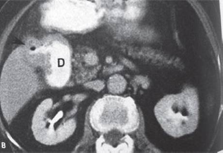
FIGURE 2 • Cholecystoduodenal fistula demonstrated via oral contrast CT Contrast in the gallbladder is indicated by the arrow and appears to communicate with the duodenum (D).
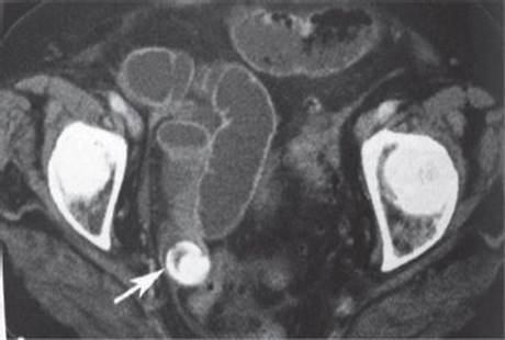
FIGURE 3 • CT scan demonstrating radiolucent calculus (gallstone, white arrow) in distal ileum.
Based on the abdominal imaging and clinical presentation, the diagnosis is of a bowel obstruction secondary to gallstone ileus. Only about 50% of cases are diagnosed preoperatively, which increases up to 77% with the addition of other imaging modalities, particularly CT, to elucidate the diagnostic picture. However, despite the improvement of radiologic imaging, clinical suspicion and upright and supine abdominal radiograph remain the most important components of the diagnostic evaluation.
Gallstone ileus accounts for < 1% to 4% of all mechanical small bowel obstructions. This incidence increases to 25% among episodes of small bowel obstruction in patients over 65 years old. The clinical picture is classically of an elderly woman presenting with recurrent intermittent episodes of obstruction. The clinical course is often subacute and indistinct, with progressive worsening over days prior to presentation at a hospital for evaluation. The average time prior to presenting at a hospital is 5 days. The average age of patients with gallstone ileus is 70 years, with women affected much more frequently; therefore, this diagnosis needs to be kept in the differential particularly for this demographic. The patient population frequently has multiple comorbidities at baseline, with 86% noted to be in the American Society of Anesthesiology classification groups 3 or 4.
Using imaging, classic findings that may be found on plain radiographs of the abdomen comprise Rigler’s triad (pneumobilia, evidence of obstruction, and ectopic gallstone). The actual triad is uncommon to see on an abdominal radiograph due to the fact that most gallstones are radiolucent (only 15% of stones are radiopaque on abdominal films). While both pneumobilia and evidence of bowel obstruction occur in about 50% of these patients each, neither are diagnostic for gallstone ileus. Ultrasound may help visualize stone impaction, fistulas, as well as gallstones in the biliary system, but often imaging is limited due to body habitus and gas in the intestinal loops. CT scan may demonstrate gallbladder thickening, pneumobilia, intestinal obstruction with an identified transition point, or visualization of an ectopic gallstone. HIDA scans may allow visualization of a gallbladder fistula or perforation, although the sensitivity is only about 50%. Despite of these investigative modalities, diagnosis at the time of surgical exploration continues to be common.
Diagnosis and Treatment
The initial treatment of gallstone ileus, much like any intestinal obstruction, begins with appropriate resuscitation and stabilization, correction of electrolyte abnormalities, and surgical exploration. The surgical decision making depends on factors revealed during the workup along with information gathered during abdominal exploration.
Our patient has been adequately resuscitated but continues to vomit, remaining uncomfortable and distended despite nasogastric decompression. Her diagnosis is explained to her caregiver, and she is scheduled for urgent surgical exploration.
Surgical Approach
The primary goal of surgical management of gallstone ileus is to relieve the obstruction quickly and safely. While there have been reports in the literature of laparoscopic exploration and treatment for gallstone ileus, the standard approach would be an open exploration (Table 2). The intestinal tract should be fully evaluated for additional nonobstructing stones as well. The common locations for stones are in the distal ileum, followed by the jejunum and gastric outlet. The colon is an uncommon site of obstruction unless a history of medical or surgical illness suggests the presence of a colonic stricture. An enterolithotomy performed proximal to the obstructing stone is used to extract the stone in retrograde fashion. If there is any suggestion of bowel injury during the extraction, a formal bowel resection is warranted.
TABLE 2. Key Technical Steps and Pitfalls
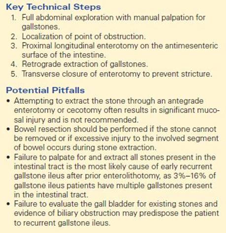
The patient is taken to the operating room where a midline laparotomy incision is used to explore the abdomen. The stone is located in the distal ileum, and an additional smaller stone is palpated in the midjejunum. Both are extracted atraumatically through an enterotomy, which is subsequently closed in a transverse orientation.
Special Intraoperative Considerations
The primary goal is to relieve the obstruction. The decision to perform a concurrent cholecystectomy and fistula closure is a controversial one as this will lead to significant increase in the operative time and increases the risk of morbidity and mortality. Therefore, addressing the obstruction alone is the most appropriate treatment strategy in the vast majority of patients. As gallstone ileus occurs most commonly in older debilitated patients, minimizing the length and complexity of the operative procedure is of paramount importance. Furthermore, the risk of recurrent gallstone ileus, or other complications related to persistence of the gallbladder and the cholecystoduodenal fistula, is relatively low.
In the unusual circumstance that gallstone ileus occurs in a stable patient without significant debilitation or comorbidity, concurrent fistula closure and cholecystectomy may be considered. Manual palpation of the gallbladder should be performed to determine if additional stones are present. The presence of stones in the gallbladder increases the risk of future ectopic stones. Patency of the extrahepatic biliary tree should also be established by preoperative laboratory studies and imaging. In general, a fistula without stones or concurrently obstructed biliary system can be expected to close spontaneously and generally should not have a concurrent cholecystectomy.
If cholecystectomy and fistula takedown are performed, the duodenal closure should be repaired using standard techniques for addressing duodenal injury. The most appropriate repair should be determined by the health of the tissue and the size of the defect. The techniques of primary repair, omental patch, pyloric exclusion, or duodenojejunostomy may each be appropriate depending on the severity of the duodenal defect, and surgeons should not approach cholecystoduodenal fistula takedown if not comfortable with these procedures. Cholecystectomy should be accomplished carefully, as the anatomy of the critical structures in the porta may be obscured by the inflammation surrounding the fistula. Closed suction drainage should be left following cholecystectomy and duodenal repair.
Postoperative Management
Postoperative care is similar to any emergency bowel operation on an elderly patient. The main source of morbidity and mortality will derive from the comorbidities of the patient and physiologic reserve to tolerate surgical exploration. Special consideration should be made to adequately resuscitate the patient. Aggressively correcting electrolyte imbalance and restoring acidbase equilibrium that may have occurred as a result of dehydration and emesis in advance of proceeding to operative intervention is preferred. A nasogastric tube for decompression until return of bowel function as well as a Foley catheter to assess adequacy of resuscitation should also be used.
In the case of patients who have undergone stone removal with or without bowel resection, the most concerning technical complication is enteric leak, and a high index of suspicion for this potentially devastating event has to be maintained in these frail and frequently malnourished patients. Prompt recognition and early surgical intervention for this complication is the only way to avoid a postoperative mortality.
In the case of patients who have undergone concurrent cholecystectomy and duodenal closure, leakage from either the cystic duct or the duodenal repair is possible. Concerning bilious drain output from operatively placed drains may be the first hint of a problem, and subsequent studies should be aimed at documenting the site of the leak. Measurement of amylase levels in the drainage may help to distinguish between a leak from the biliary tree versus the duodenum, although unfortunately, this is not a test with perfect accuracy. An upper gastrointestinal imaging (GI) with water-soluble contrast should be the first test, as a documented duodenal leak will likely require additional surgical intervention. If there is no duodenal leak, the cystic duct is the likely source. If the leak is of significant volume to prompt intervention, a percutaneous transhepatic cholangiogram and biliary drain placement should be considered, rather than attempting endoscopic retrograde cholangiography in the presence of a recent duodenal repair.
Once the patient has resumed bowel function and a diet, the drains can be removed provided there is no change in the quality and volume of effluent.
If cholecystectomy was not performed at the primary operation, interval cholecystectomy may be considered in patients who recover fully and remain reasonable operative candidates. An interval of at least 4 to 6 weeks following the initial surgery is recommended.
TAKE HOME POINTS
· Consider the diagnosis of gallstone ileus in an elderly patient with signs of bowel obstruction.
· No imaging gold standard exists for diagnosis although abdominal films, ultrasound, and CT scan are all useful adjuncts to clinical suspicion and serial examination. Rigler’s triad, although diagnostic, is exceedingly uncommon.
· Surgical exploration should aim to clear all stones from the intestinal tract in order to treat the current obstruction and prevent early recurrent obstruction.
· Cholecystectomy and fistula takedown are rarely indicated at the time of the operation for bowel obstruction.
SUGGESTED READINGS
Ayantunde AA, Agrawal A. Gallstone ileus: diagnosis and management. World J Surg. 1994;31:1292.
Clavien PA, Richon J, Burgan S, et al. Gallstone ileus. Br J Surg. 1990;77:737–742.
Muthukumarasamy G, et al. Gallstone ileus: surgical strategies and clinical outcome. J Dig Dis. 2008;9:156–161.
Reisner RM, Cohen JR. Gallstone ileus: A review of 1001 reported cases. Am Surg. 1994;60:441.
Rigler LG, Borman CN, Noble JF. Gallstone obstruction: pathogenesis and roentgen manifestation. J Am Med Assoc. 1941;117:1753.
Rodriguez-Sanjuan JC, Casado F, Fernandez MJ, et al. Cholecystectomy and fistula closure versus enterolithotomy alone in gallstone ileus. Br J Surg. 1997;84:634–637.