
Lee A. Zimmer
INTRODUCTION
Masses, both benign and malignant, arising from the sinonasal tract are uncommon and provide a substantial challenge to head and neck surgical oncologists. Often, these tumors erode or infiltrate the adjacent orbit and anterior cranial fossa. A complete history, physical examination, and imaging studies provide important information for surgical planning. Although a diagnosis can readily be made with nasal endoscopy, the open, transfacial approaches have provided the access for traditional surgical resection. Recent advances in the understanding of nasal endoscopic anatomy and technical advances in surgical instrumentation have allowed for the removal of these lesions via a purely endoscopic approach.
HISTORY
Both benign and malignant tumors of the sinonasal tract are listed in Table 14.1. Malignant tumors of the sinonasal tract make up about 3% of tumors in the head and neck. Exposure to nickel refining, wood dust, industrial fumes, and leather tanning has been implicated in the etiology of sinonasal malignancies. Industrial exposures such as radium dial paints, soldering and welding, lacquer paint, isopropyl oils, chromium, and mineral oils have also been implicated. There is also a higher incidence of malignancies in smokers as compared to nonsmokers. Benign vascular tumors are found in teenage males.
Table 14.1 Tumors Involving the Orbit
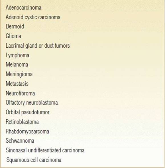
Tumors arising in the nasal cavity and sinuses often present as large lesions involving one or more sinuses. This is due to the relatively large intranasal space allowing growth prior to the onset of symptoms. Early symptoms include nasal obstruction, headache, nasal discharge, epistaxis, and anosmia. Patients with large tumors present with exophthalmos, diplopia, chemosis, supraorbital swelling, and edema of the eyelid. The absence of pain with ophthalmologic symptoms does not rule out malignancy, though deep-seated facial pain and facial paresthesia raise the index of suspicion for malignant tumors. The presence of epiphora suggests obstruction or destruction of the nasolacrimal duct. Loose-fitting dentures or loss of teeth suggests invasion of the floor of the maxilla. As tumors progress and invade the anterior cranial fossa and frontal lobe, subtle changes such as reduced speech, reduced verbal fluency, and altered expressive language may occur. The patient may begin to show signs of impaired insight and judgment while maintaining cognitive ability and memory.
PHYSICAL EXAMINATION
The physical examination should include careful examination of the sinonasal region, orbit, cranial nerves, middle ear, and neck and should include bilateral nasal endoscopy. Examination begins with an otomicroscopic examination; fluid in the middle ear suggests compression, invasion, or blockade of the eustachian tube in the nasopharynx, posterior to the pterygopalatine fossa, or the infratemporal fossa. Eye findings will suggest compression and/or invasion of orbital contents. Examination using a nasal speculum may reveal the physical nature of the tumor and suggest the vascularity of the neoplasm. Evaluation of the oral cavity, particularly of the upper alveolar ridge and teeth, will suggest the effect of the neoplasm on the floor of the maxillary sinus and nasal cavity. Evaluation of the neck is vital since lymphadenopathy would suggest a malignant process. Finally, a complete evaluation of the cranial nerves is performed looking for focal deficits.
INDICATIONS
Tumors involving the orbit that do not involve the anterior wall of the maxillary sinus or anterior table of the frontal sinus, the optic nerve, and ophthalmic artery, with little involvement of the superior orbital bone may be accessed through an endonasal approach. Tumors located between the medial and inferior rectus and medial to the ophthalmic artery and optic nerve are readily resectable. In more experienced hands, tumors extending into the medial orbital apex and medial temporal lobe may be resected.
CONTRAINDICATIONS
Although there are a few solid contraindications for the transorbital approach, some contraindications may be relative based upon the experience and skill of the surgeon. Removal of the anterior table of the frontal sinus involved by tumor is not achievable by a totally endoscopic approach to ensure negative margins. Involvement of the most lateral extent of the frontal sinus cannot be obtained by a purely endoscopic, endonasal approach. Excision of malignant tumors invading the globe through an endonasal approach is also contraindicated and requires an open orbitotomy. Endonasal excision of benign tumors lateral to the optic nerve and carotid artery is also contraindicated if the patient';s vision is intact. Posterior invasion of the carotid artery or cavernous sinus by malignant tumors greatly reduces the possibility for surgical resection with negative margins. In this situation, subtotal surgical resection followed by chemotherapy and radiation is recommended.
PREOPERATIVE PLANNING
Imaging Studies
Tumors involving the sinonasal tract require imaging using both computed tomography (CT) and magnetic resonance (MR) imaging with contrast. Contrast allows for an estimate of tumor vascularity. Image-guidance protocols should be performed if endoscopic approaches are entertained. CT imaging provides valuable information as to the integrity of the skull base, orbit, and sinonasal bony skeleton. MR allows for evaluation of soft tissue invasion, neural invasion, and entrapment of mucus in the sinuses versus tumor. If malignancy is confirmed, a positron emission tomography–CT scan should be performed for staging.
Nasal Endoscopy with Biopsy
I do not recommend in-office biopsy of sinonasal tumors, especially vascular tumors when the control of epistaxis can be very difficult to handle in the office setting. Patients are taken to the operating room for bilateral nasal endoscopy with biopsy. Definitive treatment is delayed until permanent pathologic diagnosis is confirmed.
Ophthalmology Evaluation
Patients with tumors of the sinonasal tract with ophthalmologic findings on physical examination or imaging undergo a complete eye examination by a neuro-ophthalmologist. Subtle findings such as ophthalmoplegia, visual field deficits, and optic nerve involvement may not be found on routine physical examination and help guide surgical planning. Furthermore, ophthalmologic assistance in tumor removal may be desirable in selected cases.
Neurosurgical Evaluation
At our institution, tumors abutting or eroding the skull base or involving or invading the brain undergo a full neurosurgical evaluation. It is my opinion that these tumors should be removed using a team approach.
Tumor Board
Once all of the preoperative evaluations, imaging, and pathology are complete, the case is presented at a multidisciplinary skull base tumor board for treatment planning. The tumor board consists of individuals from neurosurgery, otolaryngology, ophthalmology, medical oncology, radiation oncology, radiology, and nurse practitioners. Treatment planning is then finalized and presented to the patient.
SURGICAL TECHNIQUE
Transconjunctival Approach
Although not a requirement, some institutions prefer to have an ophthalmologist perform a transconjunctival approach in concert with an endoscopic transorbital approach to improve orbital retraction and allow retraction on the middle and inferior rectus muscles. As this is not uniformly established, the details of this approach are not described in this chapter (see Suggested Readings).
Transnasal Approach
The patient is placed in a supine position on the operating room table. The septum, middle turbinate, uncinate process, and anterior wall of the sphenoid sinus are injected with 1% lidocaine with 1:100,000 epinephrine bilaterally. Afrin- or cocaine-soaked pledgets are placed in the middle meatus and nasal cavity. While the decongestion process is taking place, the image-guidance system preferred by the surgeon is loaded and calibrated. The patient is then draped with clear tape over the eyes to protect the cornea. The eyes should be easily seen throughout the procedure for early recognition in the event of proptosis secondary to an orbital hematoma.
A wide maxillary antrostomy and total ethmoidectomy are performed on the side of the lesion. If the lesion extends to the sphenoethmoid recess, a sphenoidotomy is performed (Fig. 14.1). The middle turbinate is resected using a pair of endoscopic scissors with special attention not to violate the cribriform plate and lateral lamella. If a binostril approach is desired or the lesion crosses the midline, an anterior ethmoidectomy is performed on the contralateral side to allow visualization of the frontal recess, and the superior anterior portion of the middle turbinate is removed to prepare for a Draf III frontal sinusotomy. A complete Draf III, which allows for binostril access to the frontal sinus, is performed. If tumor is present in these regions, it is removed up to the frontal sinus and lamina papyracea with frozen section analysis of margins.

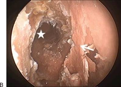

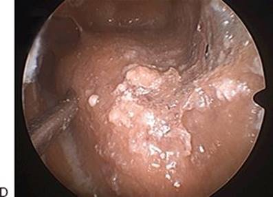
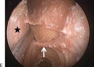
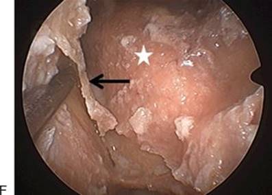


FIGURE 14.1 Cadaveric dissection demonstrating a unilateral, endonasal transorbital approach to the anterior cranial fossa. All images are 0-degree endoscopy unless otherwise noted. A. Right anterior ethmoidectomy and middle turbinate resection (arrow) have been performed showing the right frontal recess with 0-degree endoscopy (star). B. The middle turbinate has been resected (arrow). A Draf II frontal sinus exploration has been completed revealing the right frontal sinus with 0-degree endoscopy (star). C. View into the right frontal sinus showing the far lateral wall of the sinus (star) using 30-degree endoscopy. The posterior table of the frontal sinus is fully visualized (arrow). D. Straight Frazier suction showing the lateral reach of straight instrumentation into the frontal sinus without the assistance of the transorbital approach. E. The right lamina papyracea has been removed (star), and the anterior ethmoid artery is identified (arrow). F. The periorbita is compressed laterally to show the edge of the orbital roof (arrow). Again not the posterior table of the frontal sinus (star). G. View into the right frontal recess after removal of the lamina papyracea and medial half of the orbital roof. Note increased lateral reach of a straight Frazier suction compared to (D). Posterior table of the frontal sinus (star). H. Removal of the posterior table of the frontal sinus showing the anterior cranial fossa parenchyma (star).
Similar to an endoscopic orbital decompression, the lamina papyracea is removed inferiorly to the maxillary antrostomy, posteriorly to the anterior wall of the sphenoid sinus, anteriorly to the nasolacrimal canal, and superiorly to the frontal recess. The nasolacrimal canal can be resected if removal allows for negative margins. A lacrimal stent is placed prior to the completion of the surgery to prevent epiphora. The bone of the lamina papyracea is easily removed with a Cottle elevator and backbiting forceps. Thicker bone is thinned with a high-speed diamond drill with continuous irrigation and then removed as above. The anterior and posterior ethmoid arteries are either clipped with endoscopic clip appliers or cauterized with pistol-grip bipolar electrocautery and cut. Thin bone of the superior orbital roof is then removed with rongeurs or a high-speed diamond drill with continuous irrigation as the periorbita is retracted. This can be carried posteriorly over the orbit and under the dura of the frontal pole for lateral extension of tumor posteriorly over the orbit. Special attention is directed to not violate the periorbita since it causes dehiscence of adipose tissue into the frontal recess or ethmoid sinuses blocking endoscopic visualization. A small amount of orbital adipose tissue may be shrunk with bipolar cautery. By removing the orbital roof, increased lateral access to the frontal sinus is achieved. Tumor eroding into or through the posterior table can now be removed under direct visualization. Dura may be removed as a posterior margin with endoscopic scissors. The removal of tumor invading the brain parenchyma endoscopically is controversial and beyond the scope of this chapter. Cerebrospinal fluid (CSF) rhinorrhea of the posterior table can be repaired through various endoscopic techniques previously published (see Suggested Readings).
If tumor extends posterior to the globe and involves or erodes the periorbita, the periorbita is removed with endoscopic scissors (Fig. 14.2) with frozen section analysis of margins. The inferior and medial rectus muscles are first identified just under the periorbita. The optic nerve and ophthalmic artery are identified lateral to and between the medial and inferior rectus muscles and preserved in cases of benign disease and removed if involved with malignant disease. Tumor involving the globe requires an open orbital exenteration. If the artery is sacrificed, a clip is placed on the ophthalmic artery posteriorly for hemostasis. Dehiscent adipose tissue posterior to the globe is cauterized with bipolar cautery to maintain a clear field of vision for the surgeon. Further invasion of tumor into the pterygopalatine fossa, infratemporal fossa, temporal lobe, and/or cavernous sinus is beyond the scope of this chapter and is described elsewhere (see Suggested Readings). Reconstruction of the periorbital defect is not necessary for small defects. Larger defects can be reconstructed with a cadaveric dermal graft (Alloderm) held in place with sutures and packing. Packing is not required after endoscopic transorbital approaches to the anterior cranial fossa. Absorbable, hemostatic materials or fibrin glues may be used to reinforce anterior cranial base reconstruction.

Figure 14.2 Cadaveric dissection demonstrating the anatomy of the right orbital apex following removal of the lamina papyracea, periorbita, and orbital fat. (OA, ophthalmic artery; MR, medial rectus; ON, optic nerve; IR, inferior rectus; MM, Muller muscle; V2, second branch of the trigeminal nerve.)
POSTOPERATIVE MANAGEMENT
If the dura is not violated, patients are observed overnight and have a new MR or CT prior to discharge. If the dura is violated and repaired during surgery with a vascular flap (i.e., nasoseptal flap), a lumbar drain is not warranted. If a vascular flap does not cover the defect or nonvascular flaps are used for dural repair, I recommend a lumbar drain for 72 hours. Nasal saline sprays are used every 2 hours while awake to prevent crusting of the nasal cavity. Sinonasal rinses are instituted 1 week after surgery until complete healing is obtained.
COMPLICATIONS
CSF Rhinorrhea
The most common morbidity after any endonasal intracranial approach to the anterior cranial fossa is postoperative CSF rhinorrhea. With the advent of pedicled rotation flaps, the occurrence is less than 5% in the literature. Close observation and immediate repair prevents poor outcomes from meningitis and pneumocephalus.
Epistaxis
Epistaxis requiring intervention occurs in about 1% of endoscopic, endonasal approaches to the cranial base. Temporary packing of the nasal cavity followed by immediate surgical re-exploration is required to prevent retro-orbital hematoma and intracranial bleeding.
Enophthalmos
Enophthalmos is a rare complication if the periorbita is left intact during the procedure. If the periorbita is removed, the dehiscence of adipose tissue into the ethmoid sinuses and middle meatus may lead to enophthalmos. Prompt ophthalmology consultation is warranted.
Diplopia
Diplopia may occur after violation of the periorbita with subsequent enophthalmos. Diplopia may also occur if the inferior or medial rectus muscle or motor nerves are violated. Prompt ophthalmology consultation is warranted.
Visual Loss
Visual loss is a real but fortunately rare complication if the optic nerve or ophthalmic artery is disrupted during surgery.
RESULTS
Unfortunately, peer-reviewed manuscripts analyzing the outcomes of the transorbital approach to the anterior cranial fossa are limited to a few isolated case reports. In my hands, patients have a short hospital stay (2 to 3 days) and little morbidity (temporary diplopia).
PEARLS
· A wide maxillary antrostomy provides helpful orientation of the orbital floor to the surgeon.
· A full Draf III approach allows binostril, endoscopic access to the orbit, frontal sinus, and anterior cranial fossa and the use of four-handed surgery.
· A 3-mm, high-speed diamond drill with continuous irrigation helps with the removal of the nasolacrimal canal, thick lamina papyracea, and posterior table.
· Early recognition and ligation of the anterior and posterior ethmoid arteries will allow vascular control and the avoidance of a retro-orbital hematoma.
· Early recognition of the medial and inferior rectus muscles guides the surgeon to the optic nerve and ophthalmic artery.
· Various methods of skull base reconstruction to separate the intracranial contents from the sinonasal tract are described. The surgeon(s) should be competent in several methods of reconstruction.
· A thorough understanding of open skull base approaches allows the conversion of selected cases from endoscopic to open to provide appropriate surgical resection in some cases.
· A multidisciplinary, skull base tumor board is essential to provide the best service to fit the individual needs of the patient.
PITFALLS
· Unrecognized injury to the ethmoid arteries can lead to a retro-orbital hematoma and blindness.
· Angled endoscopy and instrumentation limits the ease of surgical dissection and reconstruction and should be avoided except in rare circumstances.
· Injury to the optic nerve or ophthalmic artery will cause permanent visual loss.
INSTRUMENTS TO HAVE AVAILABLE
· Standard endoscopic sinus surgery instruments
· Endoscopic bipolar electrocautery
· High-speed endonasal drill with 3 and 4 mm coarse diamond bits
· Silastic lacrimal stent tubing
SUGGESTED READING
Fortes FS, Sennes LU, Carrau RL, et al. Endoscopic anatomy of the pterygopalatine fossa and the transpterygoid approach: development of a surgical instruction model. Laryngoscope 2008;118:44–49.
Abuzayed B, Tanriover N, Gazioglu N, et al. Endoscopic endonasal approach to the orbital apex and medial orbital wall: anatomic study and clinical applications. J Craniofac Surg 2009;20:1594–1600.
Zimmer LA, Hart C, Theodosopoulos PV. Endoscopic anatomy of the petrous segment of the internal carotid artery. Am J Rhinol Allergy 2009;23:192–1966.
McKinney KA, Snyderman CH, Carrau RL, et al. Seeing the light: endoscopic endonasal intraconal orbital tumor surgery. Otolaryngol Head Neck Surg 2010;143:699–701.
Patel MR, Stadler ME, Snyderman CH, et al. How to choose? Endoscopic skull base reconstructive options and limitations. Skull Base 2010;20:397–404.
Theodosopoulos PV, Guthikonda B, Brescia A, et al. Endoscopic approach to the infratemporal fossa: anatomical study and case report. Neurosurgery 2010;66:196–202.
Ramakrishnan VR, Suh JD, Chiu AG, et al. Addition of a minimally invasive medial orbital approach in the endoscopic management of advanced sino-orbital disease: cadaver study with clinical correlations. Laryngoscope 2011;121:437–441.