
Richard G. Ellenbogen and Kris S. Moe
INTRODUCTION
Surgical approaches to the skull base have evolved significantly to minimize patient morbidity while allowing effective treatment of the underlying pathology. Technologic advances in optics and materials have mirrored and been critical in enabling this progress. Transnasal endoscopic approaches have been widely expanded and highly refined; concurrently, supraorbital “keyhole” approaches have been described that provide excellent access to the anterior cranial fossa (ACF) through the frontal bone. More recently, transorbital approaches have been described that access the anterior and middle cranial fossae through the thin bone of the orbit, using transcutaneous (upper blepharoplasty/superior lid crease [SLC]) or transconjunctival (precaruncular [PC], inferior transconjunctival [ITC], or lateral retrocanthal [LRC]) incisions.
The rationale for considering the orbit as a surgical portal was based on the dimension, location, and composition of the bone. The bone of the orbits borders the majority of the ACF and the anterior aspect of the middle cranial fossa (MCF); the roof is also among the thinnest bones in the cranium. In addition, the dimensions of the orbit are relatively wide and shallow. These characteristics offer the potential for excellent access to structures in or adjacent to these regions.
When considering means to reduce the surgical trauma of skull base procedures, it is helpful to divide the procedure into three conceptual components: (1) creation of a surgical pathway, (2) manipulation of the surgical target, and (3) reconstruction of the defect. It is difficult to reduce the morbidity of target manipulation since the therapy may require circumferential access and dissection, as well as tissue ablation. Typically, reconstruction of the defect does not create a significant amount of trauma per se, unless a donor site is created. Furthermore, depending on the location of the pathology, reconstruction may not be required if an endoscopic approach is used. Creation of the surgical pathway, however, is often a source of significant morbidity, especially when open surgical techniques are used. Since the approach is merely the means of accessing the pathology, any trauma caused in its creation is, in theory, “collateral” damage. Thus, creation of the surgical path is perhaps the component of the procedure that provides the greatest opportunity to reduce surgical morbidity, and every effort should be made to reduce this trauma to insignificance.
In order to minimize pathway trauma and optimize surgical efficacy, the approach should be short and direct. It should provide ample access for instrumentation and an unobstructed view of the lesion but avoid trauma to critical neurovascular structures. At times, two or more pathways may be required to optimize visualization and facilitate endoscopic instrumentation angles for pathology abutting critical structures. Addition of another pathway can improve visualization around critical structures and bring the line of sight out of the obstructing vector of instrumentation. Combining two or more pathways, referred to as multiportal surgery, allows four- to six-handed surgery with freedom for each hand to maneuver without collisions (Fig. 15.1). Multiportal surgery can also help to overcome the geometrically limiting funnel effect that can occur when attempting to place instruments, suction, and an endoscope through a monoportal pathway.
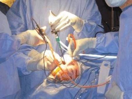
FIGURE 15.1 Four- to six-handed surgical technique. By combining pathways of different length and position, increased space between hands and instruments is provided.
The decision on the pathway(s) to use should be based on the location and characteristics of the target pathology, preferably without preexisting bias for a given approach. By performing preoperative pathway analysis using navigation software, the spectrum of approaches (monoportal or multiportal) offered by various surgical trajectories can be analyzed and optimized for each patient's individual pathology. A combination of approaches can be selected that will avoid endangering critical structures and optimize the surgeon's ability to visualize and access challenging lesions using minimally disruptive technique. Virtual endoscopy can then be performed before surgery to confirm the appropriateness of the approaches and visualize the critical structures that will be encountered.
The techniques that we use for these approaches are transorbital, not transpalpebral. The term transorbital refers to approach vectors that access or traverse the orbit without removing bone of the orbital rim or adjacent structures. The term transpalpebral refers to approaches that employ incisions that partly or completely transect an eyelid. Thus, supraorbital craniotomies can be performed with a transpalpebral incision, though the approach is not transorbital. The SLC incision described herein is a transpalpebral incision, but the approach is strictly transorbital. We have found that removing the bone of the orbital rim and adjacent structures requires more extensive incisions, creates more morbidity, and prolongs the recovery period. Furthermore, we have found from computer analysis, cadaver studies, and clinical experience that the surgical approach vector is typically not improved by excising more bone.
This chapter describes our system of transorbital approaches to the ACF and suggests ways of combining these and other approaches in multiportal technique to maximize the surgeon's ability to visualize and instrument these complex surgical targets with minimal disruption. Our transorbital approaches to the MCF are described in Chapter 34 of this book.
HISTORY
The history of patients presenting with ACF lesions is highly dependent on the type of pathology. When the orbit is involved, pain, diplopia, diminished visual acuity, proptosis, and ptosis are common presenting symptoms. A prior history of ophthalmologic problems and treatment should be noted. If the pathology involves the nasal cavity or sinuses, drainage, epistaxis, and loss of olfaction are common. Headache is a common nonspecific symptom, while decreased facial sensation may indicate involvement of the trigeminal nerve by tumor. Trismus may signify spread of tumor into the pterygomaxillary space or infratemporal fossa.
PHYSICAL EXAMINATION
A complete head and neck and neurologic examination is mandatory, with special attention to the cranial nerves. Regional lymph nodes should be examined for signs of metastatic cancer. For pathology that might encroach on the nasopharynx or nasal cavity, fiberoptic nasopharyngoscopy should be undertaken. The globes and orbits should be examined, and visual acuity should be checked. Asymmetry of orbital volume, position of the globe, size and reactivity of the pupils, and position of the eyelid should be noted. Normal extraocular muscle function should be confirmed. Strong consideration should be given to preoperative evaluation by an ophthalmologist to search for subclinical disease or other contraindications before transorbital surgery is undertaken.
INDICATIONS
Transorbital endoscopic surgery may be indicated for the treatment of pathology involving structures within or adjacent to the orbit. It can also be used as a pathway to more distant structures when it lies in the vector between the pathway entry point and the surgical target. It can be used as a single approach or combined with transnasal, transmaxillary, or supraorbital portals. It may also be used as an adjunct to a traditional craniotomy or subfrontal craniectomy. As part of a multiportal approach, the transorbital pathway can be used for visualization (endoscopy), instrumentation, or both to provide the best possible means of safely manipulating the surgical target.
A more recent application of the transorbital approaches is the surgical treatment of orbital, frontal, sinus, and intracranial manifestations of sinogenic infections such as epidural abscesses (see Suggested Readings). The indications of these procedures are expanding with further work in the laboratory and improvements in surgical technology.
CONTRAINDICATIONS
The two absolute contraindications to transorbital endoscopic surgery are found in trauma patients: a ruptured globe or hyphema (blood in the anterior chamber of the eye). Relative contraindications to undertaking these approaches are noted below, and these patients should be managed in consultation with an ophthalmologist.
· Intraocular surgery within the last 6 months (e.g., cataract, retinal, glaucoma, or corneal transplantation surgery); consult with the ophthalmic surgeon before proceeding.
· Orbital infection (risk of posterior spread of the infection).
· Severe orbital inflammation or congestion (decreased space for retraction within the orbit may lead to increased pressure on the globe and optic nerve).
· Patients with diminished corneal sensation from previous laser corneal surgery or other cause may be at increased risk for postoperative complications.
· Other orbital pathology that creates a mass effect or alters anatomy in a manner that could obstruct endoscopic access.
Glaucoma and dry eye do not appear to be contraindications to transorbital pathways, though increased care should be given to lubrication of the ocular surface during and after surgery. Consultation with the ophthalmologist is an important aspect in the care of patients with these conditions.
PREOPERATIVE PLANNING
Full radiographic imaging should be obtained to completely evaluate and characterize the pathology in question including its exact location, extent, and vascularity when indicated. Imaging for intraoperative surgical navigation should be undertaken and should include computed tomography (CT) and magnetic resonance imaging (MRI) so that fusion guidance can be obtained as needed. Preoperative analysis of the navigation images on a computer planning station is helpful to investigate the ideal surgical approach or approaches to the target. Depending on the software, this may include highlighting the pathology, analyzing the target three-dimensionally, and performing a vector analysis of the possible approaches with virtual endoscopy.
Among the factors to consider in choosing a surgical approach are the following:
· Critical structures involved with or adjacent to the pathology.
· Adequacy of exposure for passage of instruments.
· Ability to visualize the pathology from the approach angle.
· Absence of impediments to visualization or manipulation of the target.
· Ability to instrument the target from the approach angle.
· Morbidity caused by creation of the pathway.
· Ability to reconstruct defects created by the pathway or target manipulation.
· The pathway should not cross critical neurovascular structures in a manner that can expose them to pressure or other trauma.
· A shorter pathway may allow increased ease of target visualization and manipulation.
· Adequacy of exposure for four- to six-handed (four to six function) surgery as needed.
· Experience of the surgical team.
· Patient preference.
For planning surgical approaches, we consider the number of surgical functions that will be required. These functions typically involve illumination/visualization (endoscopy), aspiration, irrigation, ablation, retraction, and manipulation. The literature on skull base surgery typically describes the need for “four-handed” surgery, that is, the ability for two surgeons to work together during the procedure. While this is a critical concept, there are times when more than four “hands” may be desirable to carry out a task. To address this, developments in surgical technology have provided instruments that can carry out multiple tasks. Thus, a single instrument such as a microdebrider may provide aspiration, irrigation, and tissue ablation, providing three functions in one “hand.” There are times when by using a multifunctional instrument, a single surgeon can perform five or more functions using two hands (illumination and visualization in one hand, with irrigation, bone aspiration, and suction in the other). We emphasize, therefore, the number of surgical functions required when planning the surgical pathway.
When possible, an approach that is coplanar with the target is beneficial (Fig. 15.2). A coplanar approach consists of an entry portal, dissection pathway, and surgical target that all lie within a single axial, sagittal, or coronal plane, such as one that might be visualized on a single CT image. For example, if a lesion is located immediately superior to the planum sphenoidale, a surgical approach that allows dissection along the ACF through a PC portal will simplify the procedure by allowing for the creation of an epidural pathway along the bone of the ACF without the need to use angled endoscopy or instrumentation.
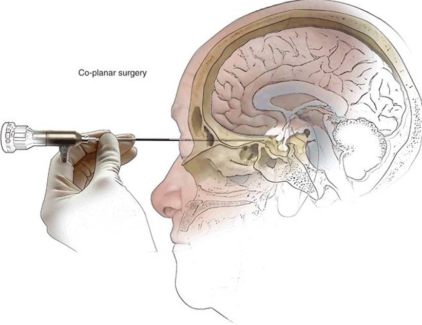
FIGURE 15.2 Coplanar surgery. The approach and target are on the same surgical plane.
Our choice of surgical portal is based on a division of the orbit into four quadrants, each of which has a specific entry portal (Fig. 15.3A). These are the superior, medial, inferior, and lateral quadrants. The approach is chosen based on the quadrant that is directly involved with the pathology or is transgressed by the optimal pathway to the surgical target (Fig. 15.3C). A schematic of the surgical portals and skull base regions of access is demonstrated in Figure 15.4. The approaches to the lateral and inferior quadrants are used primarily for access to the MCF and adjacent structures––these are described in Chapter 34on transorbital approaches to the MCF.
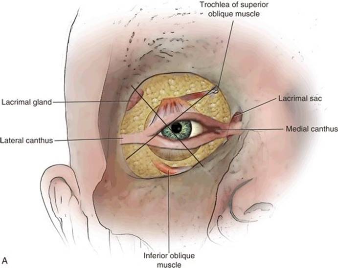


FIGURE 15.3 A. The four quadrants of the orbit. The surgical approach is chosen by the quadrant of the orbit that is most involved by the underlying pathology. The superior quadrant is accessed by the superior lid approach (SLC) approach; the medial quadrant is entered through a precaruncular (PC) incision. The inferior quadrant approach is made through an inferior transconjunctival (ITC) dissection, and the lateral quadrant is reached through a lateral retrocanthal (LRC) gateway; these are discussed in Chapter 37. B. Surgical anatomy of the orbit. The optic nerve is located in the medial orbital wall, medial and slightly superior to the junction of the superior and inferior fissures. Note that the anterior and posterior ethmoid arteries are coplanar with the optic nerve and demonstrate the level of the skull base. Dotted lines/arrows demonstrate approximate borders between the superior lid crease (SLC), precaruncular (PC), and inferior transconjunctival (ITC) approaches. 1, medial wall; 2, orbital roof; 3, lateral wall; 4, floor. C. Left. Green lines demonstrate approximate region that is accessed in SLC approach. Right. Approximate region that is accessed in PC approach (note the level of the ACF that is visible through the dehiscent lamina). Part or all of this bone can be removed as a pathway to adjacent targets.
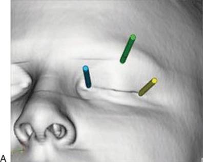
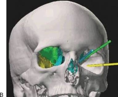
FIGURE 15.4 Transorbital neuroendoscopic portals. CT scan with soft tissue (A) and bone density (B). A. Entry points of PC (blue), SLC (green), and LRC (yellow approaches).B. Soft tissue removed, demonstrating approach angles and approximate areas of the orbit accessed by each approach. Note that there is significant overlap in the area that can be accessed by each approach. Endoscopes are illustrated to scale at 4 mm.
The superior quadrant is bounded laterally by the superior orbital fissure and its anterior extension to the orbital rim and medially by the anterior and posterior ethmoid arteries (Fig. 15.3B and C). The entry portal is created through a transcutaneous superior lid crease approach. The trochlea is typically the medial extent and the lacrimal gland the lateral extent of this portal, though the trochlea and the periosteum to which it attaches can be lifted from the bone of the orbit to extend the approach if needed. Extension of this transcutaneous approach into a lateral transconjunctival approach requires a lateral canthotomy and superior cantholysis, though this is rarely necessary.
The medial quadrant is bounded superiorly by the anterior and posterior ethmoid arteries at the skull base and ends inferiorly at the orbital floor (Fig. 15.3B and C). The portal of entry is through a precarunculartransconjunctival incision. The superior limit of this incision is the medial horn of the levator aponeurosis and muscle. There is no inferior anatomic limit as it can be continued inferolaterally into a pre-septal or ITC incision through the lower eyelid conjunctiva.
The surgical approach is chosen by orbital quadrant as noted above. In addition, the surgeon must decide whether an extracranial (subcranial) or intracranial approach will be used. If an intracranial pathway is to be used, an extradural or intradural dissection or combination thereof must be chosen. In addition, the plan must include whether the target will be manipulated by monoportal or multiportal technique and whether the approach will be ipsilateral or contralateral. Options for reconstruction should be planned before the operation begins.
The patient should be engaged in the choice of surgical pathway(s). The various possible surgical approaches should be thoroughly discussed, and detailed informed consent should be obtained. It is also important during preoperative planning to consider any adjuvant therapies such as tumor embolization that should be coordinated with other members of the team. Anticoagulant medications should be stopped as indicated.
SURGICAL TECHNIQUE
The procedure begins with the administration of a general anesthetic. If indicated, a lumbar drain is placed. If intrathecal fluorescein is going to be used for localization of a cerebrospinal fluid (CSF) leak, this should be given as early as possible to allow diffusion of the dye. The table is then rotated 180 degrees, with the anesthesia equipment and anesthesiologist at the foot of the table as demonstrated in Figure 15.5. The patient's head is placed in pins or on a circular gel headrest as preferred by the surgical team––fixation prevents alteration of the angulation of the head; changing this position may be beneficial at different stages of the operation. The head is placed in 15 degrees of retroflexion to allow the frontal lobes to relax away from the ACF if dissection is planned in this region. Less than 1 mL of 1% lidocaine with epinephrine 1:100,000 is then injected into the periorbital operative site. If a multiportal approach with transnasal portals is used, local anesthetic is infiltrated intranasally as well. The operating table is inclined 10 to 15 degrees to aid in hemostasis.
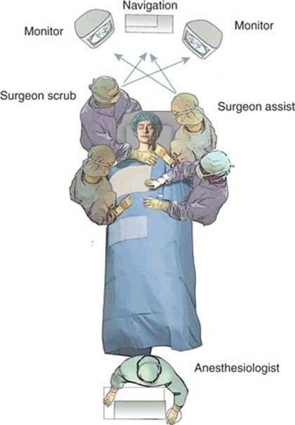
FIGURE 15.5 Operating room schematic. Monitors and navigation suspended from ceiling, angled for ergonomic viewing by all participants.
The navigation mask is placed, and the system is registered and registration accuracy confirmed. The surgical pathway vector from the planned portal to target is then analyzed on the patient using the navigation system, and the final choice of entry portal(s) is made (Fig. 15.6). The patient's face is prepared and draped in the sterile fashion preferred by the surgical team. Ophthalmic Betadine should be used instead of regular Betadine to avoid irritation of the eye. Alcohol products and other caustic prepping agents should not be used around the eyes.
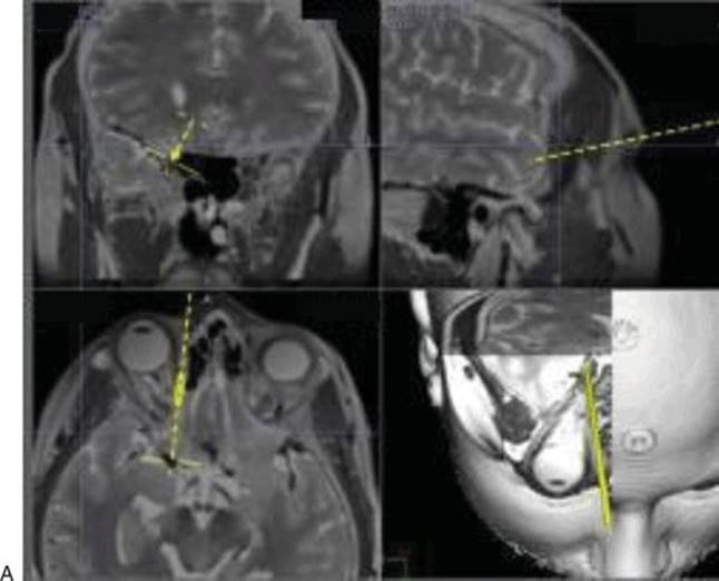
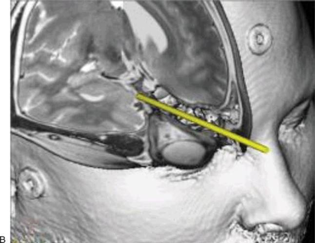
FIGURE 15.6 Preoperative surgical planning, PC approach. A. Navigation to right lateral cavernous sinus. B. Enlarged navigation planning image demonstrating pathway.
There are four primary transorbital endoscopic approaches (superior, medial, inferior, and lateral). The lateral and inferior approaches are used primarily to access the MCF and are described in Chapter 37. The approaches we use most commonly to the ACF are the superior and medial, as described below.
Medial Quadrant: Precaruncular Approach
The PC approach through the medial quadrant provides effective access above and/or below the ACF to structures in the central corridor and medial orbit roof. This includes the cavernous sinus, cavernous carotid arteries, and optic nerve (Figs. 15.7 and 15.8).
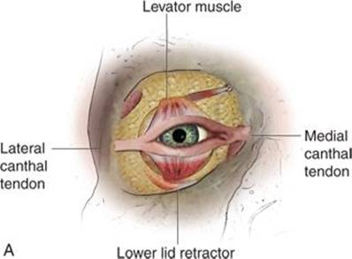
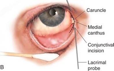

FIGURE 15.7 PC approach. A. Primary support system of eyelids: medial canthal tendon, lateral canthal tendon, levator aponeurosis and muscle, lower lid retractor. B. Lacrimal probes in place, incision between apex of medial canthus and caruncle. C. Dissection follows posterior limb of medial canthal tendon to lamina papyracea.

FIGURE 15.8 Photo of PC approach.
The approach is begun by placing a lubricated protector over the cornea. Probes are then placed in the lacrimal canaliculi to prevent inadvertent damage. These may be taped to the adjacent skin to aid in retraction. An incision is then made with a small scissors through the conjunctiva at the apex of the medial canthus, between the caruncle and skin. The incision is extended superiorly and inferiorly in the conjunctiva. The avascular plane is entered deep to the posterior limb of the medial canthal tendon, and that tendon is followed to the posterior lacrimal crest. The periorbita is incised vertically posterior to this structure and lifted laterally off the bone of the medial orbit (lamina papyracea). Dissection is continued posteriorly (using a suction elevator under visualization with a 4-mm 0-degree endoscope) between the periorbita and medial orbital wall. A malleable brain retractor is used to gently displace the orbital contents during the dissection. The anterior and posterior ethmoid arteries are cauterized with a bipolar cautery before sharply transecting them. Before cauterizing the posterior ethmoid artery, navigation is used to confirm that a safe distance is maintained from the optic nerve. The optic nerve is then visualized at the orbital apex; the nerve is located in the most posterior portion of the medial orbital wall, at the approximate level of the ethmoid arteries. The skull base lies immediately superior to the ethmoid arteries and can be seen as more dense, opaque bone than that of the lamina papyracea. At this point, the dissection proceeds along the trajectory of the planned pathway toward the target.
If the dissection is to proceed intracranially, the craniectomy is created in the superior medial orbit and medial roof. The appropriate site is confirmed with navigation. The bone can be removed with a diamond burr or an ultrasonic bone aspirator. We prefer the latter, as the instrument does not skip off the bone and appears to cause less damage to the adjacent dura if the dura is contacted. Furthermore, the bone aspirator both irrigates and aspirates through a single instrument, simplifying the maneuver.
Intracranial dissection can then proceed intra- or extradurally until the target is reached in accordance with the surgical plan.
If the optic nerve is to be decompressed, we prefer to do this by infracturing the medial and inferior aspects of the bone canal away from the nerve, using a fine periosteal elevator. This avoids the heat transmission and risk of injury that can occur from using a drill. If the bone of the canal is to be infractured, an ultrasonic bone aspirator or drill with a diamond burr may be used. The decompression is undertaken over the entire canal to the dura anterior and to the optic chiasm. The intracranial course of the nerve can be followed to the chiasm as needed. Figure 15.6 demonstrates a typical surgical navigation image of this approach.
Medial Quadrant: Precaruncular Approach to Contralateral ACF
At times, a contralateral approach to the target may be beneficial (Fig. 15.9). Pathology on the lateral wall of the sphenoid sinus/medial wall of the MCF can be difficult to visualize from an anterior approach without heavily angulated endoscopy, and instrumentation under these conditions can be challenging. Occasionally, there may be unrelated pathology that blocks an ipsilateral transorbital or transnasal approach that can be circumscribed by a contralateral approach. An example of this is shown in Figure 15.10, a patient referred with a persistent right ACF CSF leak after craniectomy and resection of a meningioma. The patient also had a massive osteoma of the right orbital roof and wall, which he declined to have resected. A contralateral (left) PC approach to the right supraorbital skull base was undertaken to repair the dura and skull base.
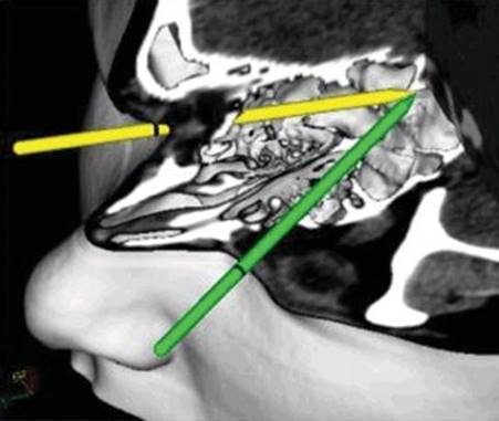
FIGURE 15.9 Schematic, ipsilateral (yellow) versus contralateral (green) PC approach to lateral aspect right MCF/sphenoid. This navigation analysis demonstrates the improved angulation to the lateral opticocarotid recess achieved by a contralateral PC approach.
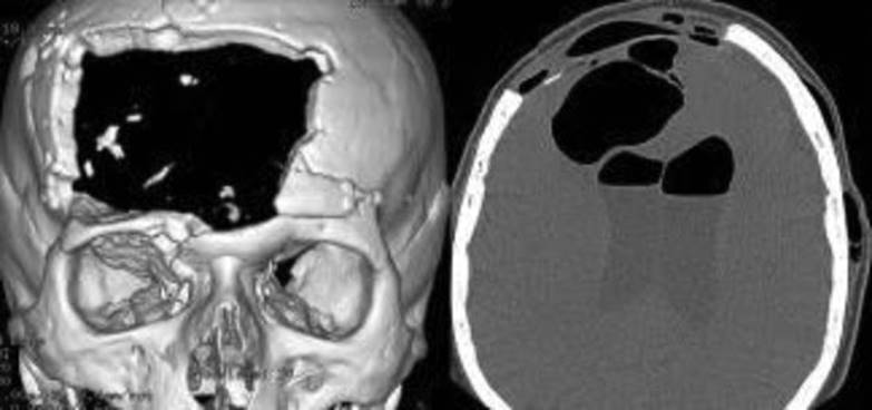
FIGURE 15.10 Patient with a right ACF CSF leak after resection of a large meningioma. Osteoma of right medial and superior orbit blocked ipsilateral PC approach; contralateral (left) PC intracranial extradural approach was used to repair the leak.
The contralateral PC approach is undertaken with the same technique as an ipsilateral PC approach. For a subcranial target, the vector from the portal to target dictates the region of the lamina papyracea that is removed. The posterior ethmoid cells are removed on the side of the approach using a microdebrider. The midline is then crossed through the perpendicular plate of the ethmoid and sphenoid rostrum. The posterior ethmoid and sphenoid sinuses are opened on the side of the pathology as dictated by the approach vector. For an intracranial target, the initial dissection is undertaken with a subcranial or intracranial dissection as directed by the approach vector. The craniectomy is created in the indicated region of the skull base, and intracranial dissection is then continued to the target zone.
Superior Quadrant: Superior Eyelid Crease Approach
Pathology involving the superior orbit, frontal sinus, supraorbital ACF, and posterior central ACF can be accessed through an SLC approach (Fig. 15.11). This approach can also be used for access to the olfactory region of the anterior interorbital skull base if, for example, the surgeon plans to resect the olfactory bulb and nerve en bloc with an esthesioneuroblastoma from an intradural approach. The incision is identical to an upper blepharoplasty, though it may be made in a more superior crease. The incision is tailored to the location of the portal as determined by preoperative path-to-target analysis. As this is a transcutaneous approach, a corneal protector is not used; a temporary tarsorrhaphy suture is placed instead. The incision is made through the skin and preseptal orbicularis oculi muscle. Deep to this, the orbital septum is identified, through which the prelevator fat can be seen. The septum and adipose tissue are not disturbed. Dissection is continued immediately posterior to the orbicularis muscle toward the superior orbital rim. When the orbital rim is identified, the periosteum is incised along its anterior border, sparing the supratrochlear and supraorbital neurovascular pedicles. A plane is created between the periorbita and the roof of the orbit using a periosteal elevator. The periorbita is dissected away from the roof of the orbit under endoscopic and navigation guidance in the manner described above. The optic nerve is identified posteriorly as indicated, and the ethmoid arteries can be visualized medially. The dissection proceeds as far laterally as necessary. The craniectomy is then performed at the point where the portal-to-target pathway meets the skull base, using an ultrasonic bone aspirator or diamond burr. The dura is then lifted off the skull base, and the intracranial dissection is continued as appropriate. A navigation image of the approach is demonstrated in Figure 15.12.
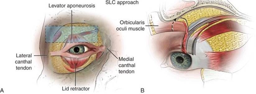
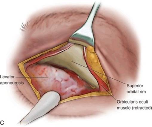
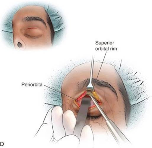
FIGURE 15.11 SLC approach. A. Region of approach highlighted. B. Oblique view, upper eyelid; dotted line demonstrates SLC dissection deep to orbicularis muscle to orbital rim, where subperiosteal plane is entered. C. Superior orbital rim exposed. D. SLC approach, retractor in place.

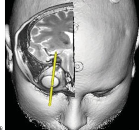
FIGURE 15.12 A. Surgical planning view, SLC approach. B. Enlargement showing anatomy of SLC approach.
The SLC approach can also be used to access the frontal sinus (Fig. 15.13). This is an excellent pathway for pathology in the lateral sinus that may be difficult to reach transnasally (e.g., obstructing osteoma). It is also applicable for treatment of orbital abscesses that stem from frontal sinusitis and which can extend to create epidural abscesses. The intersinus septum can be opened with this technique, allowing frontal sinus drainage through the contralateral frontal recess. By using an SLC approach, the orbital abscess can be drained, the frontal sinus abscess can be treated, and the epidural space can be reached for exploration or drainage of purulence. The frontal sinus is entered after the SLC approach has been completed. Navigation is used to choose the appropriate vector to the target, and the region for opening into the floor of the sinus is marked. The bone is then removed as described above. A 0-degree endoscope is used. To inspect the frontal recess from its superior aspect looking inferiorly, a 30-degree endoscope will be beneficial.
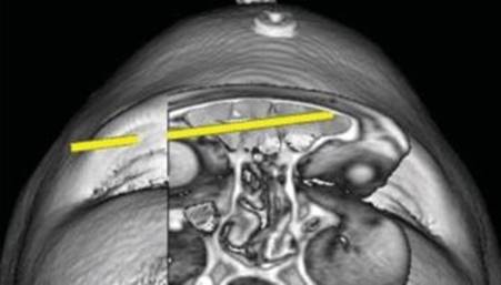
FIGURE 15.13 SLC approach to frontal sinus. The intersinus septum can be opened to allow drainage of an abscess through the contralateral frontal recess.
Reconstruction
Reconstruction is performed as needed, in a manner tailored to the pathology and type of craniectomy. Reconstruction of the medial wall is not necessary if the defect is small, there is no enophthalmos, and the periorbita is intact so that the medial rectus muscle will not catch on bone edges. If reconstruction is indicated, this is performed by placing a thin orbital fracture implant across the defect, sandwiched between the periorbita and bone superior and inferior to the defect. The navigation probe is then scanned across the reconstruction to assure that it conforms to the position of the normal medial orbital bone visible on the preoperative CT scan.
The same principles hold true for reconstruction of the bone of the roof of the orbit. If a dural defect is present, it should be repaired. Typically, the superior craniectomy does not need reconstruction. If none is performed, the globe may be noted to pulsate somewhat with the heartbeat for 1 to 2 weeks postoperatively. This generally resolves spontaneously. If reconstruction of the bone is deemed necessary, it can be performed with the technique used for the medial orbit. A short screw can be placed posterior to the orbital rim to hold the implant in place.
The SLC incision is loosely closed with 5-0 absorbing sutures through the orbicularis and 6-0 sutures placed through the skin so that fluid can drain, but the skin is approximated. We do not generally close the conjunctival incisions.
POSTOPERATIVE MANAGEMENT
The postoperative management for patients who have undergone transorbital endoscopic procedures is dictated by the target pathology being treated. The morbidity occurring from creation and reconstruction of the pathway is similar to or less than that experienced by a patient undergoing a repair of a single-wall orbital fracture. At times, patients are overly eager to be discharged since they awaken from surgery without having had a bifrontal craniotomy or procedure with a large incision and extensive bone resection. They may have to be reminded they have undergone treatment of a major ACF or MCF lesion. For those who have had a significant component of intracranial surgery and for all who have had intradural surgery, a postoperative noncontrast CT scan is obtained, and the patient is maintained in the intensive care unit overnight.
If a lumbar drain was indicated for treatment of the target pathology, this is maintained postoperatively as it would for other approaches. Pain is usually managed with oral medications. No dressings are applied; for transconjunctival incisions, ocular lubricants are used liberally for 1 week after surgery, and antibiotic ointment is applied to cutaneous incisions for 48 hours. Patients are given oral antibiotics for 5 to 7 days. The patient is seen as an outpatient in clinic at postoperative day 7, 14, and 28 and then followed as indicated for the underlying pathology. For patients who travel long distances for their surgery, postoperative care is turned over to their local surgeon after day 14. If a patient was seen by an ophthalmologist preoperatively, a postoperative visit is requested approximately 1 month after surgery, unless there is an indication for earlier evaluation.
COMPLICATIONS
To date, we have had no significant complications from the SLC or PC approaches to the ACF. The absence of large incisions used with typical craniotomies and subcranial approaches should not lead the patient or surgeon to think that these are minor procedures, however. Patients must be counseled on the risks inherent with operating on or near the eyes and critical neurovascular structures. Great care must be taken to protect the cornea and globe from injury, and the nursing staff should be informed of the significant extent of these surgical procedures to aid in appropriate postoperative monitoring.
RESULTS
We have published evaluations of our outcomes and experience using this system of transorbital approaches to orbital and skull base lesions (see Suggested Reading). We have found these techniques to be highly effective, with an excellent safety record. A primary concern with transorbital approaches is the potential exertion of pressure on the globe and optic nerve during procedures that may last 4 hours or more. While we have not had any known cases of diminished visual acuity after a transorbital endoscopic approach, we take great care to protect the ocular surface during surgery and regularly check the pupils for enlargement or irregularity that might suggest elevated intraocular pressure. If this occurs, instruments are removed from the orbit until the pupil returns to normal. The safety of these procedures was demonstrated in our evaluation of over 100 cases in which no significant complications occurred.
Patients have been pleased with these procedures. The pain has tended to be less than they expected, the recovery period is brief, and there have been no visible scars or cosmetic complaints.
PEARLS
· Obtain complete imaging preoperatively including an MRI and CT scan under the navigation protocol. Consider angiography with embolization for vascular lesions. Consult and collaborate with an ophthalmologist, especially for patients with underlying ocular pathology.
· Study the lesion preoperatively including 360-degree analysis of adjacent anatomy. Evaluate all possible angles of approach to the target, and consider multiple pathways as needed. Individualize the approach for each target, optimizing the ability to visualize and instrument the lesion.
· Work in a team consisting of a neurosurgeon and otolaryngologist, with close collaboration of specialists in other disciplines including ophthalmology, radiation oncology, and neuroradiology.
PITFALLS
· Failure to perform cadaver dissections may make it especially challenging when trying to translate these surgical concepts to patients.
· Performing these procedures without surgical navigation is potentially dangerous, particularly when performing surgery on or near the optic nerve or intracranially. If you wander in this region, the consequences are often neurologically serious.
· Check the size and shape of the pupil every 20 to 30 minutes. If the pupil begins to dilate, remove instruments from the orbit for a short time to allow the pupil to return to normal.
· Protect and hydrate the cornea to prevent abrasions or drying.
· Careful selection of the patient is essential. If you cannot reach the pathology with navigation, it is best to terminate the procedure and attempt an open approach.
INSTRUMENTS TO HAVE AVAILABLE
· Complete endoscopic skull base instrument set
· Oculoplastic set with retractors, corneal protectors, lacrimal dilator, and probes
· High-quality endoscopes (0 and 30 degree) with high-resolution monitors, preferably suspended from ceiling in ergonomic positions
· Endoscope irrigation system
· Endoscopic microdebrider
· Drill with diamond burr or, preferably, ultrasonic bone aspirator (Sonopet)
· Radiofrequency soft tissue aspirator (Coblator)
· Surgical navigation system with vector analysis and lesion highlighting (segmentation) software
· Intraoperative CT scanner (useful but not critical)
SUGGESTED READING
Ciporen JN, Moe KS, Lopez S, et al. Multi-portal endoscopic approaches to the central skull base: a cadaveric study. World Neurosurg 2010;73(6):705–712.
Moe KS, Bergeron CM, Ellenbogen RG. Transorbital neuroendoscopic surgery. Neurosurgery 2010;67(3):16–28.
Moe KS, Kim LJ, Bergeron CM. Transorbital endoscopic repair of complex cerebrospinal fluid leaks. Laryngoscope 2011;121:13–30.
Lim J, Sardesai M, Ferreira M, et al. Transorbital neuroendoscopic management of sinogenic complications involving the frontal sinus, orbit and anterior cranial fossa. J Neurol Surg B Skull Base 2012;73(6):394–400.
Moe KS, Balakrishnan K. Transorbital endoscopic surgery of the skull base and sinuses. In: Simmen D, Jones N, eds. Manual of Endoscopic Sinus Surgery, 2nd Ed. New York, NY: Thieme, 2013.
Balakrishnan K, Moe KS. Transorbital endoscopic surgery of the skull base and sinuses. In Simmen D, Jones N, eds. Manual of Endoscopic Sinus Surgery, 2nd Ed. Thieme, Stuttgart. 2014:424–431.